Shock

Published on 06/06/2015 by admin

Filed under Pediatrics

Last modified 22/04/2025

Print this page

rate 1 star rate 2 star rate 3 star rate 4 star rate 5 star
Your rating: none, Average: 0 (0 votes)

This article have been viewed 1713 times

2 Shock

Shock is an acute clinical syndrome of circulatory dysfunction in which there is failure to deliver sufficient oxygen and substrate to meet metabolic demand. All practitioners who care for children must understand and identify shock promptly to initiate an effective treatment plan. This, in turn, can help prevent the progression and poor outcomes that characterize the natural clinical course of shock. The goal is to prevent end-organ damage; failure of multiple organ systems; and, ultimately, death.

Etiology and Pathogenesis

Normal circulatory function is maintained by the interplay between the heart and blood flow with the purpose of delivering oxygen and nutrients to the tissues. Cardiac output is calculated by multiplying the stroke volume (volume of blood ejected by the left ventricle in a single beat) by the heart rate (ejection cycles per minute). Stroke volume is dependent on the filling volume of the ventricle (preload), resistance against which the heart is pumping blood (afterload), and myocardial contractility. During childhood, the heart rate is faster, and the stroke volume is smaller than during adulthood. In children, increasing the heart rate is the primary means to increase the cardiac output.

Shock develops as the result of conditions that cause decreased intravascular volume, abnormal distribution of intravascular volume, or impaired cardiovascular function. Children effectively compensate for circulatory insufficiency by increasing their heart rate, systemic vascular resistance (SVR), and venous tone. Children can therefore maintain normal blood pressures despite significantly compromised tissue perfusion. Thus, in pediatric patients, it is especially important to recognize that hypotension is not part of the definition of shock.

The clinical manifestations of shock can be directly related to the abnormalities seen on the tissue, cellular, and biochemical levels. Microcirculatory dysfunction; tissue ischemia; and release of biochemical, vasoactive, and inflammatory mediators are all part of the spectrum of pathophysiologic aberrations seen in shock. Poor perfusion of vital organs results in impaired function. For example, inadequate perfusion of the brain and kidneys results in depressed mental status and decreased urine output, respectively. As poorly perfused cells switch to anaerobic metabolism to generate energy, lactic acid accumulates resulting in a metabolic acidosis that further interferes with cell function. Hypoperfusion also initiates inflammatory events, such as the activation of neutrophils and release of cytokines, that cause cell damage and microischemia.

The prevalence of causes of shock varies by patient age, as well as region of the world. Hypovolemic shock from diarrheal illness is the leading cause of pediatric mortality worldwide, but is very rare in the United States. Congenital lesions (including heart disease) and complications of prematurity are most common in neonates and infants. Malignant neoplasms (for whom infectious complications are prevalent), infectious causes, and unintentional injuries are more common in older children and young adolescents. Injury, homicide, and suicide become more prevalent in older adolescents.

Classification of Shock

Septic Shock

Sepsis is defined as the presence of the systemic inflammatory response syndrome (SIRS) caused by a presumed or confirmed infection (Box 2-1). Sepsis may occur because of bacterial, viral, fungal, or parasitic infections. Septic shock is defined as sepsis and cardiovascular dysfunction. Classifying septic shock may be difficult because of the developmental variability in physiologic response to sepsis. A clinical picture consistent with hypovolemic, distributive, or cardiogenic shock may be present in a child with sepsis. Additionally, studies have demonstrated that the cardiovascular pathophysiology of children with sepsis can evolve over time, and the adjustment of hemodynamic therapy is commonly necessary.

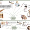
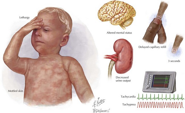
Clinical Manifestations and Evaluation

Shock remains a clinical diagnosis (Figure 2-1). Early recognition of the clinical signs of shock (including familiarity with normal ranges for vital signs by age; see Chapter 1) should lead to directed management. An accurate history should be obtained from the family and, if possible, the child, simultaneously with treatment initiation.

A history of fluid loss, as with a gastrointestinal bleed, gastroenteritis, or diabetic ketoacidosis, is consistent with hypovolemic shock. A detailed trauma history is useful because an injured child may have hypovolemic shock from hemorrhage (i.e., with blunt abdominal trauma), neurogenic shock with spinal cord injury, or obstructive shock from tension pneumothorax. A child who has had fever or is immunocompromised may have features consistent with septic shock. Exposure to an allergen, such as a food or an insect bite, could suggest distributive shock caused by anaphylaxis. A history of ingestion or medications should always be included when speaking to the family because shock may be attributable to toxin exposure. Patients with underlying heart disease may present in cardiogenic shock. Patients with a history of adrenal insufficiency (i.e., chronic steroid therapy, congenital adrenal hyperplasia, or hypopituitarism) can present with adrenal crisis and shock.

A complete physical examination should be performed, including vital signs and pulse oximetry. When a child presents in shock, it is sometimes difficult to obtain an accurate weight, which can be essential for determining fluid requirements and medication doses. If the patient’s weight cannot be measured, one may be estimated using a length-based tape system (e.g., the Broselow tape) or the child’s age.

Children in shock tend to be tachypneic, as well as tachycardic. Blood pressure should be monitored closely. Remember, children with shock may have normal blood pressures. Narrow pulse pressure may occur as a result of a compensatory increase in SVR, as in hypovolemic or cardiogenic shock. Widening of the pulse pressure can be seen as the result of decreased SVR, as can occur with distributive shock. The child’s temperature should also be measured because fever—or in young infants, hypothermia—may suggest septic shock.

When first examining an ill child, one should do a rapid assessment of mental status. Change in the level of consciousness of a child may indicate decreased cerebral oxygenation or perfusion. Signs of diminished perfusion to the brain include confusion, irritability, lethargy, and agitation.

Examining the child’s skin is another way to assess perfusion and the degree of shock. A child with normal cardiorespiratory function should have warm and pink nailbeds, mucous membranes, palms, and soles. As shock progresses and poor perfusion develops, the skin may become cool, pale, or mottled. Capillary refill, although limited by clinician variability as well as ambient temperature and the child’s body temperature, can help to evaluate children in shock. Light pressure is applied to blanch the fingernail bed. The pressure is released, and the amount of time until color returns is measured. Normal is less than 2 seconds; volume depletion or poor perfusion can increase this time to greater than 3 seconds.

The evaluation of a child with poor perfusion and shock should always include an assessment of pulses. This includes the rate, strength, and regularity of the central and peripheral pulses. In healthy children, the carotid, brachial, radial, femoral, dorsalis pedis, and posterior tibial pulses are readily palpable. A rapid pulse is a nonspecific clinical sign of distress. An irregular pulse is a warning of cardiac dysrhythmia. A weak pulse raises the concern for shock and a severe hypovolemic state. An absence of central pulses indicates ineffective or absent cardiac contractions and signifies the need for immediate resuscitative action.

After the initial evaluation of airway, breathing, and circulation (the ABCs), a complete physical examination can help elucidate the type of shock. For example, central cyanosis, a gallop rhythm, crackles on lung examination, hepatomegaly, or heart murmur may indicate an underlying cardiac condition. Children with stridor, wheeze, urticaria, or edema may have anaphylactic shock. Purpura or petechiae can be seen in children with septic shock. Bruises and abrasions can be seen with traumatic injury and may give a clue to underlying hemorrhagic shock.

Management

General Principles

Early recognition of compensated shock is critical to ensuring appropriate and expedient therapy. Initial therapy of shock is universal, regardless of the cause of the shock state, with the goals of optimizing blood oxygen content, improving cardiac output, reducing oxygen demand, and correcting metabolic abnormalities (Figure 2-2). General principles of resuscitation should be applied immediately on presentation to medical care (see Chapter 1). Ultimately, after initial management has commenced, correction of the underlying cause is essential (e.g., stopping blood loss in hemorrhagic shock, antibiotics for shock caused by bacterial infection).
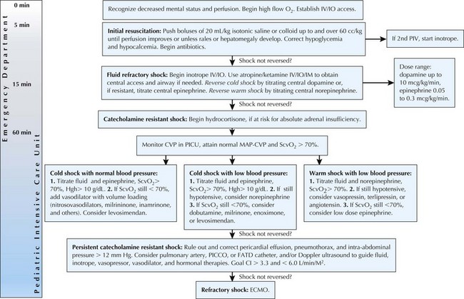
image

Figure 2-2 Algorithm for management of pediatric septic shock.

From Brierley J, Carcillo J, Choong K, et al: 2007 American College of Critical Care Medicine clinical practice parameters for hemodynamic support of pediatric and neonatal septic shock. Crit Care Med 37(2):666-688, 2009.

Immediate attention to the ABCs is mandatory. Maintenance of a patent airway with positioning or endotracheal intubation should be performed immediately if airway compromise is present. Hypoxemia should be corrected without delay; all patients with compromised perfusion should receive supplemental oxygen at 100% FiO2 (fraction of inspired oxygen). Insufficient respiratory effort should be addressed with positive-pressure ventilation.

Imminently life-threatening causes of shock should be identified and corrected. For example, a child with upper airway obstruction from anaphylaxis should receive epinephrine. If a child has severe respiratory distress, asymmetric breath sounds, and poor perfusion, a tension pneumothorax might need to be decompressed.

Vascular access is indicated in all cases. If possible, large-bore intravenous (IV) catheters should be inserted in peripheral veins. If an IV line is unable to be placed promptly, intraosseous (IO) cannulation should be performed. IV fluid boluses of 20 mL/kg of isotonic saline should be given rapidly and repeated as needed with reassessment occurring simultaneously. Rapid fluid administration should be actively performed using either a pressure bag or a push–pull system rather than using passive gravity flow for administration. IV fluids should be given with care in cardiogenic shock, so as to not worsen associated pulmonary edema.

Adequate hemoglobin is essential for optimal oxygen carrying capacity. Thus, in cases of hemorrhagic shock, O-negative packed red blood cells should be rapidly administered early in the resuscitation. Even in cases of nontraumatic shock, children who have cyanotic heart disease or neonates may require less fluid and higher hematocrit percentages to ensure adequate oxygen carrying capacity. Life-threatening metabolic abnormalities should be identified and corrected early. Hypoglycemia should be treated with 0.5 to 1 g/kg of IV dextrose. Hypocalcemia (especially decreased ionized calcium) is common in septic shock and can occur as acidosis resolves; it should be corrected with either calcium gluconate or calcium chloride.

Circulatory Support

Depending on the cause, patients in shock may require large volumes of fluid as well as vasoactive medications. Clinical studies of septic shock in children have demonstrated an association between higher volumes of fluid administration and survival. Current guidelines for septic shock recommend up to and over 60 mL/kg in the first 15 to 60 minutes. Every hour that goes by without implementation of this therapy is associated with a 1.5-fold increase in mortality. A retrospective chart review of 90 children with septic shock showed that those who received less than 20 mL/kg of fluid within the first hour had a mortality rate of 73%. Early fluid resuscitation was associated with a threefold reduction in the odds of death.

Vasoactive agents help improve cardiac output through their effects on myocardial contractility, heart rate, and vascular tone. These drugs target at least three types of receptors. The β1-receptors mediate inotropic (contractility), chronotropic (rate), and dromotropic (increased conduction velocity) activity. The β2-receptors mediate vasodilatation and smooth muscle relaxation in blood vessels and bronchial tree. The α-receptors mediate arteriole constriction systemically and bronchial muscle constriction. The dopaminergic receptors mediate smooth muscle relaxation and increase renal blood flow and sodium excretion.

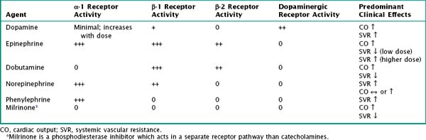
Table 2-1 outlines commonly used vasoactive agents, their targeted receptors, and their hemodynamic effects. Data are varied on the choice of initial agent; current recommendations from the American College of Critical Care Medicine state that dopamine, epinephrine, or norepinephrine may be appropriate first-line therapy for septic shock, provided they are administered through a central venous catheter. Dopamine or low-dose epinephrine may be given via a peripheral vein while central venous access is obtained.

Special Circumstances: Shock in Neonates

Neonatal physiology poses specific challenges regarding the management of shock. As mentioned previously, optimal oxygen-carrying capacity may demand a higher hematocrit in a newborn. Hypoxemia may occur more readily because of the presence of smaller and fewer alveoli, absent collateral channels of ventilation, and poor chest wall compliance; uncorrected hypoxemia may result in bradycardia in neonates. Additionally, myocardial performance is more drastically affected by acidosis and hypocalcemia in neonates. Prompt correction of hypoxemia, acidosis, and hypocalcemia is essential. Neonates are also more prone to hypoglycemia, which should be looked for and treated appropriately.

When faced with a neonate with shock, early consideration should be given to ductal-dependent congenital heart disease. Lesions marked by ductal-dependent systemic blood flow, such as aortic stenosis, hypoplastic left heart syndrome, coarctation of the aorta, and interrupted aortic arch, may present as shock in the neonatal period (see Chapter 44). Infants with these lesions depend on blood flow from the pulmonary artery across the ductus arteriosus into the aorta for perfusion of all or part of the systemic circulation. Although this is deoxygenated blood, the oxygen content is sufficient to meet the metabolic demands of the tissues. Therefore, when the ductus arteriosus closes, circulatory failure and tissue hypoxia occur.

Prostaglandin E1 (PGE1 or alprostadil) is the definitive initial therapy for neonates with ductal-dependent congenital heart disease who have not yet undergone surgical palliation or correction. An infusion at 0.1 µg/kg/min is required to reopen a closing ductus arteriosus. Side effects of prostaglandin include flushing, hypotension, pyrexia, bradycardia, seizures, and apnea. Emergent evaluation by a pediatric cardiologist should be pursued, but initiation of PGE1 therapy should not be delayed pending the evaluation.