Sexual and reproductive health

Published on 09/03/2015 by admin

Filed under Obstetrics & Gynecology

Last modified 22/04/2025

Print this page

rate 1 star rate 2 star rate 3 star rate 4 star rate 5 star
Your rating: none, Average: 0 (0 votes)

This article have been viewed 2636 times

Sexual and reproductive health

Roger Pepperell

Contraception and termination of pregnancy

The ability to control fertility by reliable artificial methods has transformed both social and epidemiological aspects of human reproduction. Family size is determined by a number of factors, including social and religious customs, economic aspirations, knowledge of contraception and the availability of reliable methods to regulate fertility.

Artificial methods of contraception act predominantly by the following pathways:

The effectiveness of any method of contraception is measured by the number of unwanted pregnancies that occur during 100 women years of exposure, i.e. during 1 year in 100 women who are normally fertile and are having regular coitus. This is known as the ‘Pearl index’ (Table 19.1).

Table 19.1

Failure rates per 100 women for different methods of contraception

  USA data used by WHO: % of women having an unintended pregnancy within the first year of usea Oxford/FPA Study (all women married and aged above 25)b
Typical use* Perfect use Overall (any duration) Age 25–34 (≤2 years use) Age 35+ (≤2 years use)
Sterilization
 Male (after azoospermia) 0.15 0.1 0.02 0.08 0.08
 Female (Filshie clip) 0.5 0.5 0.13 0.45 0.08
Subcutaneous implant 0.05 0.05
 Nexplanon®          
Injectable (DMPA) 3 0.3
Combined pills
 50 µg oestrogen 8 0.3 0.16 0.25 0.17
 <50 µg oestrogen 8 0.3 0.27 0.38 0.23
Evra® patch 8 0.3
NuvaRing® 8 0.3
Cerazette® progestogen-only pill   0.17
Old-type POP 8 0.3 1.2 2.5 0.5
IUD
 Levonorgestrel-releasing intrauterine system (LNG-IUS) 0.2 0.2
T-Safe® Cu 380 A 0.8 0.6
 Other >300 mm copper-wire IUDs (Nova-T 380®, Multiload® 375, Flexi-T® 300) ≈1 ≈1
Male condom 15 2 3.6 6.0 2.9
Female condom 21 5
Diaphragm (all caps believed similar, not all tested) 16 6 1.9 5.5 2.8
Withdrawal 27 4 6.7
Spermicides alone 29 18 11.9
Fertility awareness 25        
 Standard days method 5 15.5
 Ovulation (mucus) method 3–4
 Persona 6
 No method, young women 80–90
 No method at age 40 40–50
 No method at age 45 10–20
 No method at age 50 (if still having menses) 0–5

image

Sources: aTrussell J (2007) Contraceptive efficacy. In: Hatcher RA, Trussell J, Nelson AL, et al. (eds). Contraceptive technology: nineteenth revised edition. Ardent Media, New York.

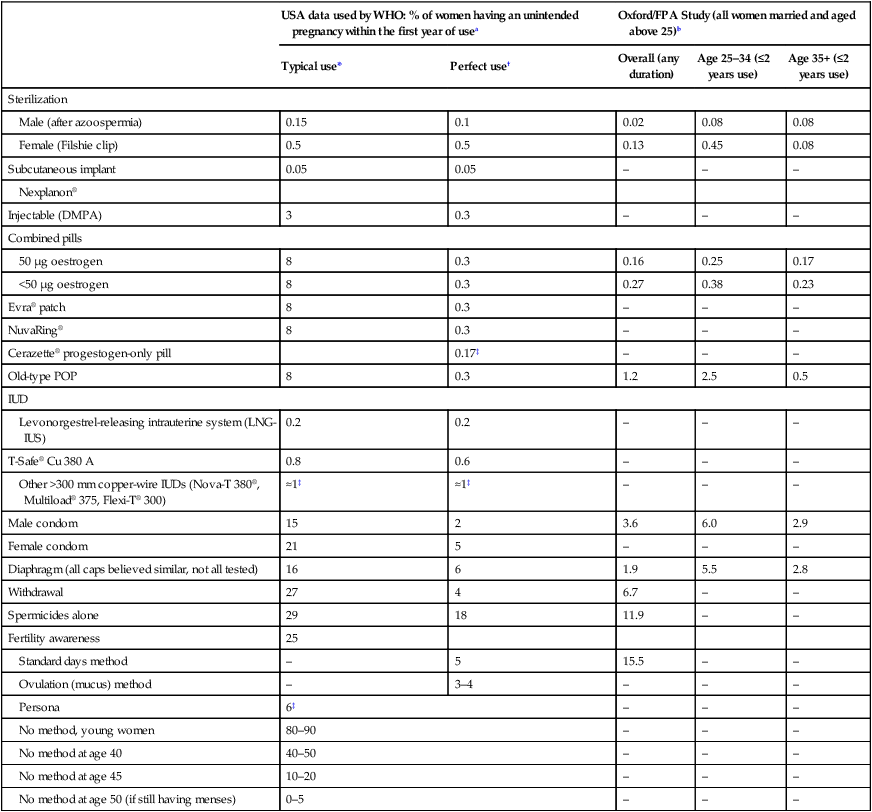
Other Notes (1) Note influence of age: all the rates in the fifth column being lower than those in the fourth column. Lower rates still may be expected above age 45. (2) Much better results also obtainable in other states of relative infertility, such as lactation. (3) Oxford/fpa users were established users at recruitment – greatly improving results for barrier methods (Qs 1.19, 4.9). (4) The Nexplanon, Cerazette and Persona results come from pre-marketing studies by the manufacturer, giving an estimate of the Pearl ‘method-failure’ rate.

bVessey M, Lawless M, Yeates D (1982) Efficacy of different contraceptive methods. Lancet 1(8276):841–842.

*Typical use: Among typical couples who initiate use of the method (not necessarily for the first time), the percentage who experience an accidental pregnancy during the first year if they do not stop use for any other reason.

Perfect use: Among typical couples who initiate use of the method (not necessarily for the first time), and who then use it perfectly (both consistently and correctly), the percentage who experience an accidental pregnancy during the first year if they do not stop use for any other reason.

Data not available from Trussell, so best alternative data given, e.g. from manufacturer’s studies.

(This Table was published in Guillebaud J, MacGregor A (2013) Contraception 6e. ©Elsevier. Reproduced from Trussell J, Wyn LL (2008) Reducing unintended pregnancy in the United States. Contraception 77(1): 1–5, with permission.)

Barrier methods of contraception

These techniques involve a physical barrier that reduces the likelihood of spermatozoa reaching the female upper genital tract. Barrier methods also offer protection against sexually transmitted infection (STIs). The relative risk of an STI-induced pelvic inflammatory disease (PID) is 0.6 for women using these methods. Women who use another method of contraception to prevent pregnancy are often advised to use a condom as well to reduce an otherwise increased risk of STI.

Male condoms

The basic condom consists of a thin, stretchable latex film, which is moulded into a sheath, lubricated and packed in a foil wrapper. The sheath has a teat end to collect the ejaculate. The disadvantages of sheaths are that they need to be applied before intercourse and that they reduce the level of sensation for the male partner. The advantages are that they are readily available, are without side effects for the female partner and provide a degree of protection against infection. They have an efficiency of 97–98% with careful use, although typical failure rates can be as high as 15 pregnancies per 100 women years. Common reasons for failure are leakage of sperm when the penis is withdrawn, putting the condom on after genital contact, use of lubricants that cause the latex to break and mechanical damage. Condoms should be unrolled completely on to the penis before genital contact occurs and held when the penis is withdrawn to avoid leakage. The penis needs to be withdrawn from the vagina before the erection is lost, or sperm will inevitably be lost from it.

Diaphragms and cervical caps

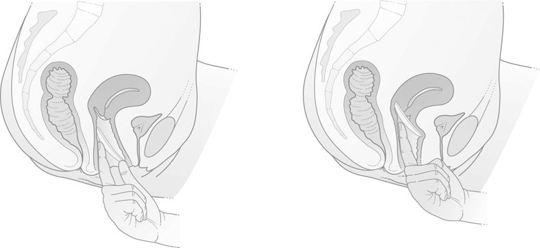
The modern vaginal diaphragm consists of a thin latex rubber dome attached to a circular metal spring. These diaphragms vary in size from 45–100 mm in diameter. The size of the diaphragm required is ascertained by examination of the woman. The size and position of the uterus are determined by vaginal examination and the distance from the posterior vaginal fornix to the pubic symphysis is noted. The appropriate measuring ring, usually between 70 mm and 80 mm, is inserted. When in the correct position the anterior edge of the ring or diaphragm should lie behind the pubic symphysis and the lower posterior edge should lie comfortably in the posterior fornix (Fig. 19.1).

The woman should be advised to insert the diaphragm either in the dorsal position or in the kneeling position while bending forwards. The diaphragm can be removed by simply hooking an index finger under the rim from below and pulling it out. The diaphragm should be smeared on both sides with a contraceptive cream, and it is usually advised that it should be inserted dome down. However, some women prefer to insert the diaphragm with the dome upwards.

The diaphragm must be inserted prior to intercourse and should not be removed until at least 6 hours later. The main advantage of this technique is that it is free of side effects to the woman, apart from an occasional reaction to the contraceptive cream. The main disadvantages are that the diaphragm must be inserted before intercourse and typical failure rates are between 6 and 16 pregnancies per 100 women years. The main reason for failure is probably that the diaphragm size chosen is actually too small and when orgasm occurs in the woman, when the vaginal size can increase dramatically, the diaphragm no longer fits adequately.

There are a variety of vault and cervical caps, which are of much smaller diameter than the diaphragm. These are suitable for women with a long cervix or with some degree of prolapse, but otherwise have no particular advantage over the diaphragm.

Spermicides and sponges

Spermicides are only effective, in general, if used in conjunction with a mechanical barrier. Pessaries or suppositories have a water-soluble or wax base and contain a spermicide. They must be inserted approximately 15 minutes before intercourse. Common spermicides are nonoxynol-9 and benzalkonium. Creams consist of an emulsified fat base and tend not to spread. Care in insertion is essential so that the spermicide covers the cervix.

Jellies or pastes have a water-soluble base that spreads rapidly at body temperature. They therefore have an advantage over creams, as they spread throughout the vagina.

Foam tablets and foam aerosols contain bicarbonate of soda so that carbon dioxide is released on contact with water. The foam spreads the spermicide throughout the vagina. Pregnancy rates vary with different agents, but average around 9–10 per 100 women years.

Sponges consist of polyurethane foam impregnated with nonoxynol-9. The failure rate is between 9% and 32%, and their use in isolation is therefore not recommended. They are inserted at least 15 minutes before intercourse and can be left in for a maximum of 12 hours.

Intrauterine contraceptive devices

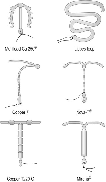
Intrauterine contraception is used by 6–8% of women in the UK. A wide variety of intrauterine devices (IUDs) have been designed for insertion into the uterine cavity (Fig. 19.2). These devices have the advantage that, once inserted, they are retained without the need to take alternative contraceptive precautions. It seems likely that they act mainly by preventing fertilization. This is a result of a reduction in the viability of ova and the number of viable sperm reaching the tube.

The first device to be widely used was the Graefenberg ring, which was made of a silver–copper alloy. Introduced in the 1930s, it ran into considerable difficulties with haemorrhage, infection, miscarriages and uterine perforation. Later, inert plastic devices such as the Lippes loop were associated with a significant increase in menstrual blood flow in many users. The development of copper IUDs has been associated with improved contraceptive efficacy and a lessening of excess menstrual blood loss.

Types of devices

The devices are either inert or pharmacologically active.

Devices containing progestogen

The levonorgestrel-releasing intrauterine system or Mirena® contains 52 mg of levonorgestrel (Fig. 19.2) which suppresses the normal build up of the endometrium so that, unlike most IUDs, it causes a reduction in menstrual blood loss. However, there is a high incidence of irregular scanty bleeding in the first 3 months after insertion of the device. Unlike previous progestogen-containing devices it does not appear to be associated with a higher risk of ectopic pregnancy. The superior efficacy of third-generation copper IUDs and the levonorgestrel-releasing system means that these are now considered the devices of choice.

Insertion of devices

The optimal time for insertion of the device is in the first half of the menstrual cycle. With postpartum women, the optimal time is 4–6 weeks after delivery. Insertion at the time of therapeutic abortion is safe and can be performed when motivation is strong. It is unwise to insert IUDs following a miscarriage because of the risk of infection. Devices may be inserted within a few days of delivery but there is a high expulsion rate.

Ideally, the woman should be placed in the lithotomy position. A cervical smear should be taken, and a swab taken for culture if there is any sign of infection. The uterus is examined bimanually and its size, shape and position are ascertained. The cervix is swabbed with an antiseptic solution and a vulsellum can be applied to the anterior lip of the cervix, although this is not essential and may cause discomfort.

The passage of a uterine sound will indicate the depth and direction of the uterine cavity and the dimensions of the cavity may be assessed by devices known as cavimeters, which measure its length and breadth. Many IUDs are available in different sizes, and cavimeters help to choose the appropriate IUD.

Insertion devices vary in construction but generally consist of a stoppered plastic tube containing a plunger to extrude the device, which may be linear or folded. The device is inserted in the plane of the lumen of the uterus and care must be taken not to push it through the uterine fundus.

Attempts at insertion of a device where the cervical canal is tight may result in vagal syncope. Acute pain following insertion may indicate perforation of the uterus. The woman should be instructed to check the loop strings regularly and to notify her doctor immediately if the strings are not palpable.

Complications

The complications of IUDs are summarized in Figure 19.3.

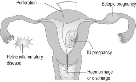
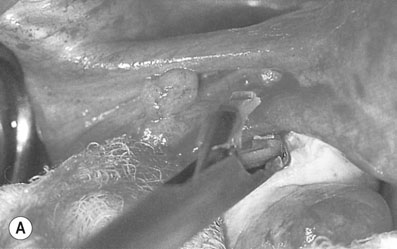
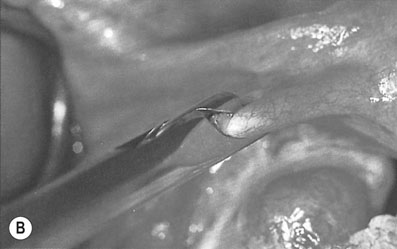
Perforation of the uterus

About 0.1–1% of devices perforate the uterus. In many cases, partial perforation occurs at the time of insertion and later migration completes the perforation. If the woman notices that the tail of the device is missing, then it must be assumed that one of the following has occurred:

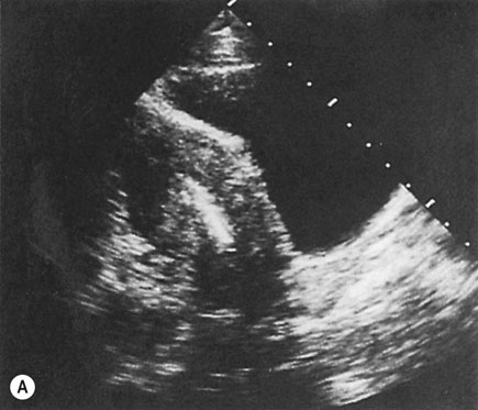
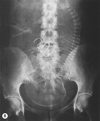
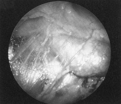
If there is no evidence of pregnancy, an ultrasound examination of the uterus should be performed. If the device is located within the uterine cavity (Fig. 19.4A), unless part of the loop or strings is visible, it will generally be necessary to remove the device with formal dilatation of the cervix under general or local anaesthesia. If the device is not found in the uterus, a radiograph of the abdomen will reveal the site in the peritoneal cavity (Fig. 19.4B). It is advisable to remove all extrauterine devices by either laparoscopy or laparotomy. Inert devices can probably be left with impunity, but copper devices promote considerable peritoneal irritation and should certainly be removed.

Pelvic inflammatory disease

Pre-existing PID is a contraindication to this method of contraception. There is a small increase in the risk of acute PID in IUD users, but this is largely confined to the first 3 weeks after insertion. If PID does occur, antibiotic therapy is commenced and, if the response is poor, the device should be removed. If the infection is severe, it is preferable to complete 24 hours of antibiotic therapy before removing the device. It is not uncommon to find evidence of Actinomyces organisms in the Pap smear routinely collected in an asymptomatic woman who has an IUD in place. This is generally not due to an actinomycotic pelvic infection, but due to the presence of these organisms on the surface of the IUD. There is no absolute consensus of what should be done if such organisms are found in the Pap smear. Some would remove the IUD, repeat the smear in 3 months and reinsert another IUD if the smear is clear, whereas others would leave the IUD in place but give a 2 week course of penicillin therapy.

Hormonal contraception

Oral contraception is given as a combination of oestrogen and progestogen, as a combined pill, or as progestogen only.

Combined pill

Most of the current combined pills contain 20–30 µg of ethinyl oestradiol and 150–4000 µg of progestogen. The progestogens used are derived from 17-hydroxyprogesterone or 19-norsteroids (Box 19.1).

The pill is usually taken for 21 days, followed by a 7-day pill-free interval during which there is a withdrawal bleed. Everyday (ED) preparations include seven placebo pills that are taken instead of a pill-free week. The concentration of the hormones may be the same throughout the 21 days (monophasic preparations) or vary across the cycle (biphasic and triphasic preparations) in order to reduce breakthrough bleeding.

Contraindications

There are various contraindications to the pill, some being more absolute than others.

The absolute contraindications include pregnancy, previous pulmonary embolism or deep vein thrombosis, sickle-cell disease, porphyria, current active liver disease or previous cholestasis (particularly where it is associated with a previous pregnancy), migraine associated with an aura or carcinoma of the breast. It is necessary to maintain a high level of vigilance in women with varicose veins, diabetes, hypertension, renal disease and chronic heart failure but none of these conditions constitutes an absolute contraindication and, in some cases, the adverse effects of a pregnancy may substantially outweigh any hazard from the pill. Women who smoke and are also over the age of 35 years have a significantly increased risk of coronary artery and thromboembolic disease.

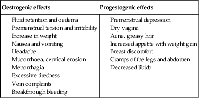
The occurrence of migraine for the first time, severe headaches or visual disturbances, or transient neurological changes are indications for immediate cessation of the pill. There are a series of minor side effects that may sometimes be used to advantage or may be offset by using a pill with a different combination of steroids (Table 19.2).

image

Major side effects

The risk of venous thrombosis is increased from 5/100 000 to 15/100 000 women per year and is further increased in smokers and women with a previous history of venous thrombosis. This compares to a risk in pregnancy and the puerperium of 60/100 000 women. Several studies have suggested that so-called ‘third and fourth-generation’ combined pills containing desogestrel, gestodene or drospirenone, are associated with a twofold greater risk of venous thrombosis than those containing other progestogens, although the risk of venous thrombosis was lower in these studies than had previously been reported.

There is an increase in arterial disease, with a 1.6 to 5.4-fold increase in stroke and 3 to 5-fold increase in myocardial infarction (although there is no significant increase in women under 25 or in non-smokers). However, both these conditions are rare in women under the age of 35 years so the overall risk remains low, with deaths from venous thrombosis attributable to the combined pill of no more than 1–2/million women years.

Although some reports have suggested there is a small increase in the relative risk of breast (relative risk 1.24) and cervical cancer (relative risk 1.5–2) in pill users, especially if it is commenced before a first pregnancy, the breast cancer increased risk is not definitely proven, and the cervical cancer risk is probably due to the incidence of wart virus infection and not the taking of the oral contraceptive pill (OCP).

There is an increase in gallstone formation and cholecystitis and an increase in glucose intolerance.

The progestogen-only pill has a higher failure rate and is more likely to be associated with irregular bleeding. If it fails there is a higher risk of ectopic pregnancy.

Beneficial effects

In addition to the prevention of unwanted pregnancy, the use of the combined pill is associated with a 30% reduction in blood loss at menstruation, a lower incidence of ectopic pregnancy (0.4/1000) and some protection against PID and benign ovarian cysts. Pill users also have a reduced risk of both endometrial and ovarian cancer of up to 50%, depending on the length of use with this benefit lasting for up to 10 years after the OCP therapy has been ceased.

imagePractical patient care of a patient requesting to use the combined OCP

It is important to obtain a complete general history and examination before prescribing the pill, and also to perform annual check-ups and cervical cytology. There are a large number of compounds commercially available, and some pills are marketed by different companies but contain the same compounds at the same concentrations. The history taken must exclude the contra-indications detailed above. Examination should include breast examination, blood pressure assessment and, except in women who have never been sexually active, speculum examination, Pap smear testing, and PV assessment. An appropriate pill for that particular patient should then be chosen, and counselling then given along the following lines.

Which pill should you choose?

In general a 30 µg ethinyl oestradiol-containing pill is usually chosen first because of its effectiveness and low cost. The 20 µg containing preparations are much more expensive but preferred by many women, and the side effects are usually less, except that breakthrough bleeding during the first few months of treatment is more common. If the woman had evidence of androgen excess, hirsutism or clinical PCOS, the OCP Diane 35 should be given because its progestogen is cyproterone acetate, an anti-androgen. If the woman has fluid retention problems, an OCP containing drospirenone is usually advisable.

If the woman has used the OCP previously and had major problems with breakthrough bleeding, has conceived when taking the pill correctly, or is on treatment with an anti-epileptic medication, it is safer to advise them to take an OCP containing 50 µg of ethinyl oestradiol.

When should it be commenced?

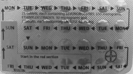
It is best commenced on day 2–3 of the next period but can be commenced at any time. Many combined pills include 7 days of placebo (‘sugar’) tablets so that the user takes a pill every day of the month and so reduces the risk of forgetting when to restart the pill after the normal 7 ‘pill free’ days each cycle (sometimes labelled ‘ED’ or everyday preparations). Each tablet including the placebos are labelled with a day of the week in these calendar packs with the placebos being a different colour (Fig. 19.5) With these pills a woman should start taking the pill on the first day of her next period starting with the inactive tablet corresponding to the current day of the week. When changing from a higher to a lower dose pill preparation women should be advised to start taking the active tablets of the new pill immediately on completing the last tablet of her previous pill, omitting the normal 7 day gap.

Interaction between drugs and contraceptive steroids

Many drugs affect the contraceptive efficacy of the pill, and therefore additional precautions should be taken (Table 19.3). Vomiting and diarrhoea also result in loss of the pill and hence the return of fertility – particularly with the low-dose pills now widely in use. Progestogen-only pills must be taken every day if they are to be effective.

Table 19.3

Interaction of various drugs with oral contraceptives

Interacting drug Effects of interaction
Analgesics Possible increased sensitivity to pethidine
Anticoagulants Possible reduction of effect of anticoagulant – increased dosage of anticoagulant may be necessary
Anticonvulsants Possible decrease in contraceptive reliability
Tricyclic antidepressants Reduced antidepressant response; increase in antidepressant toxicity
Antihistamines Possible decrease in contraceptive reliability
Antibiotics Possible decrease in contraceptive reliability
Possibility of breakthrough bleeding (this is most likely with rifampicin)
Hypoglycaemic agents Control of diabetes may be reduced
Antiasthmatics Asthmatic condition may be exacerbated by concomitant oral contraceptive
Systemic corticosteroids Increased dosage of steroids may be necessary

Injectable compounds

There are currently two main types – Depo-Provera® and Implanon®. Depo-Provera contains 150 mg of medroxyprogesterone acetate and is given as a 3-monthly intramuscular injection. Implanon is a single Silastic® rod containing etonogestrel that is inserted subdermally in the upper arm and is effective for up to 3 years. An earlier type of implant, the levonorgestrel-releasing Norplant® Silastic rod, has been discontinued but some women may still have this in place. Each of these injectable preparations works by making the cervical mucus hostile, the endometrium hypotrophic and by also suppressing ovulation.

Failure rates are low, at less than 0.1/100 women years in the first year rising to 3.9/100 over 5 years. Failures mostly relate to women already pregnant at the time of injection of the Depo-Provera or insertion of the Implanon device, so it is essential that these methods are commenced at the time of a pregnancy termination or within the first 5 days of menstruation.

Parenteral progestogen-only contraceptives are long-acting but easily reversible, effective, avoid first-pass effect liver metabolism, require minimal compliance and avoid the side effects associated with oestrogens. However, they may cause irregular bleeding or amenorrhoea, which can be a source of anxiety because of the possibility of pregnancy. Removal of the implants may be difficult and should only be carried out by a doctor trained in the procedure. Some women will experience systemic progestogenic effects such as mood changes and weight gain or develop symptoms of oestrogenic deficiency

Emergency contraception

After unprotected intercourse, missed combined pill or a burst condom, a single 750 mg levonorgestrel tablet is taken within 72 hours of intercourse, followed by a second dose exactly 12 hours later. The levonorgestrel-only method has fewer side effects than the previously used combined method and, in some countries, is available to women over the age of 16 years directly from pharmacists. Side effects include mild nausea, vomiting (an additional pill should be taken if vomiting occurs within 2–3 hours of the first dose) and bleeding. The woman should be advised that:

If the next period is more than 5 days overdue, pregnancy should be excluded. Emergency contraception prevents 85% of expected pregnancies. Efficacy decreases with time from intercourse.

If the woman concerned does not attend until more than 72 hours after the sexual activity occurred, levonorgestrel therapy is ineffective, however an IUD can be inserted if it is still before the time implantation of any embryo produced would have occurred.

Non-medical methods of contraception

The most fertile phase of the menstrual cycle occurs at the time of ovulation. In a 28-day cycle, this occurs on day 13 or 14 of the cycle. The fertile phase is associated with changes in cervical mucus that a woman can learn to recognize by self-examination and hormone changes that can be measured by home urine testing kits. Avoidance of the fertile period can be an extremely effective method in well-motivated couples.

Natural methods of family planning include the following:

• The rhythm method: Avoiding intercourse mid-cycle and for 6 days before ovulation and 2 days after it. The efficacy of this method depends on being able to predict the time of ovulation. If a regular 28 day cycle occurs, ovulation is predicted for day 14, and abstinence should be from days 8 to 16. If the cycles are very variable, varying between 24 and 32 days, the earliest ovulation would be on day 10 and the latest on day 18, so abstinence would be required between days 4 and 20.

• The ovulation method: This method takes into account the ability of a woman to recognize the increase in vaginal wetness due to cervical mucus productionin the phase before ovulation, and abstaining from sex during that time and for 2 days after the peak wetness has been observed. This method is much better than the rhythm method, but many women only get 4 days advanced warning of the time of ovulation, so intercourse on the preceding 2 days can result in a pregnancy.

• Coitus interruptus (withdrawal): A traditional and still widely used method of contraception that relies on withdrawal of the penis before ejaculation. It is not a particularly reliable method of contraception, because the best sperm often reach the tip of the penis before the male experiences the imminent ejaculation, or he forgets in the ‘heat’ of the moment.

• Lactational amenorrhoea method: Breastfeeding has historically been the most important means of family ‘spacing’. Ovulation resumes on average 4–6 months later in women who continue to breastfeed. During the first 6 months after birth this is an effective method of contraception in mothers providing they are fully breastfeeding, not giving the baby any non-breast milk or other food, AND have remained amenorrhoeic, with failure rates as low as 1/100 women being seen.

Sterilization

Contraceptive techniques have the major advantage that they are easily reversible and provide a high level of protection against pregnancy. They have the disadvantage that they require a conscious act on behalf of the individual before intercourse. When family size is complete or there is a specific medical contraindication to continuing fertility, sterilization becomes the contraceptive method of choice. Around 30% of couples use sterilization for contraception and this increases to 50% in those aged over the age of 40 years.

Counselling

It is essential to counsel both partners about the nature of the procedures and their implications and to discuss whether it is better for the male or female partner to be sterilized. In many cases, only one partner will be seeking sterilization, in which case only one point of view needs to be considered. It is important, however, to ensure that there is a full discussion of the alternatives.

Counselling should include reference to the intended method, its risks and failure rates (1/200 for female sterilization, 1/2000 for male sterilization). Women should be warned of the increased risk of ectopic pregnancy in the event of failure.

With the improvements brought about by microsurgery, it is no longer acceptable to say that sterilization is irreversible and the patient should be counselled according to the technique to be used. The partner to be sterilized will be a matter of choice and motivation. If one partner has a reduced life expectancy from chronic illness, then that partner should be sterilized.

Women should be advised to continue to use other contraception until the period occurs following the sterilization procedure. Men should be advised to use alternative contraception until they have had two consecutive semen analyses showing azoospermia 2–4 weeks apart, with these analyses not done until at least 10 ejaculations have occurred.

Techniques

Female sterilization

The majority of procedures involves interruption of the Fallopian tubes but may vary from the application of clips on the tubes to total hysterectomy. In general terms, the more radical the procedure the less likely there is to be a failure. However, very low failure rates can now be achieved using methods with high reversibility prospects and these should be the methods of choice.

Laparoscopic sterilization

The use of the laparoscope for sterilization procedures has substantially reduced the duration of hospital stay. This is the method of choice in most developed countries, but an open approach through a mini-laparotomy may be more appropriate in countries where endoscopic facilities or training are limited.

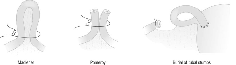
• Tubal clips. This is the most widely used method of sterilization in the UK and Australia. The clips are made of plastic and inert metals and are locked on to the tube (Fig. 19.6). They have the advantage of causing minimal damage to the tube, but their disadvantage is a higher failure rate. Failures may be due to application on the wrong structure, extrusion of the tube from the clip, recanalization or fracture of the clip so that it falls off the tube. The Filshie clip, which has a titanium frame lined by silicone rubber, has the lowest failure rate (0.5%) and is easier to apply. Yoon or Fallope rings are applied over a loop of tube and are similar to a Madlener procedure (see below). This technique is associated with considerably greater abdominal pain postoperatively, and the failure rates vary between 0.3% and 4%. The rings are not suitable for application to the tubes in the puerperium when the tube is swollen and oedematous.

• Tubal coagulation and division. Sterilization is effected by either unipolar or bipolar diathermy of the tubes in two sites 1–2 cm from the uterotubal junction. A considerable amount of tube can be destroyed with this technique. Division of the diathermied tube is said to reduce the risk of ectopic pregnancy. The failure rate depends on the length of tube destroyed. Because of the risk of thermal bowel injury with subsequent leakage and faecal peritonitis, diathermy should not be used as the primary method of sterilization unless mechanical methods of tubal occlusion are technically difficult or fail at the time of the procedure.
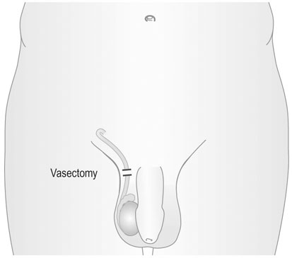
Tubal ligation (Fig. 19.7)

These procedures are usually performed through a small abdominal incision (mini-laparotomy) or at the time of caesarean section. They are less widely used with the increase in laparoscopic procedures. Even when laparoscopy is contraindicated for some reason it is still more common now to use clips to occlude the tubes.

The most basic form of the procedure involving simple ligation of the tube is known as the Madlener procedure but the failure rate may be up to 3.7%. The Pomeroy technique is the same, but the loop of tube is excised and absorbable suture material is used for the ligation. There are several variations of this technique, including the separation of the cut ends of the tubes on contralateral sides of the broad ligament. The excised segments should be examined histologically to confirm that the tube has been excised.

Vasectomy

This procedure is generally performed under local anaesthesia. Two small incisions are made over the spermatic cord and 3–4 cm of the vas deferens is excised (Fig. 19.8). The advantage of the technique is its simplicity. The disadvantages are that sterility is not immediate and should not be assumed until all spermatozoa have disappeared from the ejaculate. On average, this takes at least 10 ejaculations.

The procedure is more difficult to reverse than most forms of female sterilization and, even when satisfactory re-anastomosis is achieved, only about 50% of patients will sire children, because of the adverse effect of the production of sperm-immobilizing and sperm-agglutinating antibodies. The failure rate is 1/2000.

Failures may follow spontaneous recanalization and excision of an inadequate length of vas deferens. The excised segments should always be examined histologically to confirm that the vas has been excised. Complications of the operation include haematoma formation, wound infection and epididymitis. Also a painful granuloma may form at the cut end of the vas as a result of a foreign body reaction induced by spermatozoa.

Psychological implications of sterilization

Sterilization in women has acquired the reputation of leading to psychiatric problems and a deterioration in sexual function. Modern studies do not confirm this reputation. Apart from the fact that modern studies tend to use prospective standardized methodologies, the population being sterilized in the 21st century is very different to that of 40 years ago. Sterilization used to be performed predominantly on older women in poor gynaecological health, with large numbers of children and living in conditions of social adversity. It was carried out on medical recommendation, frequently shortly after childbirth, abortion or some other gynaecological procedure. Nowadays, sterilization is a widely accepted form of contraception used by women of all ages and social classes. They are, therefore, more representative of the population as a whole. Sterilization usually takes place at the request of the woman as an interval procedure unrelated to either childbirth or abortion. Women being sterilized have fewer children and are in better general health than previously.

The rate of psychiatric disorder following sterilization is, in general, no higher than that in the general female population. However, for women who are sterilized immediately following childbirth, there is an increased risk of suffering from postnatal depression. A previous psychiatric history and ambivalence or uncertainty about sterilization are risk factors for psychiatric disorder. Postpartum status, previous psychiatric history, ambivalence and marital discord are also risk factors for deterioration of psychosexual functioning and regret. Some authors have also suggested that, in cultures where femininity is strongly associated with fertility and where there is guilt and shame about contraception, great care should be exercised to ensure that patients are properly prepared for sterilization. Regret, often measured by a request for reversal of sterilization, appears to be most strongly predicted by marital breakdown and subsequent remarriage.

Termination of pregnancy

In the UK this is carried out in approved centres under the provisions of the Abortion Act 1967. This requires that two doctors agree that either continuation of the pregnancy would involve greater risk to the physical or mental health of the mother or her other children than termination, or that the fetus is at risk of an abnormality likely to result in it being seriously handicapped (Box 19.2). The most recent amendment to the Act (1991) set a limit for termination under the first of these categories at 24 weeks, although in practice the majority of terminations are carried out prior to 20 weeks.

All terminations carried out in the UK must be notified. Annual abortion numbers peaked in the UK in 1990 at 170 000 and declined after that until the scare over the risk of venous thrombosis with the ‘third-generation’ pills in 1996.

Methods

All women undergoing termination of pregnancy should be screened for STIs and/or offered antibiotic prophylaxis. Following termination, anti-D immunoglobulin should be given to all rhesus negative women. All women should be offered a follow-up appointment to check that there are no physical problems and that contraceptive measures are in place.

Medical termination

This is the method most commonly used for pregnancies after 14 weeks and is increasingly being offered as an alternative to surgical termination in first trimester pregnancies up to 9 weeks gestation. The standard regimens for first-trimester termination use the progesterone antagonist mifepristone (RU 486) given orally, followed 36–48 hours later by prostaglandins administered as a vaginal pessary. There are several different regimens, but all have a success rate of greater than 95%. Second trimester terminations can also be performed using vaginal prostaglandins given 3-hourly or as an extra-amniotic infusion through a balloon catheter passed through the cervix. Pretreatment with mifepristone significantly reduces the time interval from induction to abortion. After delivery of the fetus, an examination under general anaesthetic may be necessary to remove the placenta.

Psychological sequelae of termination

The majority of women who find themselves with an unwanted pregnancy are very distressed. Despite this, evidence shows that the majority of women do not experience medium- to long-term psychological sequelae, nor is there any evidence of an increase in the rate of psychiatric morbidity. The available evidence is that the rate of psychiatric morbidity following termination of pregnancy is less than if the pregnancy was allowed to proceed.

Later terminations of pregnancy

The number of women having terminations of pregnancy after 12 weeks for psychosocial reasons is falling. Second trimester terminations now account for fewer than 8% of all therapeutic terminations of pregnancy. A minority of these women are having a therapeutic abortion for psychosocial reasons; the majority for fetal abnormality.

Unlike first trimester abortions, later terminations of pregnancy are associated both with marked psychological distress and an increased rate of psychiatric disorder. Some 39% of women having an abortion for fetal abnormality are depressed at 3–9 months, although the rates fall to normal at 1 year. For women undergoing this procedure for psychosocial reasons, the cause for the increased rate of distress and morbidity is likely to be found in the delay in presenting for termination. The very young, the mentally handicapped and the chronically mentally ill may be found in this group, as well as those who have experienced marked ambivalence about their pregnancies.

The situation for women having a termination of pregnancy because of fetal abnormality is different. These are usually older women who have a much wanted pregnancy and whose problem has been diagnosed either because of a previous experience or as the result of screening. The decision to terminate the pregnancy is usually reached only after much thought and anguish. The consequence of termination is, therefore, very much like the spontaneous loss of a more advanced pregnancy, that is to say, a grief reaction. Their psychosocial recovery may be assisted by granting them the dignity of a naming and burial. Most late terminations of pregnancy involve the induction of labour and a prolonged process of giving birth. This can be a distressing and traumatic experience, and psychological recovery will be improved by sensitive and compassionate handling by the doctor and nursing staff.

Contraception following termination

Referral for termination should also be an opportunity to discuss future contraception and to ensure that adequate provision is made for this after the termination. The procedure can be combined with sterilization. This has the advantage of preventing further terminations for the woman who is certain that she has completed her family. There is little evidence that this is associated with an increase in the rate of complications or later contraceptive failure. However, because of the increase in the ‘regret rate’ for the sterilization, an interval procedure is generally recommended. IUD insertion can be carried out at the same time as termination and is not associated with an increased risk of perforation or failure. If the oral contraceptive is being used, this can be started on the same or following day.

Genital tract infections

The female genital tract provides direct access to the peritoneal cavity. Infection may extend to any level of the tract and, once it reaches the Fallopian tubes, is usually bilateral.

The genital tract has a rich anastomosis of blood and lymphatic vessels that serve to resist infection, particularly during pregnancy.

There are other natural barriers to infection:

imageTaking a sexual history

Taking an accurate sexual history is essential to the management of genital tract infections, and aspects of sexual history are relevant to a range of other presentations including subfertility, pelvic pain and disorders of sexual function. A concise sexual history will help to:

Patients (and students!) are often anxious so it is important to create a relaxed and friendly environment and have a respectful and a non-judgemental attitude. Introducing self and role, maintaining eye contact and having appropriate body language are important aspects of good communication when obtaining a sexual history. The confidential nature of the consultation should be explained. It is important etc to use language that is understandable and does not use labels or make judgements. Ask general questions first, using open ended questions. Move on to the exploration of reasons for presentation and more closed ended questions (see below). Explain there are some ‘universal’ questions that are explicitly asked of everyone to assess risk and avoid making assumptions about sexual orientation based on appearance.

Specific questions

Reason for attendance: the problem/issue, including symptoms

Direct questions about symptoms may include:

Sexual behaviour risk assessment:

STI and blood-borne virus (BBV) risk assessment: additional questions to assess timing of tests and other risks to inform testing and management planning:

Other relevant information: to identify issues that may be associated with or influence client management:

(Reproduced from NSW Sexually Transmissible Infections Programs Unit 2011. NSW Health Sexual Health Services Standard Operating Procedures Manual 2011.)

Lower genital tract infections

The commonest infections of the genital tract are those that affect the vulva and vagina. Infections that affect the vagina also produce acute and chronic cervicitis.

Symptoms

Swelling and reddening of the vulval skin is accompanied by soreness, pruritus and dyspareunia. When the infection is predominantly one of vaginitis, the symptoms include vaginal discharge, pruritus, dyspareunia and often dysuria. Cervicitis is associated with purulent vaginal discharge, sacral backache, lower abdominal pain, dyspareunia and dysuria. The proximity of the cervix to the bladder often results in coexistent trigonitis and urethritis, particularly in the case of gonococcal infections.

Chronic cervicitis is present in about 50–60% of all parous women. In many cases, the symptoms are minimal. There may be a slight mucopurulent discharge, which is not sufficient to trouble the woman and may simply present as an incidental finding that does not justify active treatment. In the more severe forms of the condition, there is profuse vaginal discharge, chronic sacral backache, dyspareunia and occasionally postcoital bleeding. Bacteriological culture of the discharge is usually sterile. The condition may cause subfertility because of hostility of the cervical mucus to sperm invasion.

Signs

These will depend on the cause. The appearance of the vulval skin is reddened, sometimes with ulceration and excoriation. In the sexually mature female, the vaginal walls may become ulcerated, with plaques of white monilial discharge adherent to the skin or, in protozoal infections, the discharge may be copious with a greenish-white, frothy appearance.

Bartholin’s glands are sited between the posterior part of the labia minora and the vaginal walls, and these two glands secrete mucus as a lubricant during coitus. Infection of the duct and gland results in closure of the duct and formation of a Bartholin’s cyst or abscess. The condition is often recurrent and causes pain and swelling of the vulva. Bartholinitis is readily recognized by the site and nature of the swelling.

In cervicitis the cervix appears reddened and may be ulcerated, as with herpetic infections, and there is a mucopurulent discharge as the endocervix is invariably involved. The diagnosis is established by examination and taking cervical swabs for culture.

Common organisms causing lower genital tract infections
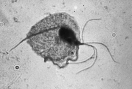
Vaginal candidiasis

Candida albicans is a yeast pathogen that occurs naturally on the skin and in the bowel. Infection may be asymptomatic or associated with an increased or changed vaginal discharge associated with soreness and itching in the vulva area. There is no evidence of male to female sexual transmission. White curd-like collections attached to the vaginal epithelium may be seen on speculum examination, although these are not present in all cases.

Candidal infections are particularly common during pregnancy, in women taking the contraceptive pill and in underlying conditions involving immunosuppression, e.g. HIV infection, diabetes or long-term steroids. In each instance, vaginal acidity is increased above normal and bacterial growth in the vagina is inhibited in such a way as to allow free growth of yeast pathogens, which thrive well in a low-pH environment. Candida hyphae and spores can also be seen in a wet preparation and can be cultured.

Genital herpes

The condition is caused by herpes simplex virus (HSV) type 2 and, less commonly, type 1. It is a sexually transmitted disease. Primary HSV infection is usually a systemic infection with fever, myalgia and occasionally meningism. The local symptoms include vaginal discharge, vulval pain, dysuria and inguinal lymphadenopathy. The discomfort may be severe enough to cause urinary retention. Vulval lesions include skin vesicles and multiple shallow skin ulcers (Fig. 19.10). The infection is also associated with an increased risk of cervical dysplasia. Partners may be asymptomatic and the incubation period is 2–14 days.

The diagnosis is made by sending fluid from vesicles for viral culture or antigen detection. After the initial infection the virus remains latent in the sacral ganglia. Recurrences may be triggered by stress, menstruation or intercourse, but are normally of shorter duration and less severe than the primary episode. Serum antibodies are raised in well-established lesions.

Syphilis

The initial lesion appears 10–90 days after contact with the spirochaete Treponema pallidum. The primary lesion or chancre is an indurated, firm papule, which may become ulcerated and has a raised firm edge. This lesion most commonly occurs on the vulva but may also occur in the vagina or cervix. The primary lesion may be accompanied by inguinal lymphadenopathy. The chancre heals spontaneously within 2–6 weeks.

Some 6 weeks after the disappearance of the chancre, the manifestations of secondary syphilis appear. A rash develops which is maculopapular and is often associated with alopecia. Papules occur, particularly in the anogenital area and in the mouth, and give the typical appearance known as condylomata lata.

Swabs taken from either the primary or secondary lesions are examined microscopically under dark-ground illumination, and the spirochaetes can be seen. The serological tests have been described in Chapter 7.

The disease then progresses from the secondary phase to a tertiary phase. It may mimic almost any disease process and affect every system in the body, but the common long-term lesions are cardiovascular and neurological.

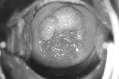
Genital warts (condylomata acuminata)

Vulval and cervical warts (Fig. 19.12) are caused by a human papilloma virus (HPV). The condition is commonly, although by no means invariably, transmitted by sexual contact. The incubation period is up to 6 months. The incidence has risen significantly over the last 15 years, particularly in women aged 16–25 years.

The warts have an appearance similar to those seen on the skin in other sites, and in the moist environment of the vulval skin are often prolific – particularly during pregnancy. There is frequently associated pruritus and vaginal discharge. The lesions may spread to the perianal region, and in some cases become confluent and subject to secondary infection. Diagnosis is usually made by clinical examination.

Treatment of lower genital tract infections

When the diagnosis has been established by examination and bacteriological tests, the appropriate treatment can be instituted. The treatment for Chlamydia and gonorrhoea is discussed below under infections of the upper genital tract. Whenever a diagnosis of sexually transmitted infection is made, it is essential to screen patients (and their partners) for other infections.

Vulval and vaginal monilial infections can be treated by topical or oral preparations. These include a single dose of clotrimazole given as a pessary or fluconazole taken orally. Recurrent infections can be treated by oral administration of ketoconazole and fluconazole. The patient’s partner should be treated at the same time, and any predisposing factors such as poor hygiene or diabetes should be corrected.

Trichomonas infections and bacterial vaginosis are treated with metronidazole 400 mg taken twice a day for 5 days, which must be taken by both sexual partners if recurrence of the infection is to be avoided. Metronidazole may be administered as a single dose of 2 g, but high-dose therapy should be avoided in pregnancy. Topical treatment with metronidazole gel or clindamycin cream is also effective for bacterial vaginosis.

If a patient is asymptomatic and there is no evidence of vaginitis on clinical examination, but trichomonal or monilial organisms are identified in a routine Pap smear, treatment of these patients is usually not required.

Non-specific vaginal infections are common and are treated with vaginal creams, including hydrargaphen, povidone–iodine, di-iodohydroxyquinoline or sulphonamide creams.

Syphilis is treated in the first instance with penicillin and, if this fails, for example in the case of coinfection, with penicillin-resistant strains of the gonococcus, doxycycline hydrochloride or other antibiotics can be used.

Infections of the vagina associated with menopausal atrophic changes are treated by the appropriate hormone replacement therapy using an oral or vaginal oestrogen preparation, or lactic acid pessaries when oestrogens are contraindicated. The same therapy may be used, with the local application of oestrogen creams, in juvenile vulvovaginitis.

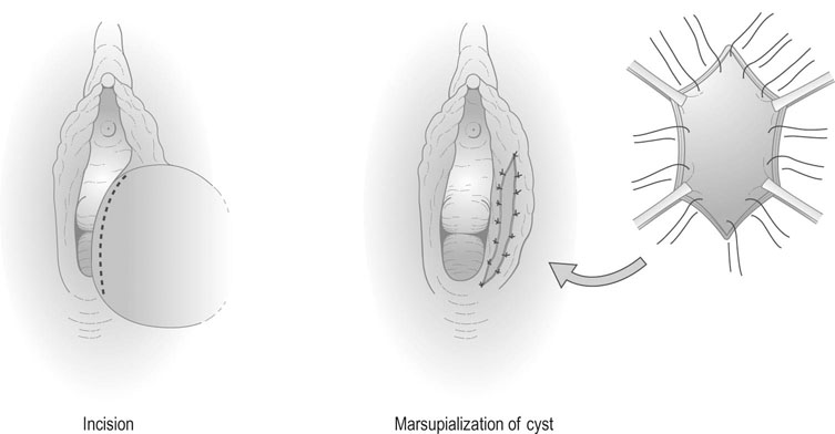
Infections of Bartholin’s gland are treated with the antibiotic appropriate to the organism. If abscess formation has occurred, the abscess should be ‘marsupialized’ by excising an ellipse of skin and sewing the skin edges to result in continued open drainage of the abscess cavity (Fig. 19.13). This reduces the likelihood of recurrence of the abscess.

Vulval warts are treated with either physical or chemical diathermy using podophyllin applied directly to the surface of the warts. Any concurrent vaginal discharge should also receive the appropriate therapy.

Herpetic infections are notoriously resistant to treatment and highly prone to recurrence. The best available treatment is aciclovir administered in tablet form 200 mg five times daily for 5 days or locally as a 5% cream.

Acute cervicitis usually occurs in association with generalized infection of the genital tract and is diagnosed and treated according to the microbiology. Medical treatment is rarely effective in chronic cervicitis because it is difficult to identify an organism and antibiotics do not penetrate the chronic microabscesses of the cervical glands. If the cervical swab is negative, the next most effective management is diathermy of the endocervix under general anaesthesia. Following diathermy, an antibacterial cream should be placed in the vagina and the woman should be advised that the discharge may increase in amount for 2–3 weeks but will then diminish. She should also be advised to avoid intercourse for 3 weeks as coitus may cause a secondary haemorrhage.

Upper genital tract infections

Acute infection of the endometrium, myometrium, Fallopian tubes and ovaries are usually the result of ascending infections from the lower genital tract causing PID.

However, infection may be secondary to appendicitis or other bowel infections, which sometimes give rise to a pelvic abscess. Perforation of the appendix with pelvic sepsis remains a common cause of tubal obstruction and subfertility. Pelvic sepsis may also occur during the puerperium and after pregnancy termination or after operative procedures on the cervix. Retained placental tissue and blood provide an excellent culture medium for organisms from the bowel, including Escherichia coli, Clostridium welchii or C. perfringens, Staphylococcus aureus and Streptococcus faecalis.

PID affects approximately 1.7% of women between 15 and 35 years of age per year in the developed world. Up to 20% of women with PID will have a further episode within 2 years. The disease is most common between the ages of 15 and 24 years, and particular risk factors include multiple sexual partners and procedures involving transcervical instrumentation. PID is an important cause of infertility. After a first episode 8% of women will have evidence of tubal infertility; subsequent episodes approximately double this figure. Women with a past history of PID are 4 times more likely to have an ectopic pregnancy when they conceive.

Symptoms and signs

The symptoms of acute salpingitis include:

The signs include:

• Signs of systemic illness with pyrexia and tachycardia.

• Signs of peritonitis with guarding, rebound tenderness and often localized rigidity. (It should be noted that guarding and rigidity rarely are seen if blood is in the peritoneal cavity, such as due to an ectopic pregnancy, whereas tenderness and release tenderness are seen even in the absence of peritonitis.)

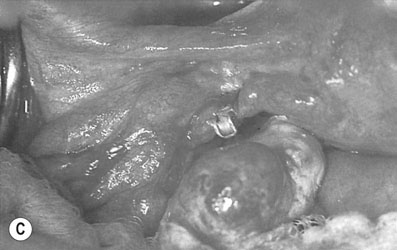
• On pelvic examination, acute pain on cervical excitation and thickening in the vaginal fornices, which may be associated with the presence of cystic tubal swellings due to pyosalpinges or pus-filled tubes; fullness in the pouch of Douglas suggests the presence of a pelvic abscess (Fig. 19.14).

• An acute perihepatitis occurs in 10–25% of women with chlamydial PID, which may cause right upper quadrant abdominal pain, deranged liver function tests and multiple filmy adhesions between the liver surface and the parietal peritoneum, and is known as the Fitz–Hugh–Curtis syndrome.

• A pyrexia of 38°C or more, sometimes associated with rigors.

Common organisms

Pelvic inflammatory disease is thought to be the result of polymicrobial infection with primary infection by Chlamydia trachomatis or Neisseria gonorrhoeae (or both) allowing opportunistic infection with other aerobic bacteria and anaerobes.

Gonorrhoea

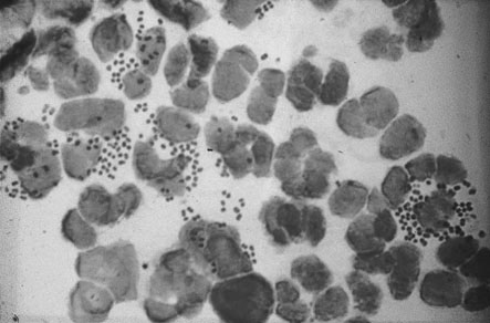
N. gonorrhoeae is a Gram-negative intracellular diplococcus (Fig. 19.15). Infection is commonly asymptomatic or associated with vaginal discharge. In cases of PID it spreads across the surface of the cervix and endometrium and causes tubal infection within 1–3 days of contact. It is the principal cause for 14% of cases of PID and occurs in combination with Chlamydia in a further 8%.

Differential diagnosis

It is often difficult to establish the diagnosis of acute pelvic infection with any degree of certainty. The predictive value of clinical signs and symptoms when compared to laparoscopic diagnosis is 65–90%. The differential diagnosis includes the following:

• Tubal ectopic pregnancy: Initially pain is unilateral in most cases. There may be syncopal episodes and signs of diaphragmatic irritation with shoulder tip pain. The white cell count is normal or slightly raised but the haemoglobin level is likely to be low depending on the amount of blood lost, whereas in acute salpingitis the white cell count is raised and the haemoglobin concentration is normal.

• Acute appendicitis: The most important difference in the history lies in the unilateral nature of this condition. Pelvic examination does not usually reveal as much pain and tenderness but it must be remembered that the two conditions sometimes coexist, particularly where the infected appendix lies adjacent to the right Fallopian tube.

• Acute urinary tract infections: These may produce similar symptoms but rarely produce signs of peritonism and are commonly associated with urinary symptoms.

• Torsion or rupture of an ovarian cyst.

Investigations

When the diagnosis of acute salpingitis is suspected, the woman should be admitted to hospital. After completion of the history and general examination, swabs should be taken from the vaginal fornices and cervical canal and sent to the laboratory for culture and antibiotic sensitivity. A midstream specimen of urine should also be sent for culture to exclude a possible urinary tract infection. An additional endocervical swab should be taken for detection of Chlamydia by enzyme-linked immunoassay (ELISA) or, preferably, polymerase chain reaction (PCR). Urethral swabs may identify chlamydial infection not detected by endocervical swabs. PCR assays of urine samples have a similar of better sensitivity (90%) compared to genital tract swabs and offer a potential means for screening for chlamydial infection in asymptomatic women.

Examination of the blood for differential white cell count, haemoglobin estimation and C-reactive protein may help to establish the diagnosis. Blood culture is indicated if there is a significant pyrexia. The diagnosis of mild to moderate degrees of PID on the basis of history and examination findings is unreliable and, where the diagnosis is in doubt, laparoscopy is indicated.

Management

When the patient is unwell and exhibits peritonitis, high-grade fever, vomiting or a pelvic inflammatory mass, she should be admitted to hospital and managed as follows:

• Fluid replacement by intravenous therapy – vomiting and pain often result in dehydration.

• When PID is clinically suspected, antibiotic therapy should be commenced. Antibiotic therapy initially prescribed for clinically diagnosed PID should be effective against C. trachomatis, N. gonorrhoeae and the anaerobes characterizing bacterial vaginosis. If the woman is acutely unwell, treatment should be started with an antibiotic such as cefuroxime and metronidazole given intravenously with oral doxycycline until the acute phase of the infection begins to resolve. Treatment with oral metronidazole and doxycycline should then be continued for 7 and 14 days, respectively.

• Pain relief with non-steroidal anti-inflammatory drugs.

• If the uterus contains an intrauterine device, it should be removed as soon as antibiotic therapy has been commenced.

• Bed rest – immobilization is essential until the pain subsides.

• Abstain from intercourse.

Patients who are systemically well can be treated as outpatients, with a single dose of azithromycin and a 7-day course of doxycycline, reviewed after 48 hours.

Chronic pelvic infection

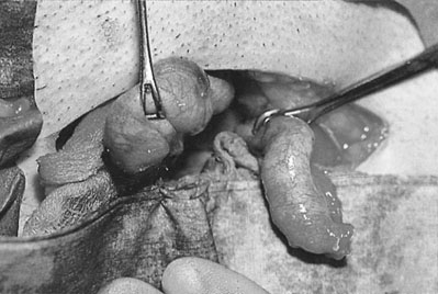
Acute pelvic infections may progress to a chronic state with dilatation and obstruction of the tubes forming bilateral hydrosalpinges with multiple pelvic adhesions (Fig.19.16).

Human immunodeficiency virus

Human immunodeficiency virus (HIV)-1 and HIV-2 are RNA retroviruses characterized by their tropism for the human CD4+ (helper) T lymphocyte. The proportion of cells infected is initially low and there is a prolonged latent phase between infection and clinical signs. Transmission occurs by sex, infected blood products, shared needles, breastfeeding and at the time of delivery. Risk groups include intravenous drug abusers and their partners, the partners of bisexual men, haemophiliacs, prostitutes and immigrants from high-risk areas. Although HIV infection is more common in men in the developed world, anonymous testing shows that 0.3% of pregnant women in London are infected and it is now the most common cause of death in African American females aged 24–35 years in the US. In parts of sub-Saharan Africa, 20–30% of all pregnant women are HIV positive. Vertical transmission rates can be reduced from 40% to less than 1% by antenatal treatment with the modern antiretroviral drugs, delivery by elective caesarean section and avoidance of breastfeeding. Although HIV infection was a life-ending sentence for most people in the past as most developed acquired immunodeficiency syndrome (AIDS) as an end result within a few years of becoming infected with the HIV, with modern continuous therapy most are able to be controlled and progression to AIDS is much less common.

The main clinical states can be identified as:

Common opportunistic infections include Candida, HSV, HPV, Mycobacterium spp., Cryptosporidium spp., Pneumocystis carinii and cytomegalovirus. Non-infective manifestations include weight loss, diarrhoea, fever, dementia, Kaposi’s sarcoma and an increased risk of cervical cancer.

The diagnosis is made by detecting antibodies to the virus, although these may take up to 3 months to appear.

Disorders of female sexual function

Disorders of sexual function are reported by up to a third of women. Sometimes they are accompanied by awareness of the underlying disturbance but often, as with other emotional difficulties, the link between cause and effect is obscure even to the sufferer. Sexual problems may therefore appear in the guise of mental or physical illness or disturbances of behaviour and relationships, and thus form a part of the working experience not only of doctors but of anyone in the ‘caring’ professions. Lack of knowledge about sex and the anatomy of the genital tract remain common and a source of anxiety.

The commonest complaints are:

Dyspareunia

Dyspareunia is defined as painful intercourse. It is predominantly but not exclusively a female problem. The aetiology is divided on the basis of whether the problem is superficial (at the entrance to the vagina) or deep (only occurs with deep penile insertion) and it is therefore particularly important to obtain a concise history.

Superficial dyspareunia

Pain felt on penetration is generally associated with a local lesion of the vulva or vagina from one of the following causes:

• Infection: Local infections of the vulva and vagina commonly include monilial and trichomonal vulvovaginitis. Infections involving Bartholin’s glands also cause dyspareunia.

• Narrowing of the introitus may be congenital, with a narrow hymenal ring or vaginal stenosis. It may sometimes be associated with a vaginal septum. The commonest cause of narrowing of the introitus is the over-vigorous suturing of an episiotomy wound or vulval laceration or following vaginal repair of a prolapse.

• Menopausal changes: Atrophic vaginitis or the narrowing of the introitus and the vagina from the effects of oestrogen deprivation may cause dyspareunia. Atrophic vulval conditions such as lichen sclerosus can also cause pain.

• Vulvodynia: This is a condition of unknown aetiology characterized by persisting pain over the vulva.

• Functional changes: Lack of lubrication associated with inadequate sexual stimulation and emotional problems will result in dyspareunia.

Deep dyspareunia

Pain on deep penetration is often associated with pelvic pathology. Any woman who develops deep dyspareunia after enjoying a normal sexual life should be considered to have an organic cause for her pain until proved otherwise. The common causes of deep dyspareunia include:

• Acute or chronic pelvic inflammatory disease: including cervicitis, pyosalpinx and salpingo-oophoritis (Fig. 19.16). The uterus may become fixed. Ectopic pregnancy must also be considered in the differential diagnosis in this group.

• Retroverted uterus and prolapsed ovaries: If the ovaries prolapse into the pouch of Douglas and become fixed in that position, intercourse is painful on deep penetration.

• Endometriosis: Both the active lesions and the chronic scarring of endometriosis may cause pain.

• Neoplastic disease of the cervix and vagina: At least part of the pain in this situation is related to secondary infection.

• Postoperative scarring: This may result in narrowing of the vaginal vault and loss of mobility of the uterus. The stenosis commonly occurs following vaginal repair and, less often, following repair of a high vaginal tear. Vaginal scarring may also be caused by chemical agents such as rock salt, which, in some countries, is put into the vagina in order to produce contracture.

• Foreign bodies: Occasionally, a foreign body in the vagina or uterus may cause pain in either the male or female partner. For example, the remnants of a broken needle or partial extrusion of an intrauterine device may cause severe pain in the male partner.

Vaginismus

Vaginismus is the symptom resulting from spasm of the pelvic floor muscles and adductor muscles of the thigh, which prevents or results in pain on attempted penile penetration. A physical barrier may be present but is not necessarily causative. The woman may be unable to allow anyone to touch the vulva. Primary vaginismus is usually due to fear of penetration. Secondary vaginismus is more likely to be the result of an experience of pain with intercourse after infection, sexual assault, a difficult delivery or surgery. Even after the condition has improved, fear of further pain may lead to involuntary contraction of the vaginal muscles, which is in itself painful, completing the vicious circle. Encouraging the patient to explore her own vagina and feel for herself that there is no abnormality or pain can help break this cycle. Resort to surgery is likely to confirm the patient’s fears of abnormality and often leaves the presenting problem unchanged

Loss of libido

Loss of desire is the commonest symptom in women complaining of sexual dysfunction. If it has always been present it may be a result of a repression of sexual thoughts as a result of upbringing or religious belief or a feeling that sex is dirty or unsuitable in some way. It may represent differences between the expectations of the couple. Loss of desire in a relationship that was previously satisfactory is more likely to be due to:

Disorders of male sexual function

Normal male sexual function is largely mediated through the autonomic nervous system. Erection occurs as a result of parasympathetic (cholinergic) outflow causing vasocongestion. Orgasm and ejaculation are predominantly sympathetic (adrenergic). Emission occurs by the sequential expulsion of fluid from the prostate gland, vas deferens and seminal vesicles into the posterior urethra. Emission and closure of the vesical neck are mediated by alpha-adrenergic systems, while opening of the external sphincter (to allow antegrade ejaculation) is mediated through the somatic efferent of the pudendal nerve. Ejaculation is stimulated by the dorsal nerve of the penis and involves contractile activity of the bulbocavernous and ischiorectal muscles as well as the posterior urethra. These responses are easily inhibited by cortical influences or by impaired hormonal, neural or vascular mechanisms.

The principal features of sexual dysfunction in men are:

All or any of these may be present from adolescence or have their onset at any time of life after a period of healthy sexuality. The causes of loss of libido have been previously described above under female sexual dysfunction.

Erectile dysfunction

Erectile dysfunction or impotence, the inability in the male to achieve erection for satisfactory penetration of the vagina, is the most common problem seen.

It is now recognized that a high proportion (50%) of such men, especially those over the age of 40 years, have an underlying organic cause. Of these diabetes is the commonest as a result of damage to the large and small blood vessels and neuropathy. Neurological impotence may also be caused by injuries to the spinal cord, brain and prostate, and multiple sclerosis. Hyperprolactinaemia is associated with erectile dysfunction as well as with loss of libido. While androgens are not essential for erection they influence it through their effects on libido and nitrogen oxide release in the cavernosum. Recreational drugs such as alcohol are known to cause erectile failure and more than 200 prescription drugs are known to have it as a side effect. The most common of these are antihypertensive and diuretic agents. Others include antidepressant and sedative medications.

In the younger age group the cause is more likely to be psychogenic. Depression, reactive or endogenous, is an important aetiological or concomitant condition. The stress provoked by timing intercourse with ovulation may result in erectile dysfunction in couples undergoing treatment for infertility.

Treatment

Mild psychogenic cases will usually respond to simple measures such as counselling, sex therapy and sensate focusing exercises.

Treatment with bromocriptine may restore sexual function in cases where prolactin levels are raised.

Intracavernous injection of prostaglandin E1 is effective in patients with both psychogenic and organic causes of erectile dysfunction, although pain and the fear of injection cause some patients to stop treatment. Sildenafil is an effective orally administered alternative, with up to 70% of attempts at intercourse being successful compared with 22% with placebo. It promotes erection by potentiating the effect of nitric oxide on vascular smooth muscle, thus increasing blood flow to the penis. Concurrent use in patients taking nitrate therapy for myocardial ischaemic disease causes significant hypotension.

Ejaculatory problems

Ejaculatory dysfunction encompasses premature, retarded, retrograde and absent ejaculation. Anejaculation and premature ejaculation are more often seen in younger patients. Retrograde ejaculation is often a result of an organic cause or after surgery, e.g. prostate operations. The diagnosis is usually made on the presenting history.

Treatment

For premature ejaculation the squeeze technique described by Masters and Johnson involves application of pressure to the top of the penis. This diminishes the urge to ejaculate, although the success rate is poor. Alternative approaches include the use of a local anaesthetic and selective serotonin-reuptake inhibitors. Anejaculation and retarded ejaculation can be treated by teaching masturbation techniques, couple counselling and sensate focus exercises. Retrograde ejaculation is regarded mainly as a fertility problem. Treatment may involve surgery or drug therapy with alpha-adrenoceptor agonists.

image   Essential information

Upper genital tract infection

• Usually from ascending lower genital tract infection

• Can follow abortion, normal delivery or an operative procedure on the cervix.

• Commonly due to C. trachomatis or N. gonorrhoeae when sexually transmitted

• Presents as pain, fever, discharge and irregular periods

• Bilateral pain on cervical excitation and raised white cell count

• Differential diagnosis includes ectopic pregnancy, urinary tract infection and appendicitis

• Management includes fluid replacement, antibiotics, analgesia and rest

• Surgery (laparoscopy or laparotomy) is indicated to confirm diagnosis if in doubt, for drainage of pelvic mass and to clear pelvis in unresponsive chronic disease

• Major cause of infertility worldwide, resulting in tubal obstruction in 40% of cases after three or more attacks