Severe dehydration in a young woman

Published on 10/04/2015 by admin

Filed under Surgery

Last modified 10/04/2015

Print this page

rate 1 star rate 2 star rate 3 star rate 4 star rate 5 star
Your rating: none, Average: 0 (0 votes)

This article have been viewed 1135 times

Problem 48 Severe dehydration in a young woman

She is of normal build and is well kept. Her temperature is 37.5°C, her pulse rate is 135 bpm and her supine blood pressure is 95/60 mmHg. She has deep sighing respirations with a respiratory rate of 25 breaths/minute. Her mouth is extremely dry and her tissue turgor is reduced. She has no other abnormalities on cardiac and respiratory examination, and she has no focal neurological signs. Her abdomen is not distended but appears guarded and she reacts as if in pain when palpated. Bowel sounds cannot be heard.

Another girl appears who says she is a companion from the hostel. The two have only known each other for 2 weeks and agreed to travel together. The girl claims her friend became unwell over the last week, particularly over the last 2 days. She complained of being very tired. The patient had become lethargic and listless. She had not wanted to eat, but had complained of being very thirsty and had been drinking large amounts of water. Today she stayed on her bunk and was unable to get up. She complained of abdominal pain and vomited three times. The friend had gone out for a few hours and on her return found the patient looking very unwell and an ambulance had been called. The patient is not known to have any major medical problems or to take medications or drugs of any kind.

There are a number of possible causes for this woman’s comatose state.

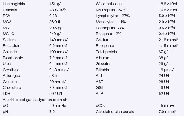
A finger-prick blood glucose measurement has been obtained using a bedside glucose meter and reads ‘high’. A urinary catheter drains 50 mL urine which, when tested with a strip, registers ketones as ++++. After initial resuscitation, results of the preliminary blood tests come back as follows:

You have a working diagnosis for this patient who is critically ill.

You institute emergency management. An intravenous line is inserted, and she is managed in the high dependency ward with close nursing care to monitor all vital signs and to keep the airway clear. The patient’s urine output is regularly charted to establish that renal function is normal and urine production is taking place. An insulin infusion is commenced.

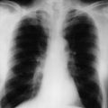
In addition to your fluid replacements, the electrolytes are checked after 2 hours to measure the potassium concentration. It is now 3.6 mmol/L and you begin to add potassium supplementation to the intravenous infusion at 13.6 mmol and later to 27.2 mmol (1–2 g KCl) per hour. You check her potassium level again at 4 hours and 8 hours. A chest X-ray is performed and urine microscopy sent off to look for any intercurrent infection. After 12 hours the patient’s blood glucose is down to 15 mmol/L. The intravenous fluid is changed to 5% dextrose 1 L over 6 hours with 2 g KCl added to each litre of fluid.

Twenty-four hours later your patient’s condition has significantly improved. She is fully conscious, no longer vomiting or nauseated and is allowed to start eating. The urine shows only a trace of ketones.

She responds well to your insulin regimen, and is stable for discharge. Further history establishes that she has previously been healthy: now she has a diagnosis of diabetes. She is frightened by what has happened, and anxious about her diabetes management.

The patient decides to curtail her backpacking holiday and return home.

Answers

A.1 Her Glasgow coma score is 11 (E4, V2, M5).

A.2 Given her young age, history of polydipsia and clinical evidence of dehydration and hyperventilation, the most likely diagnosis is diabetic ketoacidosis. Abdominal pain is quite common in this condition and does not indicate any acute abdominal problem (although intra-abdominal problems must be considered in the differential diagnosis). Other diagnoses to consider include poisoning (alcohol, food or drug-related, particularly street drugs) and infection (e.g. meningitis).

A.3 As part of the immediate management, the following must be undertaken:

A.4 The patient has a severe metabolic acidosis as characterized by the low serum pH and low bicarbonate level. The low pCO2 is due to respiratory compensation for the metabolic acidosis. This explains the hyperventilation.

The increased anion gap is due to the presence of anions not measured in the calculated anion gap. This ‘hidden’ anion here is ketone bodies.

Metabolic acidosis can be divided into:

The apparent hyperkalaemia is due to the shift of potassium from the intracellular space to the extracellular space caused by the acidosis (potassium in exchange for hydrogen ions). In spite of the elevated serum potassium, these patients are usually depleted of total body potassium. This is an important point to grasp. Large amounts of potassium are lost via the kidneys as a result of the glucose diuresis and also through vomiting. The serum potassium level will need careful monitoring as it will drop rapidly when treatment with insulin is commenced and the acidosis starts to correct, placing the patient at risk of hypokalaemia and arrhythmias.

Buy Membership for Surgery Category to continue reading. Learn more here