115
Seborrheic keratosis
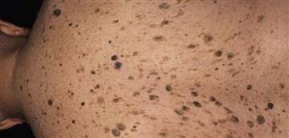
Diffuse yellow and brown waxy papules and patches over the back of this elderly man.
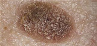
Large seborrheic keratosis waxy plaque with a sharp border and verrucous changes centrally.
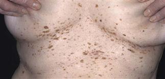
Lesions are often concentrated on the back, upper chest, neck and inframammary areas. Rubbing and chafing from clothing or from maceration in this area can cause these lesions to be inflamed.
Smooth-surfaced lesions contain dark or light round horn pearls embedded in the lesion or protruding from the surface. Seborrheic keratoses may mimic warts, nevi and melanoma.
DESCRIPTION
Common, benign, persistent, waxy brown epidermal lesion with various clinical appearances. One of the most common benign growths of the skin. Often confused and misdiagnoses with other benign and malignant lesions.
HISTORY
• Most people develop at least one or more seborrheic keratosis in their lifetime. • Males and females equally affected. • Lesions may be localized to areola in men and women. • Tendency to develop multiple seborrheic keratoses may be inherited.
PHYSICAL FINDINGS
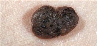
• Seborrheic keratoses are typically multiple and can arise at any site except palms and soles. • Size and surface appearance of lesions vary considerably. Most are between 0.2 and 2.0 cm. Lesions may be flat or raised significantly. Surface may be smooth, velvety, rough or verrucous. Retained keratin cysts may be seen just under the surface within clefts. Color of lesions extremely variable, including white, pink, brown, and black; color may vary within single lesion. Lesions tend to be oval, sharply demarcated, and often oriented along skin cleavage lines. Most have ‘stuck on’ appearance and waxy texture. Surface tends to crumble when inflamed or picked. • Raised or pedunculated seborrheic keratoses may be indistinguishable from skin tags and compound melanocytic nevi. Flat seborrheic keratoses may mimic spreading pigmented actinic keratosis or superficial spreading melanoma. If diagnostic doubt exists, perform a skin biopsy. • Dermatosis papulosa nigra is term used to describe seborrheic keratoses of face seen more commonly in African-Americans. Dermatosis papulosa nigra lesions are 1–2 mm, dark-brown keratotic papules concentrated around eyes and upper cheeks. • Stucco keratoses describe the small, whitish seborrheic keratoses more commonly found on lower legs, ankles and feet of elderly white people. The sign of Leser–Trélat is the sudden explosive onset of numerous seborrheic keratoses in association with internal malignancy, usually gastrointestinal malignancy.
TREATMENT
• Treatment may be indicated for symptomatic lesions which are inflamed, irritated or bleeding. Lesions may be removed when they are symptomatic; this usually occurs when they are located in an area of friction and frequent trauma. • Removal often requested for cosmetic reasons. Patients should be informed that cosmetic removal of seborrheic keratoses is not usually covered by medical insurance. • Cryosurgery is effective for flat to minimally raised lesions. Thicker lesions best removed by cautery and curettage under local anesthesia. • Hypopigmentation or hyperpigmentation are possible side effects of cryotherapy or any method of removal. Residual scarring, if any, is minimal. Applying gentle pressure to the surrounding skin often provides enough tension to allow for easy curettage of lesions.


