124
Sebaceous hyperplasia
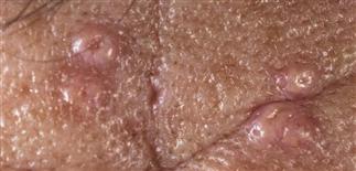
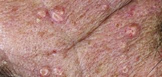
Sebaceous hyperplasia is a common benign condition consisting of prominently enlarged sebaceous glands on the face.
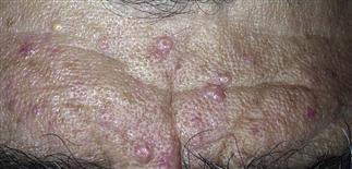
Yellow-white papules are commonly found on the forehead and cheeks. The central pore is almost a constant feature.
Sun damage has been suggested as a contributing factor. Genetic factors probably play a significant role as well.
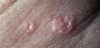
The lesion begins as a 1- to 2-mm, soft, pale yellow to skin-colored minimally elevated papule. Enlarges over time, and may be confused with basal cell carcinoma.
DESCRIPTION
Common benign condition consisting of prominently enlarged sebaceous glands on the face.
HISTORY
• Occurs in both men and women. • Papules rarely appear before age 30 but become increasingly more common with advancing age. Roughly 80% of patients over age 70 have at least one such lesion. • Most lesions represent a single, hypertrophied sebaceous gland with multiple lobules arranged around a central enlarged sebaceous duct. • Lesions occur in all skin types but are more easily seen in lighter skin. • Etiology unknown. Genetic inheritance plays a large role. Sun damage has been suggested as a contributing factor. • Lesions are entirely asymptomatic but persistent. • Papules can become disfiguring and are mostly of cosmetic concern. Patients are typically concerned that the lesions represent basal cell carcinoma or other type of skin cancer.
PHYSICAL FINDINGS
• Lesion begins as a 1- or 2-mm, soft, pale yellow to skin-colored, minimally elevated papule. With time, lesion attains a maximum size of 3–4 mm and develops a central umbilication. • Mature papules possess a distinctly yellow-orange color and are more sharply defined from the surrounding skin. • Papules may be solitary but are more commonly multiple and scattered randomly on the forehead, eyelids, nose, and cheeks. • Papules may yield sebum from the central umbilication with palpation. • An orderly array of fine telangiectasias may radiate outward from the umbilication toward the periphery of the papule. • Individual lesions may be confused with basal cell carcinoma, small keratoacanthoma, or molluscum contagiosum.
TREATMENT
• Treatment is not required but may be requested for cosmetic reasons. • Cryosurgery, carbon dioxide laser, electrodesiccation and curettage, and trichloroacetic acid are all effective in ablating individual lesions. • The sebaceous lobules located within the superficial dermis must be destroyed for the treatment to be successful. • Care must be taken to avoid over-treatment so as to minimize the risk of permanent scarring. • Reassurance is often all that is needed for the patient with sebaceous hyperplasia.


