Screening for adult disease

Published on 11/04/2015 by admin

Filed under Surgery

Last modified 22/04/2025

Print this page

rate 1 star rate 2 star rate 3 star rate 4 star rate 5 star
Your rating: none, Average: 0 (0 votes)

This article have been viewed 1154 times

6

Screening for adult disease

Principles of screening

Introduction

Medical screening is a public health activity that involves examining or testing asymptomatic, apparently healthy people to detect disease at an early stage. Measures can then be taken to prevent the disease (if there is a precursor stage), treat it early (hoping for improved cure rates), or at least offer treatment to delay advanced disease. For example, colonic screening can detect adenomas and carcinomas; removing adenomas prevents the well-recognised adenoma–carcinoma sequence, and actual cancers detected are often earlier and at a more curable stage. Unfortunately, for cancers without an easily detected early precursor stage such as breast or prostate, beneficial outcomes are elusive.

Screening can detect disorders that predispose to other diseases, for example, hypertension or elevated cholesterol levels, to discover people at increased risk of atherosclerotic heart disease and stroke. Screening is also useful for infection control, e.g. preoperative screening of patients from residential homes for meticillin-resistant Staphylococcus aureus (MRSA) carrier status to enable elimination therapy before operation.

An entire population can be screened (mass screening) but more usually it is targeted at at-risk groups. Selection might be by age, gender or cardiovascular risk factors, for example (Box 6.1). Opportunistic screening involves a more random approach, such as screening patients who happen to attend a particular clinic.

Assessing the potential benefits of screening

Many subscribe to the simplistic view that screening must be ‘a good thing’. These include the lay public, people associated with distressing diseases, populist politicians and people with vested financial interests. Poorly conceived screening, however, may consume massive resources to identify just a few new cases with little clinical benefit, e.g. CT scanning for lung cancer. Worse still, early diagnosis of a condition where early intervention brings no advantage may cause suffering. These people can be prematurely placed into an anxiety-provoking sick role and given unrealistic expectations. They may also be subjected to unnecessary treatments with potentially severe side-effects, e.g. some breast or prostate cancers that might never progress to invasive disease.

As with any public health measure, medical and social benefits accruing from any screening programme need to be rigorously evaluated and the process separated entirely from the incentive to screen for profit. Whole-body scanning by CT or MRI is currently strongly marketed on the basis that a scan will show unsuspected abnormalities and allow early treatment. Abnormalities are bound to be discovered by such extensive screening, but it is difficult to reliably determine which signify serious disease and which (if any) should be treated. Doctors should not perform unvalidated screening tests any more than they should use unproven drugs, and should resist patient pressure for inappropriate screening.

Premature introduction of screening: Politicians can play a part in initiating inappropriate screening programmes. UK prime minister, Margaret Thatcher sanctioned nationwide breast screening in 1988, 2 weeks before a general election; some believe this was to garner the women’s vote. The decision was premature and based on insufficiently validated evidence from the Swedish two counties study and the UK Forrest report. In the 1970s, screening for cancer of the uterine cervix was widely introduced, also before its efficacy had been fully evaluated. Fortunately, it has proved successful despite the difficulty of engaging women at high risk. Sadly, the natural history of untreated dysplastic cervical cellular abnormalities was not properly established before the impact of widespread screening made this ethically impossible. This severely hampered scientific study of the disease, its early diagnosis and best treatment.

Criteria for assessing a screening programme: Many years ago the World Health Organization (WHO) realised that even beneficial screening could be expensive, unpleasant, inaccurate and unproductive, and could adversely affect psychological or physical well-being. In 1968, they published a list of criteria for effective screening programmes (Box 6.2) including attributes of the disease, the test and the treatment. These principles are still relevant today and have been added to by the UK National Screening Committee and other groups. Box 6.3 shows a summary of these modified criteria.

Criteria for an effective screening programme

In order to initiate a new national screening programme, certain criteria must be fulfilled. There must be a perceived need in the medical community or in the wider public. Pilot studies are then carried out. If outcomes are promising, large scale prospective randomised controlled trials (RCTs) need to be performed, seeking robust evidence for implementation. This is critical because it is politically difficult to stop a screening programme, even when evidence shows little benefit, e.g. breast screening (Nordic Cochrane Collaboration 2001 and 2006).

The disease: The screened condition should be an important health problem either because it is common (such as lung or prostate cancer) or has serious but preventable consequences such as carotid artery disease or abdominal aortic aneurysm (AAA). The prevalence (proportion of cases already in a population) and the incidence (the proportion of new cases) of the disease in the population at risk are discovered from pilot studies. There should be a truly early stage where treatment outcomes are better than at a late stage. Colorectal adenomas and early cancers are good examples.

The biological behaviour or natural history of the disease should be well understood, including how latent disease progresses to clinical disease, and the disease course should be reasonably predictable. For example, AAAs are known to expand smoothly for the most part and rarely rupture until they are large. The risks of untreated disease also need to be understood and there should be a long period between the first detectable stages and overt disease.

The diagnostic test: The test must be valid, i.e. reliable in detecting the disease. This is defined by sensitivity and specificity. Sensitivity is the capability of the test to identify affected individuals in the screened population, i.e. the proportion of people who have the disease and are detected. A test with many false negative results is insensitive and unreliable. A UK appeal court ruled that sensitivity is paramount in (cervical) screening and awarded damages to women with missed diagnoses at screening. Specificity is the degree to which a positive test can be relied upon to prove the disease is present; in other words, the higher the false positive rate, the lower the specificity.

The test must be simple and cheap and it must identify the disease by a reliable, validated and reproducible method. The distribution of test values should be understood well enough to define normality or relative risk associated with particular stages, e.g. for an AAA, the diameter with a high risk of rupture. Intervals for repeating the test should be worked out for normal subjects and for those with positive results near the threshold.

The complete screening programme must be clinically safe and acceptable socially and ethically to health professionals and the public. This includes the test and any diagnostic procedures, treatments or interventions screening initiates. For example, if a test is perceived as unpleasant, e.g. colonoscopy, uptake is low and the benefits are proportionately smaller.

The overall benefits should be greater than the risks; this includes any physical and psychological harm caused by the test, diagnostic procedures and treatment.

Diagnosis and treatment: Cases identified by the test must be amenable to effective, acceptable and safe diagnostic procedures and the potential benefits of medical or surgical intervention prompted by earlier diagnosis need to be understood.

There should be clear evidence from high-quality randomised controlled trials (RCTs) that early treatment produces better outcomes than treatment at later stages, i.e. ‘cure’ should be more likely, survival longer, or earlier treatment easier. Treatment should have minimal side-effects. There also needs to be agreement in advance about who should be offered treatment and its nature; this evidence may emerge from RCTs.

The overall benefits of screening must outweigh the risks. This includes any physical and psychological harm caused by the test, the diagnostic procedures and the treatment. Treatment facilities must be adequate with the capacity to deal with the extra workload from screening.

Screening overall must be cost effective compared with other health care interventions and needs. Costs include the testing, further diagnostics and treatment, administration, staff training and quality assurance. For example, because of its high unit cost, CT screening is unlikely to ever be implemented unless proved extremely effective in early diagnosis of a common, highly remediable life-threatening condition. In a world of competing public health measures, debate continues about what is an acceptable cost per life year saved: £100—£5000—£35 000?

Any screening programme must be sustainable in terms of management, monitoring and quality standards. High-quality, realistic, unbiased information needs to be offered to potential participants about the consequences of testing, investigation and treatment to help them decide whether to go ahead. This is currently a subject of debate regarding breast screening in the UK.

Ideally, any primary prevention interventions for the disease should be implemented before or in parallel with the screening programme.

Limitations of screening: Screening is conceptually and ethically different from usual clinical practice as the process is aimed at the whole population, and achieved by dealing with apparently healthy individuals. Participants expect the diagnosis of the presence or absence of disease will be accurate, and that if disease is found, the outcome will be favourable. However, there is no guarantee of protection because there will always be false positive and negative results, however good the test. Also, the disease may appear or progress unexpectedly rapidly between screenings. This emphasises the importance of good population education and properly informed consent for individuals engaging in the programme. Participating in screening must be a free choice and it may or may not have health benefits and significant adverse effects.

Bias: A number of phenomena can lead to mistaken claims for efficacy of a screening programme:

Lead time bias—screening relies on the principle that a serious or fatal disease can be diagnosed at an early stage, and that doing so will definitively improve morbidity and mortality. However, if it does not alter the disease course, earlier diagnosis gives the statistical illusion of prolonged survival. It also makes affected patients acutely aware of the presence of their disease for longer.

Selection bias occurs if the more health conscious people, often at lower risk of the disease, undergo screening.

Length bias occurs when screening detects less aggressive variants than those the screening programme was set up to discover. Length bias is frequently cited in the context of breast cancer screening.

Other aspects of screening:

Screening for cancer

Early detection of cancer

As a principle, the earlier in its natural history that malignancy is diagnosed and treated, the better the prognosis. The ideal would be to detect cancer before invasion or metastasis had occurred, i.e. during the pre-invasive stage. However, many cancers invade and spread before they reach a detectable size or produce tumour markers. Where true early detection is possible, health education can alert the public to early symptoms and warning signs. In skin and testicular tumours, this should include regular self-examination. Self-examination is still promoted for breast cancer but large trials have shown it to be ineffective and that it causes harm with more biopsies.

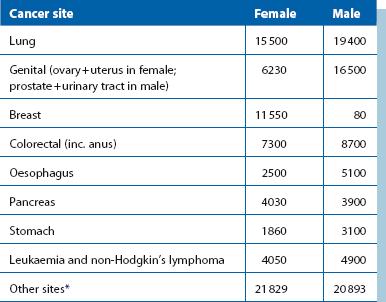
The common cancer killers are shown in Table 6.1, with bronchus still leading the field. Unfortunately, this disease does not fit criteria for screening, having no detectable early stage; attempts at screening have been uniformly ineffective.

In women, breast cancer is a huge public health problem, followed by carcinoma of the cervix and ovary. In men, prostate cancer is a large and growing problem. Colorectal cancer is common and evenly matched in frequency in both sexes in the UK; 30 000 new cases are detected each year and 16 000 die of it. Gastric and pancreatic cancers are also big killers but screening is of little value except in areas of exceptionally high incidence. In China, high-risk areas for oesophageal cancer have been identified and brush cytology without gastroscopy has proved beneficial.

Several genetic predispositions to cancer have been identified, e.g. polyposis coli for colorectal cancer and BRCA1 and BRCA2 for breast and other cancers. Genetic screening is not covered in this chapter but individual disorders are described in other chapters.

Cervical cancer

Poorly organised screening trials using cervical smears and Papanicolaou staining began in the UK in the mid-1960s and national screening started in 1988. Nearly all reports show early detection and treatment prevents 80–90% of invasive cervical cancers and has greatly reduced cervical cancer mortality. The International Agency for Research on Cancer (IARC) indicated that yearly screening between the ages of 25 and 64 reduces invasive cancer by 94%, 3-yearly screening reduces it by 91%, 5-yearly by 84%, and 10-yearly by 64%. These figures are the basis for the present UK policy—that yearly screening is unnecessarily frequent and 3- or 5-yearly screening is best.

In the UK, 4 million women are screened annually and 82% of the 14 million eligible women have been screened over the previous 5 years. The programme costs £150 million a year, amounting to £37.50 per screen. Both the number of invasive cancer cases and deaths from it halved between 1988 and 2005, taking the disease from sixth most common cancer in women to 13th.

British data show that about a quarter of all cervical cancers occur in each of the four age groups 25–39, 40–54, 55–69 and 70+ years but there are problems with recruiting young women and women from lower socio-economic strata. Both groups have been shown to be at higher risk. In a study from Hawaii in 2003, only 1 in 12 eligible women had not been screened in the preceding 5 years, but this small group accounted for two-thirds of the invasive cancers in the community. Thus there are real concerns that those at greatest risk are not being tested. In addition, those with positive results may not be treated effectively. Recent recommendations in the UK are that women should first be invited for screening at age 25. They should then be screened 3-yearly until 49 and 5-yearly from 50 to 64. Women of 65+ only need screening if they have not been screened since age 50 or a recent abnormality has been found.

A recent cost effectiveness study from Peru, India, Kenya, Thailand and South Africa indicates that a single screen (and treatment if necessary), employing testing for human papillomavirus (HPV) in cervical cells or visual inspection of the cervix after swabbing with acetic acid rather than a cervical smear is a cheap and effective way to reduce a woman’s lifetime risk by 25–36%. Types 16 and 18 HPV cause most cervical cancer worldwide and effective vaccines are now available. Two rounds of screening at 35 and 40 years could reduce lifetime risk by a further 40%. If screening were introduced across the developing world then the global incidence of cervical cancer could fall by about 50%. Ideally, all young women should be vaccinated against HPV with modern effective quadrivalent vaccines; this would virtually eliminate cervical cancer and the need for screening.

Breast cancer

Mammographic screening for breast cancer was introduced nationally in the UK in the late 1980s following the Forrest report of 1986. Most developed countries followed suit after results from the HIP study of New York, the Swedish two-county study and the Canadian National Breast Screening Study. These appeared to demonstrate a 30% reduction in mortality from breast cancer in screened women. In the Swedish study, there was also a significant 13% reduction in all-cause mortality.

Mammographic screening detects breast cancers of smaller size than those presenting clinically, with around 30% either carcinoma in situ or invasive cancers less than 0.5 cm in diameter. A high proportion are node negative—only about 20% have axillary spread compared with 40% for symptomatic cancer. By detecting small lesions, screening substantially increases the reported incidence of invasive breast cancer. This might be expected to mean fewer new cases in later years, as prevalent cases would disappear from the population. In Norway and Sweden this has proved untrue, suggesting that screening is not detecting most of the clinically important cases that progress to become invasive or metastasise.

In any population of women with breast cancer, lesions will be at different stages of development and pathological potential. These may be grouped as follows:

Large trials have concluded that as many as one-third of cancers detected by screening would never have presented clinically.

The sensitivity of screening for clinically significant cancers is poor; in particular, lobular or mucinous cancers and some rapidly proliferating, high-grade tumours may not be detectable. This is illustrated by the high proportion of interval cancers presenting clinically between screening visits. In one representative series, 38% of all breast cancers presented as interval cancers. These tended to occur in younger patients with dense breasts and with a higher usage of hormone replacement therapy (HRT) or the oral contraceptive pill. A study in New South Wales estimated that screening over-diagnosed invasive cancer by 30–42% in women aged 50–69 and other studies have confirmed this. A Dutch study showed that false positives adversely affect quality of life for at least a year.

On the basis of tumour doubling times, breast cancers detected clinically have been present for an average of 8 years, whereas mammographically detected lesions have been present for about 6 years—a long period in which to metastasise. This may explain the failure of screening to increase the cure rate for clinically significant cancers, and also calls into question the political pressure for patients with suspected breast cancers to be evaluated within a very short time.

Effectiveness of mammographic screening: Mammographic screening every 2 years has been estimated to avert only 2 deaths in 1000 women aged between 50 and 59 over a period of 10 years. To achieve this requires 5000 screens and 242 recalls, and for 64 women to have at least one biopsy. Five women will have ductal carcinoma in situ detected, some of which may never progress to invasive cancer. Less than 1% of women invited for screening will benefit; a much larger percentage have to endure false alarms, unnecessary surgery and inappropriate labels of cancer.

In the USA, the independent Health Services/Technology Assessment Texts (HSTAT) reviewed published clinical trials and concluded that ‘in absolute terms, the mortality benefit shown with mammography screening was small enough that biases in the trials could erase or create the observed mortality reduction’.

The Cochrane view of breast screening: The Cochrane Collaboration is an international non-profit organisation that rigorously and dispassionately reviews published research evidence and provides up-to-date information about the effects of health care. Over 11 000 articles have been published in 20 years on breast screening. The Nordic Cochrane Centre reviewed all RCTs in 2001 and found that astonishingly few were of sufficient rigour to reliably determine whether screening reduced morbidity and mortality. Only seven RCTs were identified, of which only two were of sufficiently high quality. Evidence from the adequately randomised Canadian and Malmö trials showed screening had no significant effect. The other five trials, in which randomisation was inadequate, found that screening decreased the risk of death by about 25% but showed a slight increase in risk for screened women for death from any cause. A further Cochrane review was published in 2006, which reanalysed data from published trials. Both reviews found little benefit from breast screening and in 2006 they stated that the absolute risk reduction for breast cancer from screening was only 0.05%. They also found that screening led to substantial over-diagnosis and over-treatment. They concluded: ‘the currently available reliable evidence does not show a survival benefit of mass screening for breast cancer, and the evidence is inconclusive for breast cancer mortality.’ These controversial conclusions imply that breast screening does no good, causes actual harm and probably should be abandoned. This, however, is unlikely to happen.

Why breast screening is claimed to improve survival: Several factors could explain how screening could appear to reduce mortality, as follows:

• Lead time bias with over-detection of clinically insignificant lesions increases the apparent number of breast cancers. These cases do well

• Mortality from breast cancer can occur over a very long period; 35 years after treatment, the commonest cause of death in a Cambridge UK series was still breast cancer. Thus screening studies need to be prolonged

• Improvements in breast cancer treatment took place over the period of the main trials. Tamoxifen and perhaps improved chemotherapy undoubtedly extended absolute survival between the early and late 1980s. Over a similar period, other cancer rates fell for largely unknown reasons: thyroid cancer fell by 12%, testis by 17% and melanoma by 23%

• Subjectivity, unrealistic optimism and perhaps vested interests may lead to misleading presentation of statistics and unsustainable claims for the industry of breast screening

Colorectal cancer

Colorectal cancer is a major health hazard that kills 16 000 people a year in the UK; only about 10% are diagnosed early. Early cancers have survival rates of better than 90% but the all-stage 5-year survival rate of 35% has hardly improved despite treatment advances, because most cases present late. Nine out of 10 cases occur in people over 50.

Screening for colonic cancer has a good chance of being effective. It fulfils many criteria required for a screening programme. In particular there is a clear sequence of adenoma progressing to adenocarcinoma. Also, early cancers are detectable and progress steadily to advanced cancers. About 75% arise sporadically, most likely in pre-existing adenomas. Thus a window of opportunity exists for detecting adenomatous polyps at a premalignant stage or cancers at an early invasive stage (i.e. pathologically less advanced than those with symptoms), where they are potentially curable. The transition phase from benign to malignant is long, shown by the cumulative risk of cancer in polyps 10 mm or larger being only 8% at 10 years.

However, detection methods are the stumbling block. Screening by symptoms alone is very unreliable. The sensitivity of guiac faecal occult blood (FOB) testing is no better than 50% and the specificity is also low. A new quantitative FOB test, faecal immunological testing (FIT) is much more sensitive and specific and is likely to replace the guiac test. With 2-yearly FOB testing alone, there are many interval cancers: 30–60% of cancers and as many as 80% of polyps are missed after three rounds of testing. Sensitivity and interval cancer rates can be improved by adding flexible sigmoidoscopy. A randomised multicentre once-only trial of flexible sigmoidoscopy in 100 000 people found it reduced the rate of colorectal cancer by 33% and deaths from it by 43%. Rectosigmoid cancers were reduced by 50% (Lancet 2010; 375; 1624–1633) but right-sided cancers are not detected. Unfortunately, both faecal occult blood (FOB) testing and flexible endoscopy are distasteful and patient participation is low. Better forms of screening would undoubtedly improve participation.

Despite the drawbacks of FOB testing, meta-analysis of four RCTs has shown that FOB screening can reduce mortality from colorectal cancer by 16% for those allocated to screening and by 23% of those actually screened. On this basis, a 2-yearly FOB screen offered to 10 000 people aged over 40 with two-thirds attending for at least one test would prevent 8.5 deaths (CI: 3.6–13.5) from colorectal cancer over 10 years, a mortality reduction of 23% (RR 0.77, CI: 0.57–0.89); 2800 participants would have a colonoscopy and there would be 3.4 major complications from this, i.e. perforation or haemorrhage (Cochrane). The cost of screening was £5290 per cancer detected and an estimated £1584 per life year gained.

Reports of pilot screening studies suggest screening could improve mortality by 33%, and national screening with FOB testing every 2 years has been approved for implementation in the UK. This started in April 2006 for men and women aged 60–69 (50–74 in Scotland). In addition, large-scale pilots of endoscopic colorectal screening are being trialled in patients in their late 50s.

Prostate cancer

Prostate cancer is the most common cause of cancer deaths in older men and 84% of deaths are in men over 70. Unfortunately, the prospects for prevention remain poor. The problem is not so much in detecting the disease but in avoiding false positives, detecting it early enough and predicting its clinical course. Many cancers remain forever dormant, as shown by post-mortem studies in men who have died of something else: foci of prostate cancer occur in 70% of 70-year-olds, 60% of 60-year-olds and 50% of 50-year-olds. These patients died with the disease rather than from it and offering radical treatment for them is clearly inappropriate. Serum prostate specific antigen (PSA) levels increase with the volume of tumour, so high levels indicate extensive disease. In one study the age-specific median concentration was 40 µg/L in men who died within 3 years, 6 in men who died in 3–6 years and 4 in those who died between 6 and 10 years. In another study, using a cut-off level of 10 µg/L, the false positive rate was 4% (similar to breast screening) but 15/16 men with prostate cancer had extra-prostatic disease. Using the common cut-off of 4, false positive rate was 18% and 22/33 had extra-prostatic disease. Thus PSA is a good test only for cancers that cause death within 3 years; after that the common cut-off of 4 detects only half of those that would cause death or serious morbidity. Unfortunately, histological grade only partly predicts clinical outcome.

In the USA, uncontrolled screening using PSA has spawned an apparent epidemic of prostate cancer. The chairman of the UK National Screening Committee stated that ‘the scientific evidence is that screening for prostate cancer does not reduce mortality, and causes actual harm by exposing people to a procedure which has side effects of incontinence and impotence and where there is no evidence that they will benefit’.

The UK government sponsored a Health Technology Assessment on prostate screening in 1997, which stated that the criteria for a population screening programme had not been met. Their findings were that:

Overall, PSA screening causes harm. Some men receive unnecessary treatment because the cancer is incurable or because it would never have presented clinically, and those treated risk infection (from biopsies), incontinence and impotence.

Screening for cardiovascular disease

Abdominal aortic aneurysm

Introduction

Ruptured abdominal aortic aneurysm (AAA) causes at least 6000 deaths each year in the UK and 1.4% of all deaths in men over 65. The peak mortality is between 65 and 85 years and the risk of rupture is roughly proportional to the diameter; when this reaches 6 cm, the risk rises sharply. A ruptured aortic aneurysm is nearly always an acute emergency and carries a very high mortality. About half the cases never reach hospital and die at home or in transit to hospital. Half of the remainder (25%) die without an operation and half undergoing operation die. Thus the true mortality rate is 85–90%. An emergency operation requires a trained vascular surgeon, ties up an emergency team for 3 or more hours, requires an intensive care bed for 3 or more days, uses large quantities of bank blood, and costs 25% more than an elective procedure, whether or not the patient survives. Detecting aneurysms before rupture means that less risky elective interventions, with a mortality of 5% or less, can be employed.

Appropriateness of screening for AAA

By WHO criteria, AAA is a near ideal candidate for screening. It is an important health problem, the natural history regarding expansion and rupture is fairly well understood, there is an easily detectable early stage, and treatment at an early stage is more beneficial than at a later stage (i.e. ruptured). Ultrasound is a suitable and highly reliable test for the early stage and it is acceptable, with an average of 80% of those invited attending. Appropriate intervals for retesting have been determined by randomised trials. There is adequate health service provision for the extra workload: a screening programme generates approximately 6 extra aneurysm repairs per year per surgeon.

Several trials have shown that the risks are less than the benefits, with up to 75% reduction in rupture rate, a low elective operative mortality, no excess psychological morbidity in those screened, and survival after operation being little different from an unaffected population. Costs appear to be balanced against benefits, with trials estimating the cost per life year saved at between zero (Huntingdon and Danish studies) and £12 500 (2006 figures from the UK Multicentre Aneurysm Screening Study).

Trials of AAA screening

Several large studies have published data, with a remarkable concordance between results. The prevalence of AAA, the age distribution, attendance rates, reduction in AAA mortality and the cost effectiveness calculations from studies in Chichester, Gloucester, Huntingdon, Denmark, Western Australia and the large UK Multicentre Aneurysm Screening Study all concur.

Only one study, from Chichester, has randomised women into screening. The prevalence of AAA was six times lower (1.3%) than in men (7.6%). Over 5- and 10-year follow-up intervals, the incidence of rupture was the same in the screened and the control groups. Screening women for AAA was considered to be neither clinically indicated nor economically viable.

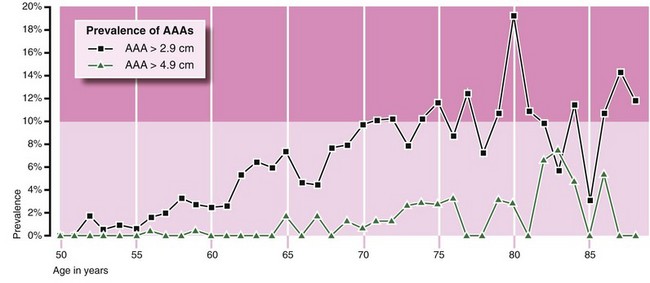
The Huntingdon study: In the Huntingdon study, 15 000 men of 50 and over were screened over 7 years: 540 were found to have an AAA larger than 2.9 cm, and 69 an AAA larger than 4.9 cm (Fig. 6.1). Very few small and no large aneurysms were found under the age of 60, an important factor when planning screening. Over the age of 70, there was virtually a 10% incidence of small AAAs. In this study, AAA mortality also fell by 75% and the number needed to be invited to save one life was 600.
The UK MASS study: Screening was undertaken in four centres and 68 000 people were randomised to screening or not, beginning in 1997. Some 98% of people in both groups were matched with national mortality statistics. After 7 years, 21% of men had died. There was a 76% attendance among those invited (27 147 were screened) and 4.9% had an aortic diameter of 3 cm or greater. All-cause mortality fell by 4% in those invited and there was a 47% risk reduction for AAA deaths in this group. The cost of screening and treating detected aneurysms was £12 500 per life year saved and no adverse effects were found on quality of life. The 30-day mortality for elective cases was 6%; for emergency cases operated upon, mortality was 37%. It was calculated that if screening were offered to a population, only 6% of AAA workload would eventually be on ruptures compared with around 30%. After 10 years, the relative risk-reduction of aneurysm-related deaths was still 49% (CI: 37–57%), the cost per man invited was £100 and the cost per life year gained was £7600. Thus the benefit of a single screen lasted at least 10 years, though the incidence of rupture rose after 8 years.