Chapter 19 Sagittal magnetic resonance scans
Anyone who has undertaken the exercises described in Chapters 17 and 18, should find it straightforward to read sagittal, magnetic resonance image (MRI) scans of the lumbar spine. Those exercises asked the reader to expect the structures that lie anterior, lateral and posterior to the lumbar vertebral column. In plain radiographs, those structures were invisible, but in MRI scans, they are actually evident. Reading sagittal MRI scans, therefore, amounts to no more than matching expectation with what is actually evident.
1 Which parts of the vertebral column will be evident?
2 Which elements of the contents of the vertebral canal will be evident?
3 Which structures anterior to the vertebral column will be evident?
4 Which structures posterior to the vertebral column will be evident?
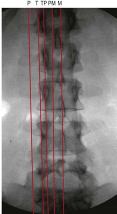
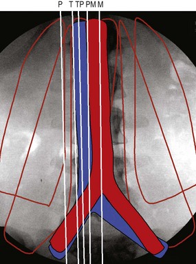
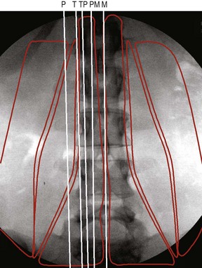
Sagittal MRI scans are typically taken across five standard planes (Fig. 19.1). Additional scans might be taken between these standard planes, or slightly displaced left or right from them; but the principles of analysis remain the same.
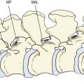
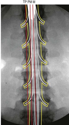
With respect to the contents of the vertebral canal, median, paramedian and transpedicular scans will intersect different components of the dural sac and its contents (Fig. 19.2). A median scan will intersect the conus medullaris of the spinal cord at upper segmental levels, and the cauda equina at lower levels. A paramedian scan may miss the spinal cord and show only the cauda equina. A transpedicular scan will intersect the spinal nerves and their dural sleeves, as they pass under the pedicles. Tangential and peripheral scans will not intersect the contents of the vertebral canal.
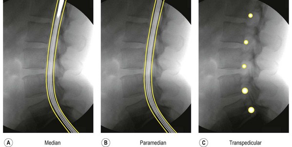
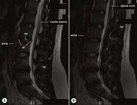
In a lateral view, the reader should, therefore, expect a median scan to show the dural sac, the conus medullaris of the spinal cord and the cauda equina (Fig. 19.3A). A paramedian scan would show only the cauda equina (Fig. 19.3B). A transpedicular scan would show only the spinal nerve roots and their sleeves (Fig. 19.3C).
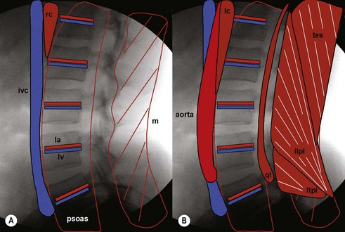
Anteriorly, different scans will variously intersect the crura of the diaphragm and the great vessels, or the psoas major and quadratus lumborum (Fig. 19.4). Posteriorly, the scans will pass through multifidus, longissimus thoracis, or iliocostalis lumborum (Fig. 19.5).
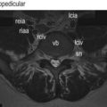
The actual appearance of individual structures: their longitudinal extent and their shape that should be expected on sagittal scans, can be derived from what the reader would expect to see on lateral radiographs. Across the concavities of the vertebral bodies, the reader would expect to encounter the lumbar arteries and lumbar veins, covered by psoas. (Fig. 19.6A). In front of the vertebral bodies, but to the right-hand side, they would expect the right crus and the inferior vena cava (Fig. 19.6A). Posteriorly, but behind the laminae, they would expect the multifidus (Fig. 19.6A). In front of the vertebral bodies, but more to the centre and to the left, the reader would expect the left crus and the aorta (Fig. 19.6B). Laterally, in front of the transverse processes they would expect the psoas major and the quadratus lumborum (Fig. 19.6B). Posteriorly, behind the transverse processes they would expect the various elements of the erector spinae (Fig. 19.6B).
Median scan
From Figure 19.1, the reader would expect that a median scan should intersect the vertebral bodies and intervertebral discs anteriorly, and the spinous processes posteriorly. Since it is a median scan, it should show the nucleus pulposus at the centre of each disc, and the anulus fibrosus anterior and posteriorly. From Figures 19.2 and 19.3, the reader would expect a median scan to intersect the conus medullaris and the cauda equina. From Figure 19.4, the reader would expect that a median scan should intersect the aorta. From Figure 19.6, the reader would expect the aorta to terminate opposite L4. It thus transpires that this is what a median MRI scan shows (Fig. 19.7).
Paramedian scan
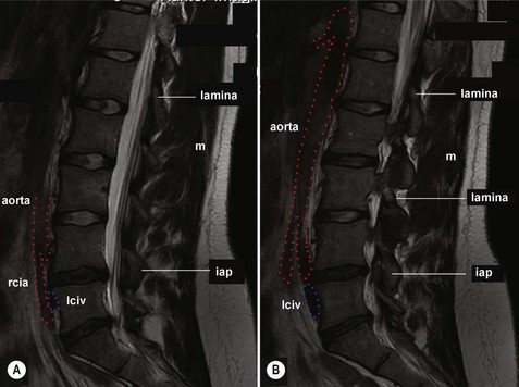
From Figure 19.1, the reader would expect that a paramedian scan should intersect the vertebral bodies and intervertebral discs anteriorly, and the laminae posteriorly (or the more medial elements of the zygapophysial joints). It should show the nucleus pulposus at the centre of each disc, and the anulus fibrosus anteriorly and posteriorly. From Figures 19.2 and 19.3, the reader would expect a paramedian scan to intersect the cauda equina. Paramedian scans that are to the right of the midline should pass near or through the right-side edge of the aorta, and in front of L5 they should pass through the right common iliac artery passing to the right, and covering the left common iliac vein passing to the left (Fig. 19.4). Paramedian scans to the left of the midline should substantially intersect the aorta. Posteriorly, the scan should pass through the multifidus. It thus transpires that this is what paramedian MRI scans show (Fig. 19.8).
Transpedicular scans
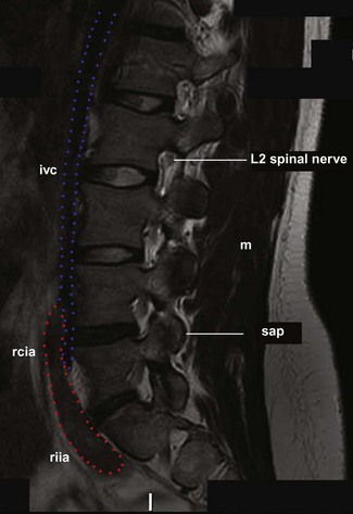
From Figure 19.1, the reader would expect that a transpedicular scan should intersect the vertebral bodies and intervertebral discs anteriorly, and the pedicles and zygapophysial joints posteriorly. From Figures 19.2 and 19.3, the reader would expect a transpedicular scan to intersect the lumbar spinal nerve in their intervertebral foramina. Transpedicular scans on the left most likely will pass lateral to the aorta, although they might intersect the more lateral segments of the aorta. On the right, transpedicular scans should intersect the inferior vena cava (Fig. 19.4). In front of L5 they should pass through the right common iliac artery (Fig. 19.4). Posteriorly, the scan should pass through the multifidus. It thus transpires that this is what paramedian MRI scans show (Fig. 19.9).
Tangential scans
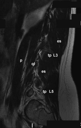
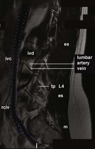
From Figure 19.1, the reader would expect that a tangential scan should pass through the edges of the vertebral bodies and intervertebral discs, and across the concavities of the vertebral bodies. From Figure 19.6, the reader would expect a transpedicular scan to intersect the lumbar arteries and veins. Transpedicular scans on the left most likely will pass lateral to the aorta, although they might intersect the more lateral segments of the aorta (Fig. 19.4). On the right, transpedicular scans should intersect the inferior vena cava (Fig. 19.4). In front of L5 they should pass through the right common iliac artery (Figs. 19.4, 19.6). Posteriorly, from Figure 19.5, the reader would expect tangential scans to intersect the erector spinae at most levels, and the multifidus at lumbosacral levels. It thus transpires that this is what paramedian MRI scans show (Fig. 19.10).
Peripheral scans
From Figure 19.1, the reader would expect that a peripheral scan should pass through the transverse process. From Figures 19.4, 19.5 and 19.6, the reader would expect a peripheral scan to intersect the psoas major and quadratus lumborum anteriorly, and the erector spinae posteriorly. It thus transpires that this is what peripheral MRI scans do (Fig. 19.11).