CHAPTER 80 Sacral Fractures
Anatomic and Biomechanical Considerations
The sacrum is the lowest functional portion of the spinal column and provides an anchor for the mobile lumbar spine. Lumbosacral motion occurs through the lumbosacral intervertebral disc and the paired zygapophyseal (facet) joints. In addition to the usual intervertebral stabilizing structures, the lumbosacral articulation is further stabilized by the iliolumbar ligaments connecting the L5 transverse processes to the crest of the ilium and the sacrolumbar ligaments that have an origin contiguous with the iliolumbar ligament and insert into the anterosuperior aspect of the sacrum and sacroiliac joint.1 These structures make the L5-S1 motion segment inherently more stable than the more cephalad lumbar motion segments.
The sacrum has an inclination that is balanced by a cascade of lumbar lordosis, thoracic kyphosis, and cervical lordosis so that a plumb line from C7 typically passes through the posterior aspect of the L5-S1 intervertebral disc. The sacral inclination angle, defined as the angle between a tangent to the dorsal surface of the sacrum and a vertical reference line, varies between 10 and 90 degrees and is usually between 45 and 60 degrees.2,3 Pelvic incidence measures the orientation of the lumbosacral junction relative to the pelvis and approximates 50 degrees in the general population.4 It is normally referred to in the context of sagittal plane malalignment in lumbar spondylolisthesis but is also valuable in determining sagittal alignment when treating sacral fractures. Sacral fractures that significantly alter spinal sagittal alignment, usually by shifting the C7 plumb line anteriorly, may result in difficulty with maintaining an erect posture, compensatory hyperlordosis, and associated functional deficits.5
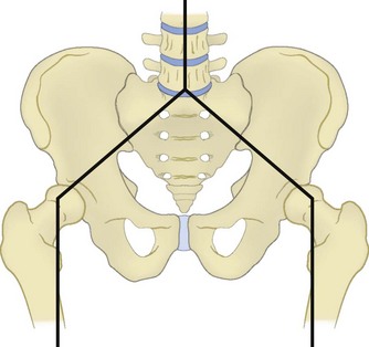
The sacrum forms the posterior segment of the pelvic ring through the sacroiliac joints. Forces transmitted through the lumbosacral articulation into the upper sacrum propagate laterally across the sacroiliac joints to the pelvis (Fig. 80–1). Because of these load-transferring properties, the sacrum has been described as the keystone of the pelvic ring.6 More specifically, the sacrum functions as a “true keystone” in the pelvic outlet plane, in which the orientation of the sacrum relative to the ilium is such that axial forces lock the sacrum into the pelvic ring and further stabilize the sacroiliac articulation. In the pelvic inlet plane the sacrum is shaped like a “reverse keystone” and its position in this plane is inherently unstable, permitting pelvic ring motion (Fig. 80–2). This pelvic ring instability necessitates substantial intrinsic and extrinsic ligamentous stabilization of the sacroiliac joints. Intrinsic stability is provided by the substantial interosseous ligaments and posterior sacroiliac ligaments, as well as the relatively weaker anterior sacroiliac ligaments. Extrinsic stability is provided by the sacrospinous and sacrotuberous ligaments in the pelvic floor.7
The sacrum itself is composed of five kyphotically aligned vertebral segments. The upper two sacral segments are similar in size to the lumbar vertebrae, whereas the lower three sacral segments rapidly diminish in size.8 Intersegmental fusion begins between the lowest two segments around the age of 15 and proceeds in a cephalad direction until the sacrum is completely fused by 25 to 30 years of age. Significant variability in upper sacral anatomy can exist in the form of transitional vertebrae and sacral dysplasia. Forms of transitional vertebrae include a sacralized lumbar segment, an incomplete congenital fusion of L5 to the sacrum that is usually unilateral, and a lumbarized sacrum that denotes a persistent articulation between the S1 and S2 segments.9 Complete or bilateral sacralization may result in a dysplastic sacral morphology with the upper sacrum located cephalad relative to the iliac crests. Because upper sacral variability results in significant alteration in the relationships among the sacrum, pelvis, and spinal column, as well as their adjacent neurovascular structures, it must be recognized during the evaluation and treatment of sacral fractures, particularly if surgical treatment is considered.10
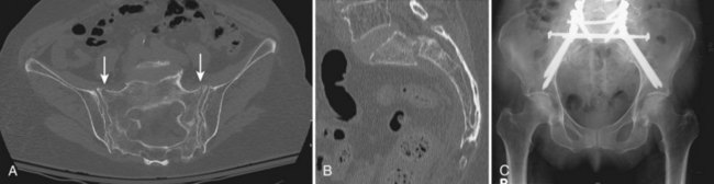
The upper sacral body, termed the base of the sacrum, consistently has the highest density of cancellous bone, with the S1 segment adjacent to the superior endplate having the greatest density overall. The ventral aspect of the upper S1 body that projects anteriorly and superiorly into the pelvis is termed the sacral promontory. The sacral ala, the lateral portion of the sacrum that articulates with the ilium through the sacroiliac joints, is largely cancellous and is formed by the coalescence of the sacral transverse processes. There is wide variability in sacral alar morphology among individuals, and the ala can be a substantial broad shoulder of bone or a narrow, partially segmented structure. The cancellous alar bone is hypodense, particularly in older individuals, and an alar void is a consistent finding in middle-aged and older adults (Fig. 80–3).11,12 The relative difference in bone density between the upper and lower sacral body predisposes this area to fracture. The hypodense ala is predisposed, particularly in the older and osteopenic person, to fracture line propagation. This effect is accentuated by the relative strength of the sacroiliac joint ligaments that stabilize and prevent their displacement or disruption. Similarly, younger individuals injured before complete ossification of the sacrum are predisposed to disruption at the S1-2 level owing to relative weakness at that interval. The relatively sparse alar bone density must be taken into consideration when planning reconstructive procedures.
The posterior surface of the sacrum is convex and is formed by the coalescence of posterior elements. Accordingly, the middle sacral crest corresponds to the spinous processes, the intermediate sacral crests correspond to the zygapophyseal joints, and the area in between corresponds to the laminae. The lowest one or two sacral segments have incompletely formed bony posterior elements, resulting in an aperture into the sacral spinal canal: the sacral hiatus. Enlargement of the sacral hiatus may relatively weaken the sacrum and predispose it to fracture.13
Sacral pedicles and laminae form the lateral and posterior borders of the spinal canal as it tapers in size distally from a triangular cross section at the S1 level to a flat and narrow cross section at the most caudal segments. The dural sac typically ends at the S2 level, and the filum terminale attaches its caudal end to the coccyx. At the junction of the body and the sacral ala are four paired ventral and dorsal neuroforamina through which the sacral nerve root ventral and dorsal rami pass, respectively. The relative space available to the sacral nerve roots in the ventral foramina is lowest at the S1 and S2 levels, where the nerve roots occupy one third to one fourth of the foraminal space compared with the S3 and S4 levels, where the nerve roots occupy one sixth of the available foraminal space. It follows that the lower sacral roots are less likely to be impinged upon in injuries involving displacement of the neuroforamina.14
Numerous structures are adjacent to the sacrum, which may be injured in sacral fracture-dislocations. The majority are neurovascular structures, with the rectum being the only visceral structure in close proximity. Below the rectosigmoid junction, located at the S3 level, the mesentery disappears and the colon lies directly adjacent to the sacrum.15
The dorsal nerve roots exit through their respective posterior neuroforamina to supply motor branches to the paraspinous muscles and cutaneous sensory branches that form the cluneal nerves. Anteriorly, the L5 nerve root is intimately associated with the sacrum as it passes underneath the inferior edge of the sacrolumbar ligament and drapes over the anterosuperior aspect of the sacral ala. It anastomoses with the L4 ventral ramus and in passing caudad adjacent to the sacral ala is joined by the exiting ventral sacral nerve roots to form the sacral plexus.16 Branches of the sacral plexus include the sciatic nerve, pudendal nerve, superior gluteal nerve, and inferior gluteal nerve. In addition, the sympathetic chain lies directly on the ventral sacrum immediately medial to the neuroforamina. At each level it distributes a gray ramus communicans to the ventral nerve root. Therefore the sacral plexus mediates major lower extremity function, as well as bowel, bladder, and sexual function. The dual innervation of the perineal structures from both the left and right sacral plexus is protective of bowel, bladder, and sexual function. These functions are largely preserved in the event of transection of the sacral nerve roots unilaterally, whereas bilateral transection causes complete loss of function.17
The presacral area has an extensive vascular network that is highly variable. The middle sacral artery typically courses ventrally along the midline of the L5 vertebral body, across the sacral promontory, and down the sacrum after branching from the aorta at the common iliac bifurcation. The common iliac arteries subsequently give rise to the internal iliac arteries that lie anterior to the sacroiliac joints and give off both superior and inferior lateral sacral arteries. The superior lateral sacral artery traverses the upper sacroiliac joint and proceeds caudad just lateral to the sacral foramina, giving off spinal arteries that pass through the S1 and S2 ventral foramina to the spinal canal. The inferior lateral sacral artery traverses the inferior aspect of the sacroiliac joint before anastomosing with the middle sacral artery and giving off spinal arteries that pass through the S3 and S4 ventral foramina.9 The internal iliac veins lie posteromedially to the internal iliac arteries and course caudad. They are located medial to the sacroiliac joint directly adjacent to the sacral ala.15 The internal iliac veins are the repository of an extensive presacral venous plexus. This plexus is formed by extensive anastomoses between the lateral and middle sacral veins and receives transforaminal veins that communicate with epidural veins in the spinal canal.1,8,18 This extensive vascular network renders anterior exposures to the sacrum impractical and perilous.
History and Classification
Bonnin reported the first classification of sacral fractures in 1945.19 He divided sacral fractures by mechanism of injury into those occurring as a result of either direct or indirect forces. Direct injuries occurred from a projectile or a fall. These were isolated injuries to the sacrum and had minimal or no impact on either spinal or pelvic instability. Conversely, with indirect injuries resulting from the transmission of forces through a disrupted pelvic ring, he described several anatomic patterns of injury including the propensity of the sacrum to fracture through the S1 and S2 neuroforamina at the junction of the ala and the body, resulting in a “broken link in the solid connections between the ilium and the vertebral column.” In addition to pelvic ring and/or spinal column instability, anatomic patterns of injury that included the neuroforamina or spinal canal were noted to be associated with possible neurologic deficit. Specifically, S1 or S2 nerve root injury was common in his cases. Bowel and bladder dysfunction as a result of lower sacral nerve root injury was sometimes also seen.19
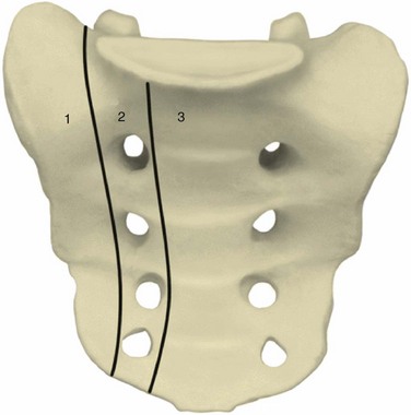
Several other classification schemes based on the identification of additional injury patterns were subsequently proposed.20,21 None was widely adopted until 1988 when Denis and colleagues,14 based on a series of 236 sacral fractures, formulated a simplified anatomic classification that correlates fracture location with the incidence of neurologic injury. This classification divides the sacrum into three zones (Fig. 80–4). Zone I, or alar zone, fractures remain lateral to the neuroforamina throughout their course. Zone II, or foraminal zone, fractures are located in the transition area between the sacral ala and body and involve one or more neuroforamina while remaining lateral to the spinal canal. Zone III, or central zone, fractures involve the spinal canal. The key relationship is that the incidence of neurologic injury increases as fractures are more centrally located. Accordingly, in the series of Denis and colleagues, zone I fractures had a 5.9% incidence of neurologic injury, primarily to the L5 nerve root as it coursed over the ala. Zone II fractures had a 28.4% incidence of associated neurologic injury occurring as a result of either foraminal displacement and resulting impingement on the exiting nerve root or the “traumatic far-out syndrome” in which that L5 nerve root is caught between the L5 transverse process and the displaced sacral ala. Zone III fractures had a 56.7% incidence of neurologic injury resulting from injury at the level of the spinal canal, with 76.1% of these individuals having bowel, bladder, and sexual dysfunction. Denis and colleagues14 noted a spectrum of zone III sacral fracture-dislocations including both transverse and longitudinal fracture line orientations and described a subtype that they termed a sacral burst fracture consisting of retropulsion of the sacral body with an intact sacral lamina.
Gibbons and colleagues22 subsequently considered 44 cases of sacral fracture according to the Denis classification system. They found a 34% incidence of neurologic injury overall. Six of 25 patients (24%) with zone I injuries had L5 and/or S1 nerve root injuries. Two of seven patients with zone II injuries similarly had L5 and S1 deficits. No patient with zone I or II injuries had bowel or bladder dysfunction. Three of five patients with zone III injuries had neurologic injury, two with bladder dysfunction. Gibbons and colleagues22 used these findings as a basis for a classification of neurologic injury from sacral fractures. Neurologic injuries were classified as 1, none; 2, paresthesias only; 3, motor loss with bowel and bladder intact; and 4, bowel and/or bladder dysfunction. They also noted that significant neurologic deficit is rare in transverse sacral fractures below the S4 level.
Huittinen,23 in a postmortem study of 42 individuals with posterior pelvic ring disruptions, studied the incidence and anatomic basis of nerve injuries. A total of 40 lumbosacral nerve injuries were identified in 20 of the autopsies. Fifty-three percent were traction injuries and 38% were ruptures. Compression injuries, the most amenable to surgical intervention, were present in only 20% of the nerve injuries.
Because of the high potential for neurologic injury, as well as spinal column instability, sacral body (Denis zone III) fractures have been specifically considered by several investigators. In 1969 Purser reported a case of transverse fracture of the sacrum across the S1-2 interval with bilateral upper sacroiliac joint disruption and L5 transverse process avulsions.24 There was anterior angulation without significant translation of the upper sacrum. Although the fracture passed through the S1 neuroforamina, there was minimal foraminal displacement and the patient was neurologically intact. Subsequently, in 1976 Bucknill and Blackburne25 reported three cases of transverse fractures of the upper sacrum all associated with neurologic deficits. All three patients had lower extremity weakness, and one had bladder dysfunction. Weaver26 and colleagues described transverse sacral fractures as analogous to the traumatic L5-S1 spondylolisthesis described by Nicoll in 194927 that resulted from a flexion moment applied in a position of hip flexion and knee extension. Schmidek and colleagues agreed that transverse sacral fractures are a variation of traumatic L5-S1 spondylolisthesis that he attributed to the relative bony weakness in the upper sacrum, particularly in skeletally immature patients before ossification is complete.21 Fountain and colleagues28 further differentiated upper transverse sacral fractures from lower transverse sacral fractures that occur from a direct fall resulting in the coccyx levering the sacrum and fracturing it at the S3 level, the apex of the sacrum, and the caudal end of the sacroiliac joint. They reported a series of six patients who had three upper and three lower sacral fractures. All six initially had neurologic dysfunction from cauda equina injury manifesting as urinary retention, decreased anal sphincter tone, or both. There have been several other case reports of transverse fractures of the sacrum. Review of these case reports is remarkable for a high association of neurologic deficit characterized as cauda equina injury affecting lower extremity, as well as bowel and bladder function.20,26,29–37
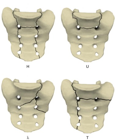
Early case reports often characterized the injury pattern as solely a transverse fracture, possibly owing to imaging limitations. Computed tomography (CT) demonstrates that most transverse fractures of the upper sacrum have complex fracture patterns. The majority of these injuries are now understood to consist of a transverse fracture of the sacrum with associated longitudinal or “vertical” injury components, usually in the form of bilateral transforaminal fractures that extend rostrad to the lumbosacral junction, the so-called “U” fracture. Variations in fracture line propagation include the “H,” “Y,” and “lambda” fracture patterns (Fig. 80–5). There is also a high incidence of L5 transverse process fractures, indicating disruption of the iliolumbar ligament.33
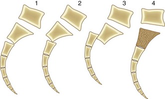
Roy-Camille and colleagues38 reviewed their experience of 13 patients with transverse sacral fractures, 11 as a result of attempted suicide by jumping. They classified the fractures as type I, flexion deformity of upper sacrum (angulation alone); type II, flexion deformity with posterior displacement of the upper sacrum on the lower sacrum (angulation and posterior translation); and type III, anterior displacement of the upper sacrum without angulation (anterior translation alone). On the basis of cadaveric studies, they hypothesized that types I and II were caused by impact with the lumbar spine in flexion, whereas type III fractures were caused by impact with the lumbar spine and hips in extension.38 Strange-Vognsen and Lebech39 subsequently reported a case of comminution of the upper sacrum without significant angulation or translation that they termed a type IV injury relative to the Roy-Camille classification. They hypothesized that in type IV injuries the lumbar spine was in the neutral position at the point of impact (Fig. 80–6).
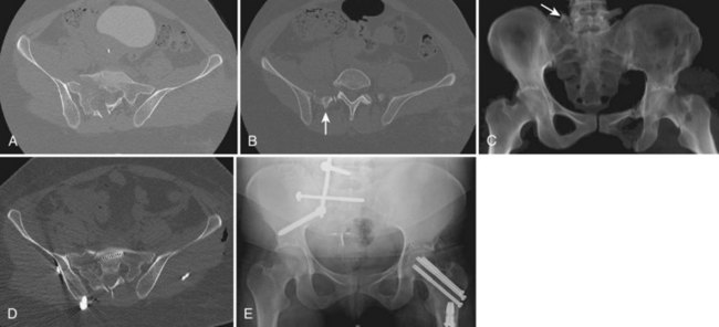
Other specific patterns of sacral fractures have been identified as resulting from specific mechanisms or having predictable patterns of associated injuries. In particular, in contrast to transverse Denis zone III sacral fractures, midline longitudinal Denis zone III sacral fractures have a low incidence of neurologic injury (Fig. 80–7).40–42 This injury, in which the sacrum is disrupted through a sagittal plane, was originally reported by Wiesel and colleagues in 1979, who suggested that these fractures may have a lower incidence of neurologic injury than transverse fractures because the nerve roots are displaced laterally instead of transected.42 Bellabarba and colleagues40 subsequently described this injury as a variant of the anteroposterior compression pelvic ring injury. In their series of 10 patients, none had neurologic deficits, in contradistinction to the high incidence of neurologic injury reported in patients with predominantly transverse sacral fractures through the spinal canal.40
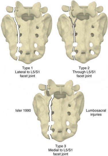
Even in the absence of a transverse fracture line, sacral fractures can be associated with spinal column instability. Isler described variations of longitudinal sacral fractures through the S1 and S2 neuroforamina that result in L5-S1 motion segment instability owing to facet joint disruption (Fig. 80–8).43 Injuries with the fracture line lateral to the S1 articular process are not associated with instability of the lumbosacral articulation because the L5-S1 articulation remains continuous with the stable fracture component of the sacrum. Fracture that extends into or medial to the S1 articular process, however, may disrupt the associated facet joint and potentially destabilize the lumbosacral junction (Fig. 80–9). Complete displacement of the facet joint can cause a locked facet joint, making sacral fracture reduction difficult with closed methods alone. Facet disruption may also cause post-traumatic arthrosis and late lumbosacral pain.
In contrast to sacral fractures that occur as a result of high-energy mechanisms of injury, the precipitating event in sacral insufficiency fractures is often not identifiable or may be related to a fall onto the buttocks from a standing or sitting position. Insufficiency fractures are stress fractures that occur in weakened bone, and these injuries typically occur in postmenopausal women due to osteoporosis. Conditions contributing to the osteoporosis such as chronic corticosteroid use or a history of radiation therapy to the pelvis are often present (Fig. 80–10).27,44 The presenting symptoms are often vague and frequently consist of poorly localized low back pain that may be exacerbated by sitting and standing. These fractures are typically oriented vertically and occur through the ala adjacent to the sacroiliac joint. There may also be a transverse component resulting in more complex “U”-fracture variants. Because of the predisposition of these patients to osteoporotic fractures, there is high association with insufficiency fractures at other sites, most commonly the pubis and thoracolumbar vertebrae. Neurologic deficits are uncommon in most series.45,46 Cauda equina dysfunction has been reported, however, and neurologic status must be carefully considered.47
Mechanical factors related to the transfer of forces from the lumbosacral spine to the pelvis may also cause sacral insufficiency fractures in the patient predisposed to osteoporosis.39 Specifically, unilateral sacral insufficiency fractures have been identified contralateral to the convex side of lumbar scoliosis.45 Pelvic stress concentration as the result of lumbosacral spine fusion has also been described,48 and sacral insufficiency fractures caudal to previous lumbar fusions are also increasingly being reported. These fractures probably represent failure of the sacrum to withstand forces concentrated there as a result of the large cephalad lever arm.49–53
Evaluation
Sacral fractures therefore occur in two distinctly different patient groups. Most commonly, sacral fractures are the result of high-energy trauma. As noted previously, they are also increasingly being seen in patients with osteopenic bone disorders that predispose to pathologic fractures. In both groups of patients, diagnosis is frequently delayed, which may result in further displacement or neurologic deterioration. In their retrospective analysis of 236 patients with sacral fractures, Denis and colleagues14 found that in the neurologically intact patient the diagnosis of sacral fractures was made during the initial hospitalization only 51% of the time. The presence of a neurologic deficit increased the diagnostic accuracy to only 70%. As in most cases of diagnostic delay, the etiology of missed sacral fractures is multifactorial and ranges from the relative difficulty in identifying these fractures on screening anteroposterior pelvis radiographs combined with the presence of distracting injuries in the trauma patient to low clinical suspicion in patients with insufficiency fractures.14,54
In the trauma patient the transfer of energy resulting in a fracture of the sacrum often causes other injuries including life-threatening head and thoracoabdominal trauma. In these patients emergent resuscitation is the top priority. Accordingly, Advanced Trauma Life Support (ATLS) protocol begins with a primary survey during which conditions that are immediately life threatening are addressed. Resuscitation is focused on maintaining cardiopulmonary and hemodynamic stability. Only after this primary goal has been achieved should the secondary survey, composed of examination of the patient to identify additional injuries, be completed.55
The secondary survey includes screening evaluation of both the spinal column and pelvic ring. Precautions to maintain spinal column integrity are necessary, and patients should be initially maintained on a flat surface and log-rolled side to side to prevent spinal column displacement. Evaluation includes inspection and palpation of the patient’s back from the occiput to the coccyx. Significant findings associated with sacral fractures are the presence of skin discoloration or laceration, palpable stepoff or crepitus, and ballotable fluid in the lumbopelvic region. Significant soft tissue contusion or internal degloving such as a Morel-Lavallee lesion can have important implications on subsequent treatment.56 Rectal and vaginal examination including the use of a speculum and proctoscope allows for detection of occult open sacral fractures.
Because pelvic ring disruption may be associated with significant intrapelvic hemorrhage, provisional methods of pelvic ring stabilization may be necessary to reduce pelvic volume and provide provisional stability. These methods include the application of a circumferential pelvic antishock sheet,10 pelvic clamp, anterior external fixator, or skeletal traction depending on the pelvic ring fracture pattern. Associated vascular injury, particularly to the hypogastric arterial system, may require embolization to adequately control arterial hemorrhage.57
Determining the patient’s neurologic status is of paramount importance in patients with sacral fractures.58–60 A rectal examination is performed early in the workup of all multiply injured patients, even in the absence of obvious sensorimotor deficits in the extremities, to evaluate perianal sensation, anal sphincter tone, and voluntary perianal contraction and to assess for presence of anal wink and the bulbocavernosus reflex. A straight-leg raise test should also be performed in the cognitively unimpaired patient to assess for entrapment of the lumbosacral plexus. Extremity motor function is graded on a scale of 0 to 5 according to the American Spinal Injury Association (ASIA) modification of the Frankel grading system, and a sensory level is obtained. Determination of level of injury by neurologic examination is limited to the L5 and S1 levels, however, and injuries to the lower sacral roots cannot be more specifically identified than obtaining a perianal sensory level.
Fracture displacement can cause neurologic injury from a variety of mechanisms including angulation, translation, and direct compression by displaced bone fragments. Potentially reversible injuries include contusion, compression, and traction. Recovery of transected or avulsed nerve roots cannot be expected. Delayed neurologic deficit can occur from epidural hematoma, late fracture displacement, or callus formation19 and should be promptly reinvestigated to determine its cause.
There are multiple adjunctive methods for the evaluation of the neurologic status in a patient with a sacral fracture. Electrodiagnostic studies are particularly valuable in the evaluation of cognitively impaired patients and are also useful in differentiating upper motor neuron injuries or spinal cord injury from cauda equina injury in patients with head or proximal spinal column injury, in the evaluation of patients with urinary tract injury, and for intraoperative monitoring.61 Conventional electromyography (EMG) and somatosensory-evoked potentials (SSEPs) are useful in the evaluation of L5 and S1 function. Pudendal SSEPs and anal sphincter EMG are necessary for evaluation of the sacral roots below the S1 level.62–65 Possible abnormalities on sphincter EMG include detrusor areflexia, uninhibited sphincter relaxation, or denervation. EMG is limited in the acute setting because these abnormalities may take weeks to emerge. For patients with neurogenic bladder, serial postvoid residuals or cystometrography is a useful diagnostic aid. In patients with elevated postvoid residuals, other causes such as impaired function due to narcotic inhibition must be considered.
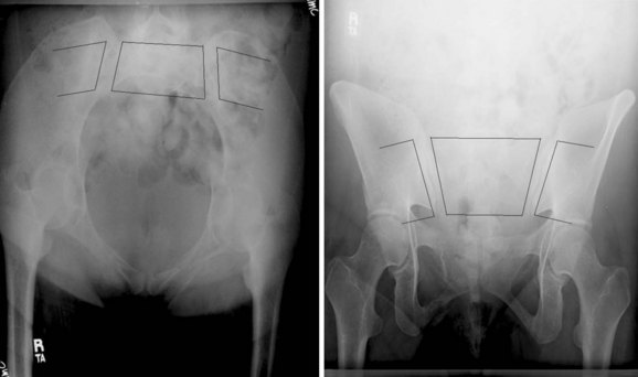
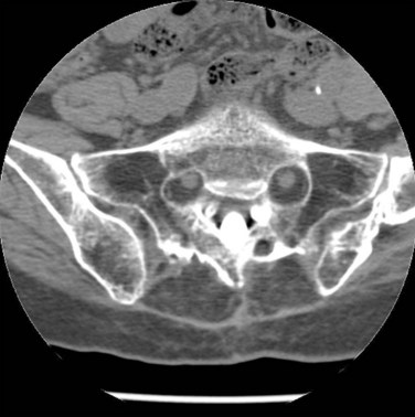
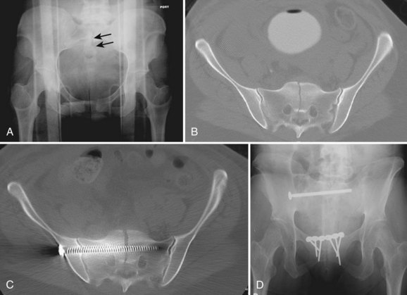
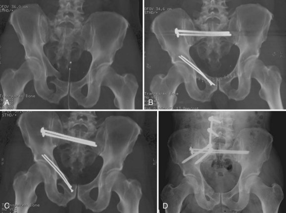
The advent of more sophisticated imaging techniques in the routine initial assessment of the trauma patient’s visceral injuries such as CT of the abdomen and pelvis has assisted the detection of previously unrecognized sacral fractures. The identification of a sacral fracture on these studies mandates complete radiographic evaluation including a dedicated CT scan of the sacrum with 2-mm or less axial cuts and sagittal and coronal re-formations to allow the detail required for determining the fracture configuration, resulting instability pattern, and extent of sacral canal and neuroforaminal compromise (Fig. 80–11).66 Three-dimensionally reformatted CT scans may add insight into fracture morphology for less experienced clinicians or in the case of highly complex fracture configurations. Magnetic resonance imaging (MRI) is not usually helpful except in cases of unclear neurologic deficits or discrepancies between skeletal and neurologic levels of injury, although it may provide early evidence of lumbosacral nerve root avulsion.67 Recent advances in MRI neurography allowing for visualization of the lumbosacral plexus injuries may be of some help in their evaluation, although at this point it is neither a reliably effective nor a practical diagnostic tool and more clinical experience with this modality is necessary.
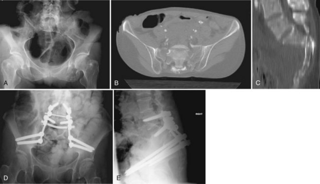
FIGURE 80–11 A to C, Sacral U fracture in suicidal jumper. D and E, After treatment with lumbopelvic fixation.
General Principles
Decision making in sacral fracture treatment is primarily based on the fracture pattern and neurologic status. Other key factors are the patient’s general medical condition and additional injuries, particularly in the trauma patient. Hemodynamic instability or compromised pulmonary function may preclude early surgical stabilization in critically injured patients. Conversely, the benefits of early mobilization in trauma patients with pulmonary injuries may make early surgical stabilization advisable.68,69 Surgical timing in patients with closed-head injuries in particular is controversial, and skilled perioperative management is essential to prevent secondary brain injury.70,71 Isolated sacral fractures in the patient who is otherwise healthy may be amenable to a period of recumbency followed by protected weight bearing. Chronic medical conditions also need to be considered and may require an initial period of nonoperative stabilization before surgical intervention while medical conditions are optimized.
Careful examination of the fracture pattern is essential in determining if the sacral fracture is associated with instability of the spinal column/pelvic ring weight-bearing axis. Unilateral longitudinal fractures through the ala or foraminal zone maintain continuity of the contralateral weight-bearing axis, allowing weight bearing on that side (Fig. 80–12, see Fig. 80–9). Bilateral displaced longitudinal fractures, as well as transverse fractures with associated bilateral longitudinal fractures through the upper sacrum, dissociate the spinal column from the pelvic ring and represent complete disruption of the weight-bearing axis.38,72–74 Weight bearing on either lower extremity may cause displacement in these instances. Conversely, transverse fractures below the sacroiliac joint have no implication on the weight-bearing axis as long as they are not associated with secondary fracture lines extending proximally.75
Neurologic deficit needs to be correlated with fracture anatomy. Residual compression of nerve roots at the level of the spinal canal or neuroforamina due to the impingement of bony fragments or malalignment of the spinal canal at the fracture site with the cauda equina draping over a kyphotic ridge should be identified. Possible neurologic deterioration from persistent fracture instability should also be considered. A review of case reports of sacral fractures with neurologic deficit indicates that the effectiveness of operative decompression in facilitating neurologic recovery is unclear, and improvement has been reported with both operative and nonoperative management* Each case needs to be individually considered, and in our opinion surgical decompression should be considered in the presence of a potentially reversible neurologic injury.
Nonoperative Treatment
Nonoperative treatment in displaced, high-energy sacral fractures can be problematic, however. Although surgical complications can be avoided by using closed treatment methods, potential disadvantages include life-threatening pulmonary and thromboembolic events that are associated with prolonged recumbency in patients with multiple injuries. Other potential complications are the development of decubitus ulcers, the inability to reliably relieve sacral canal and neuroforaminal compression, and the potential for late instability causing neurologic deficits.76
Nonoperative treatment is most attractive in unilateral fractures that are minimally displaced and have no associated neurologic deficits. Mobilization is usually with a walker or crutches enabling a toe-touch weight-bearing status on the injured side. These patients must be followed carefully for displacement, which may warrant surgical stabilization. Insufficiency or stress fractures are also usually amenable to nonoperative treatment in spite of frequent bilateral involvement. Along with activity modification and correction of underlying metabolic conditions, nonoperative treatment has a high likelihood of success in the majority of insufficiency and stress fractures and is usually the treatment of choice in these fractures.47
Neurologic Decompression
Decompression of the neural elements can be achieved by either direct or indirect means. Indirect decompression can sometimes be achieved simply with fracture reduction. An attempt at indirect decompression is best accomplished before consolidation of the fracture hematoma.77 If neural impingement persists and is associated with a neurologic deficit, direct decompression should be considered.
In patients with sacral root deficits, direct decompression alone by laminectomy has been advocated as a method for providing sacral root decompression and therefore theoretically enhancing the possibility of neurologic recovery while minimizing the potential for complications associated with more extensive dissection and surgical stabilization.28 Surgical exposure is preferably performed through a straight posterior midline approach. The parasagittal approach is not recommended because it limits exposure to the spine and may result in the need for bilateral parasagittal exposures, which are even less desirable owing to the potential for soft tissue necrosis. Intraoperative fluoroscopy is useful for orientation and to assess alignment and decompression of the spinal canal. Decompression can be performed focally for selective ventral foraminal impingement or to achieve a more comprehensive decompression of the S1-4 neural elements. In the instance of L5 root entrapment between the L5 transverse process and an alar bone fragment, decompression is performed by following the root laterally onto the shoulder of the ala and removing the offending fragment.
Surgical Stabilization Techniques
Surgical stabilization is performed to avoid prolonged recumbency in the critically injured patient, as an adjunct to neurologic decompression, and as the most effective method to correct or prevent displacement that might adversely affect postural alignment or predispose to chronic pain or nerve compression. An association between residual displacement and chronic pain is debatable, however, and its cause in the trauma patient may be mostly due to associated injuries. Recently published functional outcome studies indicate that a minority of sacral fracture patients returned to their preinjury vocational status over a year after injury. Although the vast majority of patients sustained long-term physical and mental impairment, the severity of these impairments did not correlate directly with fracture characteristics but was more a function of associated injuries and sacral root function.78,79 It has been theorized that restoration of appropriate sagittal alignment of the sacral fracture leads to decreased pain by allowing for a more physiologic alignment of the lumbar spine and, more specifically, preventing compensatory lumbar hyperlordosis.5 Pelvic incidence can therefore be used as an intraoperative guide to lumbopelvic alignment and, therefore, adequacy of reduction.
Roy-Camille38 described a technique for direct osteosynthesis of sacral fractures with sacral alar plates. The plates are placed lateral to the dorsal foramina and oriented vertically. The orientation of these plates is theoretically optimal to allow for compression loading across a transverse fracture, but, in the more common multiplanar, comminuted fractures, the construct is often compromised by poor screw purchase in comminuted or osteopenic bone. The construct also cannot effectively address associated nontransverse fracture planes and does not provide adequate strength to neutralize the enormous forces across the lumbopelvic junction, requiring a 2- to 4-month period of restricted weight bearing. Because of these limitations, it is our opinion that sacral alar plating alone has few clinical applications. Its utility is usually in combination with other methods of surgical stabilization in the treatment of sacral fractures involving the weight-bearing axis.80 Direct plating alone, however, may be potentially useful in maintaining alignment of transverse sacral fractures below the sacroiliac joints because these fractures are not subject to the high loads seen with fractures involving the weight-bearing axis, and the goal in their treatment is primarily to avoid pain due to prominence or nonunion.75
Anterior pelvic ring stabilization, whether by open reduction and internal fixation or external fixation, is inadequate to stabilize an unstable fracture of the sacrum.81,82 Sacral fractures treated by pelvic ring stabilization require posterior stabilization. Various methods of posterior pelvic ring fixation including sacral bars, tension band plating, and iliosacral screws have all been described as methods to stabilize the posterior pelvic ring.61,83 Simonian and Routt,82 in a biomechanical study of cadaveric specimens, found no difference in the resulting pelvic ring stability between the various constructs. Sacral bars and tension band plating are both predisposed to soft tissue problems, owing to their location superficial to the nearly subcutaneous dorsal sacrum.83 The potential for dorsal soft tissue compromise is minimized by the use of iliosacral screws, and this method has become widely used for posterior pelvic ring fixation. In all methods of posterior pelvic ring stabilization, however, fixation is perpendicular to the weight-bearing axis and does not provide adequate stabilization for immediate weight bearing.84,85
Iliosacral screw fixation is usually performed in a percutaneous manner and, consequently, blood loss is minimal. The insertion is dependent on the surgeon’s ability to achieve an anatomic reduction under adequate fluoroscopic visualization; otherwise, in addition to the problem of malreduction, a safe trajectory for iliosacral screw placement cannot be reliably established.61,86 It can be performed with the patient in either a supine or prone position assisting either concurrent anterior pelvic ring stabilization or open decompression of foraminal debris. In spite of these advantages, iliosacral screw fixation has several potential pitfalls. In the instance of comminuted longitudinal fractures through the neuroforamina, overcompression of the foramina can occur if the screws are placed using interfragmentary compression, which can potentially lead to nerve root injury.81 However, in experienced centers the reported rate of neurologic injury is low, even without the use of electrodiagnostic monitoring.87 Like other methods of posterior pelvic ring stabilization, percutaneous iliosacral screw fixation carries the disadvantage of not allowing for reduction of sacral angulation. It is also dependent on screw purchase into the iliac wing and sacrum that may be decreased in instances of comminution or osteoporosis with the risk of fixation failure by the screws displacing within the sacral body. As mentioned previously, the orientation of the screw perpendicular to the fracture’s deforming forces may also contribute to a higher likelihood of fixation failure (see Fig. 80–12). Nevertheless, iliosacral screw fixation has been shown to be an effective method of stabilization in unstable longitudinal sacral fractures.88 A 3-month period of protected weight bearing is recommended with iliosacral screw fixation.
Contrary to sacral bars and posterior tension band plating, which are largely ineffective in stabilizing multiplanar sacral fractures with a transverse component, iliosacral screws can also be used successfully in patients with minimally displaced sacral “U” fracture variants. In a series of 13 patients without cauda equina deficits who had fractures with minimal sacral angulation and displacement allowing for in situ screw placement, Nork and colleagues89 found bilateral percutaneous iliosacral screw fixation along with an 8- to 12-week period of immobilization in a thoracolumbosacral-hip orthosis to be safe and effective in treating this type of injury. However, this minimally invasive method does not allow for reduction of fracture angulation. Moreover, if a near-anatomic reduction cannot be achieved, the safe zone for iliosacral screw trajectory may be small or absent, thus also precluding its use in more complex and highly displaced injuries.86 Iliosacral screw fixation alone is therefore not recommended for patients with displaced sacral “U” fractures and their variants if they are irreducible by closed manipulation or if neural decompression is required.
Lumbopelvic fixation provides the biomechanically strongest fixation of sacral fractures.90 This technique was derived from the Galveston method of anchoring the caudal end of long thoracolumbar rod constructs into the ileum. Sacral fracture fixation is obtained rostrally by segmental pedicle screw fixation in the lumbosacral spine and caudally by long screw fixation in the ileum.91–102 The construct spans the sacrum and mimics the normal load transfer from the lumbar spine to the pelvis.53,90 The strength of the construct permits immediate weight bearing without the use of external bracing (see Fig. 80–11).
Lumbopelvic fixation is performed through a dorsal approach with the patient in the prone position. The longitudinally oriented constructs are placed laterally, adjacent to the posterior iliac wings. The lateral placement also allows for posterior decompression of the neural elements, as well as open reduction of the sacral fracture. Screw placement is performed under fluoroscopic guidance to ensure correct screw orientation. A thorough understanding of pelvic anatomy and facility with fluoroscopic positioning is necessary for verification of correct screw placement. Screw malposition can be catastrophic, potentially injuring neurovascular structures in the sciatic notch or the pelvic viscera or penetrating the acetabulum. If placed correctly the hardware is low profile. Infection and wound-related problems are common, however, and have been found to approach 20%.92,97 A formal sacroiliac joint arthrodesis is not usually performed, and in many instances the rod will break from fatigue failure, an expected consequence of sacroiliac joint motion. Late rod fracture is asymptomatic in the majority of cases, and the need for routine hardware removal is questionable (see Fig. 80–11).92,97
The optimal stabilization of complex sacral fractures may require the use of multiple methods of fixation. For instance, sacral “U” or “H” fractures can be stabilized by the combined use of sacral alar plates for the transverse fracture component and iliosacral screw fixation for the longitudinal fracture component. Lumbopelvic fixation can also be combined with iliosacral screw fixation, the so-called triangular osteosynthesis technique, to obtain optimal fixation in the vertical direction along the weight-bearing axis and the horizontal direction stabilizing the pelvic ring.90,96 Lumbopelvic fixation of sacral fractures requiring neural decompression may benefit from sacral plating, which in this case is used solely to fine-tune fracture realignment and prevent recurrent displacement of bone into the spinal canal, allowing the lumbopelvic fixation to neutralize the bulk of the loads being transferred across the sacrum.
Summary
Pearls
Key Points
1 Denis F. Sacral fractures. An important problem-a retrospective analysis of 236 cases. Clin Orthop. 1988;227:67-81.
2 Gibbons KJ, Soloniuk DS, Razack N. Neurological injury and patterns of sacral fractures. J Neurosurg. 1990;72:889-893.
3 Huittinen VM. Lumbosacral nerve injury in fracture of the pelvis. Acta Chir Scand Suppl. 1972;429:7-43.
4 Peretz AM, Hipp JA, Heggeness MH. The internal bony architecture of the sacrum. Spine. 1998;23:971-974.
5 Routt MLC, Nork SE, Mills WJ. Percutaneous fixation of pelvic ring disruptions. Clin Orthop. 2000;375:15-29.
6 Schildhauer TA, Ledoux WR, Chapman JR, et al. Triangular osteosynthesis and iliosacral screw fixation for unstable sacral fractures: A cadaveric and biomechanical evaluation under cyclic loads. J Orthop Trauma. 2003;17:22-31.
7 Bellabarba C, Schildhauer TA, Vaccaro AR, Chapman JR. Complications associated with surgical stabilization of high-grade sacral fracture dislocations with spino-pelvic instability. Spine. 2006;31(11 Suppl):S80-S88.
8 Schildhauer TA, Bellabarba C, Nork SE, et al. Decompression and lumbopelvic fixation for sacral fracture-dislocations with spino-pelvic dissociation. J Orthop Trauma. 2006;20:447-457.
1 Williams PL, Warwick R, Dyson M, et al, editors. Gray’s Anatomy, American ed. New York: Churchill Livingstone, 1989.
2 Jackson RP, McManus AC. Radiographic analysis of sagittal plane alignment and balance in standing volunteers and patients with low back pain matched for age, sex, and size. Spine. 1994;19:1611-1618.
3 Wiltse LL, Winter RB. Terminology and measurement of spondylolisthesis. J Bone Joint Surg Am. 1983;65:768-772.
4 Legaye J, Duval-Beaupere G, Hecquet J, et al. Pelvic incidence: a fundamental pelvic parameter for three dimensional regulation of spinal sagittal curves. Eur Spine J. 1998;7:99-103.
5 Hart RA, Badra MI, Madala A, Yoo JU. Use of pelvic incidence as a guide to reduction of H-type spino-pelvic dissociation injuries. J Orthop Trauma. 2007;21:369-374.
6 Schmidek HH, Smith D, Kristiansen TK. Sacral fractures: Issues of neural injury, spinal stability, and surgical management. In: Dunsker SB, et al, editors. The Unstable Spine. New York: Harcourt, 1986.
7 Tile M. Fractures of the Pelvis and Acetabulum, 2nd ed. Baltimore: Williams & Wilkins; 1995.
8 Morris H, Lond MB, editors. Human Anatomy. Philadelphia: P. Blakiston’s Son & Co, 1989.
9 Williams P, Warwick R, Dyson M, editors. Gray’s Anatomy, 37th (British) ed, Edinburgh: Churchill Livingstone, 1989.
10 Routt ML, Simonian PT, Agnew SG, Mann FA. Radiographic recognition of the sacral alar slope for optimal placement of iliosacral screws: A cadaveric and clinical study. J Orthop Trauma. 1996;10:171-177.
11 Peretz AM, Hipp JA, Heggeness MH. The internal bony architecture of the sacrum. Spine. 1998;23:971-974.
12 Smith SA, Abitbol JJ, Carlson GD, et al. The effects of depth of penetration, screw orientation and bone density on sacral screw fixation. Spine. 1993;18:1006-1010.
13 Carter SR. Stress fracture of the sacrum: Brief report. J Bone Joint Surg. 1987;69:843-884.
14 Denis F, Davis S, Comfort T. Sacral fractures: An important problem-a retrospective analysis of 236 cases. Clin Orthop. 1988;227:67-81.
15 Mirkovic S, Abitbol JJ, Steinman J, et al. Anatomic consideration for sacral screw placement. Spine. 1991;16:S289-S294.
16 Esses SI, Botsford DJ, Huler RJ, Rauschning W. Surgical anatomy of the sacrum: A guide for rational screw fixation. Spine. 1991;16:S283-S288.
17 Gunterberg B. Effects of major resection of the sacrum. Acta Orthop Scand Suppl. 1976;162:1-38.
18 Grant’s Atlas of Anatomy, 8th ed. Baltimore: Williams & Wilkins. 1983.
19 Bonnin JG. Sacral fractures. J Bone Joint Surg Am. 1945;27:113-127.
20 Sabiston CP, Wing PC. Sacral fractures. Classification and neurologic implications. J Trauma. 1986;26:1113-1115.
21 Schmidek HH, Smith DA, Kristiansen TK. Sacral fractures. Neurosurgery. 1984;15:735-746.
22 Gibbons KJ, Soloniuk DS, Razack N. Neurological injury and patterns of sacral fractures. J Neurosurg. 1990;72:889-893.
23 Huittinen VM. Lumbosacral nerve injury in fracture of the pelvis. Acta Chir Scand Suppl. 1972;429:7-43.
24 Purser DW. Displaced fracture of the sacrum: Report of a case. J Bone Joint Surg Br. 1969;51:346-347.
25 Bucknill TM, Blackburne JS. Fracture-dislocations of the sacrum: Report of three cases. J Bone Joint Surg Br. 1976;58:467-470.
26 Weaver EN, England GD, Richard DE. Sacral fracture: case presentation and review. Neurosurgery. 1981;9:725-728.
27 Nicoll EA. Fractures of the dorsolumbar spine. J Bone Joint Surg Br. 1949;31:376.
28 Fountain SS, Hamilton RD, Jameson RM. Transverse fractures of the sacrum: A report of six cases. J Bone Joint Surg Am. 1977;59:486-489.
29 Carl A, Delman A, Engler G. Displaced transverse sacral fractures: A case report, review of the literature, and the CT scan as an aid in management. Clin Orthop. 1985;194:195-198.
30 Ebraheim NA. Zone III fractures of the sacrum. Spine. 1996;21:2390-2396.
31 Fardon DF. Displaced fracture of the lumbosacral spine with delayed cauda equina deficit: Report of a case and review of literature. Clin Orthop. 1976;120:155-158.
32 Ferris B. Anteriorly displaced transverse fracture of the sacrum at the level of the sacroiliac joint: A report of two cases. J Bone Joint Surg Am. 1983;65:407-409.
33 Fisher RG. Sacral fracture with compression of cauda equina: Surgical treatment. J Trauma. 1988;28:1678-1680.
34 Phelan ST, Jones DA, Bishay M. Conservative management of transverse fractures of the sacrum with neurological features: A report of four cases. J Bone Joint Surg Br. 1991;73:969-971.
35 Rao SH. Traumatic transverse fracture of sacrum with cauda equine injury: A case report and review of literature. J Postgrad Med. 1998;44:14-15.
36 Rodriguez-Fuentes AE. Traumatic sacrolisthesis S1-S2. Spine. 1993;18:768-771.
37 Yasuda T, Shikata J, Iida H, Yamamuro T. Upper sacral transverse fracture: A case report. Spine. 1990;15:589-591.
38 Roy-Camille R, Saillant G, Gogna G, Mazel C. Transverse fracture of the upper sacrum: Suicidal jumper’s fracture. Spine. 1985;10:838-845.
39 Strange-Vognsen HH, Lebech A. An unusual type of fracture in the upper sacrum. J Orthop Trauma. 1991;5:200-203.
40 Bellabarba C, Stewart JD, Ricci WM, et al. Midline sagittal sacral fractures in anterior-posterior compression ring injuries. J Orthop Trauma. 2003;17:32-37.
41 Hatem SF, West OC. Vertical fracture of the central sacral canal: Plane and simple. J Trauma. 1996;40:138-140.
42 Wiesel SW, Zeide MS, Terry RL. Longitudinal fracture of the sacrum: Case report. J Trauma. 1979;19:70-71.
43 Isler B. Lumbosacral lesions associated with pelvic ring injuries. J Orthop Trauma. 1990;4:1-6.
44 Herman MP, Kopetz S, Bhosale PR, et al. Sacral insufficiency fractures after preoperative chemoradiation for rectal cancer: incidence, risk factors, and clinical course. Int J Radiat Oncol Biol Phys. 2009;74:818-823.
45 Gotis-Graham I, McGuigan L, Diamond T, et al. Sacral insufficiency fractures in the elderly. J Bone Joint Surg Br. 1994;76:882-886.
46 Weber M, Haster P, Gerber H, et al. Insufficiency fractures of the sacrum: Twenty cases and review of the literature. Spine. 1993;18:2507-2512.
47 Jacquot JM, Finiels H, Fardjad S, et al. Neurological complications in insufficiency fractures of the sacrum: Three case reports. Rev Rhum Engl Ed. 1999;66:109-113.
48 Wood KB, Geissele AE, Ogilvie JW. Pelvic fractures after long lumbosacral spine fusions. Spine. 1996;21:1357-1362.
49 Elias WJ, Shaffrey ME, Whitehill R. Sacral stress fracture following lumbosacral arthrodesis: Case illustration. J Neurosurg. 2002;96(1 Suppl):135.
50 Fourney DR, Prabhu SS, Cohen ZR, et al. Early sacral stress fractures after reduction of spondylolisthesis and lumbosacral fixation: Case report. Neurosurgery. 2002;51:1510-1511.
51 Klineberg E, McHenry T, Bellabarba C, et al. Sacral insufficiency fractures caudal to instrumented posterior lumbosacral arthrodesis. Spine. 2008;33:1806-1811.
52 Mathews V, McCance SE, O’Leary PF. Early fracture of the sacrum or pelvis: An unusual complication after multilevel instrumented lumbosacral fusion. Spine. 2001;26:E571-E575.
53 Wood KB, Schendel MJ, Ogilvie JW, et al. Effect of sacral and iliac instrumentation on strains in the pelvis. Spine. 1996;21:1185-1191.
54 Laasonen EM. Missed sacral fractures. Ann Clin Res. 1977;9:84-87.
55 ATLS manual. In Crosby LA, Lewallen DO, editors: Emergency Care and Transportation of the Sick and Injured, 6th ed, American Academy of Orthopedic Surgeons, 1995.
56 Kellam JF, McMurtry RY, Paley D, Tile M. The unstable pelvic fracture: Operative treatment. Orthop Clin North Am. 1987;18:25-41.
57 Ben-Menachem Y, Coldwell DM, Young JW, Burgess AR. Hemorrhage associated with pelvic fractures: Causes, diagnosis, and emergent management. AJR Am J Roentgenol. 1991;157:1005-1014.
58 Byrnes DP, Russo GL, Duckert TB, Cowley RA. Sacrum fractures and neurological damage: Report of two cases. J Neurosurg. 1977;47:459-462.
59 Goodell CL. Neurological deficits associated with pelvic fractures. J Neurosurg. 1966;24:837-842.
60 Lam CR. Nerve injury in fractures of the pelvis. Ann Surg. 1936;104:945-951.
61 Routt MLJr, Simonian PT. Closed reduction and percutaneous skeletal fixation of sacral fractures. Clin Orthop. 1996;329:121-128.
62 Cohen BA, Major MR, Huizenga BA. Pudendal nerve evoked potential monitoring in procedures involving low sacral fixation. Spine. 1991;16:S375-S378.
63 Helfet DL, Koval KJ, Hissa EA, et al. Intraoperative somatosensory evoked potential monitoring during acute pelvic fracture surgery. J Orthop Trauma. 1995;9:28-34.
64 Kothbauer K, Schmid UD, Seiler RW, Eisner W. Intraoperative motor and sensory monitoring of the cauda equina. Neurosurgery. 1994;34:702-707.
65 Slimp JC. Electrophysiologic intraoperative monitoring for spine procedures. Phys Med Rehabil Clin North Am. 2004;15:85-105.
66 Kuklo TR, Potter BK, Ludwig SC, et al. Spine Trauma Study Group: Radiographic measurement techniques for sacral fractures consensus statement of the Spine Trauma Study Group. Spine. 2006;31:1047-1055.
67 Sasaka KK, Phisitkul P, Boyd JL, et al. Lumbosacral nerve root avulsions: MR imaging demonstration of acute abnormalities. AJNR Am J Neuroradiol. 2006;27:1944-1946.
68 Bone LB, Johnson KD, Weigelt J, Scheinberg R. Early versus delayed stabilization of femoral fractures: A prospective randomized study. J Bone Joint Surg Am. 1989;71:336-340.
69 Johnson KD, Cadambi A, Seibert GB. Incidence of adult respiratory distress syndrome in patients with multiple musculoskeletal injuries: Effect of early operative stabilization of fractures. J Trauma. 1985;25:375-384.
70 Jaicks RR, Cohn SM, Moller BA. Early fracture fixation may be deleterious after head injury. J Trauma. 1997;42:1-5.
71 Scalea TM, Scott JD, Brumback RJ, et al. Early fracture fixation may be “just fine” after head injury: No difference in central nervous system outcomes. J Trauma. 1999;46:839-846.
72 Marcus RE, Hansen ST. Bilateral fracture-dislocation of the sacrum: A case report. J Bone Joint Surg Am. 1984;66:1297-1299.
73 Pennal GF, Tile M, Waddell JP, Garside H. Pelvic disruption: Assessment and classification. Clin Orthop. 1980;151:12-21.
74 Wild J, Hanson GW, Tullas HS. Unstable fractures of the pelvis treated by external fixation. J Bone Joint Surg Am. 1982;64:1010-1020.
75 Sommer C. Fixation of transverse fractures of the sternum and sacrum with the locking compression plate system: two case reports. J Orthop Trauma. 2005;19:487-490.
76 Latenser BA, Gentilello LM, Tarver AA, et al. Improved outcome with early fixation of skeletally unstable pelvic fractures. J Trauma. 1991;31:28-31.
77 Pohlemann T, Angst A, Schneider E, et al. Fixation of transforaminal sacrum fractures: A biomechanical study. J Orthop Trauma. 1993;2:107-117.
78 Tötterman A, Glott T, Søberg HL, et al. Pelvic trauma with displaced sacral fractures: functional outcome at one year. Spine. 2007;32:1437-1443.
79 Tötterman A, Glott T, Madsen JE, Røise O. Unstable sacral fractures: associated injuries and morbidity at 1 year. Spine. 2006;31:E628-E635.
80 Templeman D, Goulet J, Dawelius PJ, et al. Internal fixation of displaced fractures of the sacrum. Clin Orthop. 1996;329:180-185.
81 Routt MLC, Simonian PT, Swiontkowski MF. Stabilization of pelvic ring disruptions. Orthop Clin North Am. 1997;28:369-388.
82 Simonian PT, Routt MLJr. Biomechanics of pelvic fixation. Orthop Clin North Am. 1997;28:351-368.
83 Suzuki T, Hak DJ, Ziran BH, et al. Outcome and complications of posterior transiliac plating for vertically unstable sacral fractures. Injury. 2009;40:405-409.
84 Suzuki K, Mochida J. Operative treatment of a transverse fracture-dislocation at the S1-S2 level. J Orthop Trauma. 2001;15:363-367.
85 Taguchi T, Kawai S, Kaneko K, Yugue D. Operative management of displaced fractures of the sacrum. J Orthop Sci. 1999;4:347-352.
86 Reilly MC, Bono CM, Litkouhi B, et al. The effect of sacral fracture malreduction on the safe placement of iliosacral screws. J Orthop Trauma. 2003;17:88-94.
87 Gardner MJ, Farrell ED, Nork SE, et al. Percutaneous placement of iliosacral screws without electrodiagnostic monitoring. J Trauma. 2009;66:1411-1415.
88 Routt MLC, Nork SE, Mills WJ. Percutaneous fixation of pelvic ring disruptions. Clin Orthop. 2000;375:15-29.
89 Nork SE, Jones CB, Harding SP, et al. Percutaneous stabilization of U-shaped sacral fractures using iliosacral screws: Technique and early results. J Orthop Trauma. 2001;15:238-246.
90 Schildhauer TA, Ledoux WR, Chapman JR, et al. Triangular osteosynthesis and iliosacral screw fixation for unstable sacral fractures: A cadaveric and biomechanical evaluation under cyclic loads. J Orthop Trauma. 2003;17:22-31.
91 Acharya NK, Bijukachhe B, Kumar RJ, Menon VK. Ilio-lumbar fixation–the Amrita technique. J Spinal Disord Tech. 2008;21:493-499.
92 Bellabarba C, Schildhauer TA, Vaccaro AR, Chapman JR. Complications associated with surgical stabilization of high-grade sacral fracture dislocations with spino-pelvic instability. Spine. 2006;31(11 Suppl):S80-S88.
93 O’Brien JR, Yu WD, Bhatnagar R, Sponseller P, Kebaish KM. An anatomic study of the S2 iliac technique for lumbopelvic screw placement. Spine. 2009;34:E439-E442.
94 Sagi HC. Technical aspects and recommended treatment algorithms in triangular osteosynthesis and spinopelvic fixation for vertical shear transforaminal sacral fractures. J Orthop Trauma. 2009;23:354-360.
95 Schildhauer TA, McCullough P, Chapman JR, Mann FA. Anatomic and radiographic considerations for placement of transiliac screws in lumbopelvic fixations. J Spinal Disord Tech. 2002;15:199-205.
96 Schildhauer TA, Josten C, Muhr G. Triangular osteosynthesis of vertically unstable sacrum fractures: A new concept allowing early weight-bearing. J Orthop Trauma. 1998;12:307-314.
97 Schildhauer TA, Bellabarba C, Nork SE, et al. Decompression and lumbopelvic fixation for sacral fracture-dislocations with spino-pelvic dissociation. J Orthop Trauma. 2006;20:447-457.
98 Schildhauer TA, Bellabarba C, Selznick HS, et al. Unstable pediatric sacral fracture with bone loss caused by a high-energy gunshot injury. J Trauma. 2007;63:E95-E99.
99 Schildhauer TA, Chapman JR, Mayo KA. Multisegmental open sacral fracture due to impalement: a case report. J Orthop Trauma. 2005;19:134-139.
100 Strange-Vognsen HH, Kiaer T, Tondevold E. The Cotrel-Dubousset instrumentation for unstable sacral fractures: Report of 3 patients. Acta Orthop Scand. 1994;65:219-220.
101 Vilela MD, Gelfenbeyn M, Bellabarba C. U-shaped sacral fracture and lumbosacral dislocation as a result of a shotgun injury: case report. Neurosurgery. 2009;64:E193-E194.
102 Lebwohl NH, Cunningham BW, Dmitriev A, et al. Biomechanical comparison of lumbosacral fixation techniques in a calf spine model. Spine. 2002;27:2312-2320.


