S
saccade See movements, fixation.
saccadic eye movement See movement, saccadic eye.
saccadic oscillation See flutter, ocular; myoclonus, ocular; opsoclonus.
saddle bridge See bridge, saddle.
safety glass See glass, safety.
sag Abbreviation for sagitta or sagittal depth; the height of a segment of a circle or sphere.
See lens measure; vertex depth.
sagittal depth See sag; vertex depth.
sagittal focus See astigmatism, oblique.
sagittal plane See plane, sagittal.
saline, physiological A 0.9% sterile solution of sodium chloride in water. This concentration of sodium chloride is considered approximately isotonic with the tears. It is used to store and rinse soft contact lenses, to irrigate the eye, etc. Syn. normal saline; NaCl 0.9%.
See eyewash; irrigation; solution, isotonic.
‘salt and pepper’ fundus See fundus, salt and pepper.
Salzmann’s nodular degeneration See degeneration, Salzmann’s nodular.
sample See sampling.
sampling The selection of a group of subjects from a population. This is usually done for the purpose of experimentation. The part of the population selected is called the sample: it is usually considered to be representative of a given population. A good sample must be random, i.e. every possible member of that population has an equal chance of being selected. Otherwise, it is said to be biased. Sampling can extend either across geographical areas (spatial sampling) or over a period of time (temporal sampling).
Sampaolesi’s line See line, Sampaolesi’s.
Sandhoff’s disease See disease, Sandhoff’s.
sarcoidosis An idiopathic, multisystem granulomatous disorder in which the eye is affected in about 20% of patients. Ocular manifestations include anterior uveitis with aqueous flare, keratoconjunctivis sicca, lacrimal gland lesion, ‘mutton-fat’ keratic precipitates, posterior synechia, iris granulomas and nodules, intermediate uveitis, posterior uveitis or optic neuropathy.
See dacryoadenitis; iridocyclitis; keratopathy, band; keratopathy, exposure; syndrome, Mikulicz’s.
sarcoma Malignant tumour formed by proliferation of mesodermal cells.
Sattler’s layer See layer, Sattler’s.
Sattler’s veil Clouding of vision accompanied by seeing coloured haloes around lights caused by corneal oedema resulting from contact lens wear, more frequently the hard lens type (PMMA). The cause has been shown to be due to a circular diffraction pattern formed by the basal epithelial cells and the extracellular spaces. Syn. Fick’s phenomenon.
See clouding, central corneal.
saturation Attribute of a visual sensation, which permits a judgment to be made of the proportion of pure chromatic colour in the total sensation. Note: This attribute is the psychosensorial correlate, or nearly so, of the colorimetric quantity purity (CIE).
scale, Snell–Sterling visual efficiency See visual efficiency scale, Snell–Sterling.
scanning laser polarimetry See polarimetry, scanning laser.
scatter illumination, sclerotic See illumination, sclerotic scatter.
scatter, Tyndall See effect, Tyndall.
scattering, Rayleigh Diffusion of radiation in the course of its passage through a medium containing particles the size of which is small compared with the wavelength of the radiation (CIE).
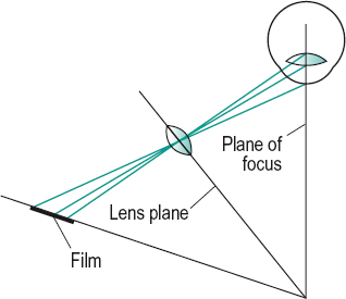
Scheimpflug photography An imaging technique that provides an assessment of the anterior segment of the eye in a sagittal plane. The principle of the technique is that the film plane, lens plane and subject plane (plane of focus) are tilted in such a way that all three planes intersect along a straight line. In this condition a subject will be completely in focus. The technique can be used to detect and monitor opacities in the media of the anterior segment (e.g. in the lens), to document these changes and to carry out biometric evaluation (e.g. depth of the anterior chamber, lens thickness measurement) (Fig. S1).
Scheiner’s disc; experiment; test See under the nouns.
schematic eye See eye, schematic.
Schiötz tonometer See tonometer, impression.
Schirmer’s test See test, Schirmer’s.
Schlemm’s canal See canal, Schlemm’s.
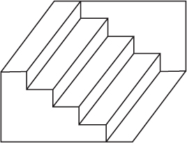
Schroeder’s staircase This is an ambiguous figure that gives rise to two different perceptions. It consists of a drawing of parallel step-like lines extending from the upper corner of a parallelogram to the lower opposite corner. One either sees a staircase from underneath or a staircase from above and, upon continuous viewing, the impression alternates (Fig. S2). Syn. Schroeder’s staircase visual illusion.
See figure, Blivet; Necker cube; Rubin’s vase.
Schwalbe, anterior limiting ring of; line See ring of Schwalbe, anterior limiting.
Schwann cell See cell, Schwann.
science of vision See vision science.
scimitar scotoma See scotoma, arcuate.
scintillans, synchisis See synchisis scintillans.
scintillating scotoma See scotoma, scintillating.
scissors movement See movement, scissors.
sclera The tough, white, opaque, fibrous outer tunic of the eyeball covering most of its surface (the cornea contributes 7% of, and completes, the outer tunic). Its anterior portion is visible and constitutes the ‘white’ of the eye. In childhood (or in pathological conditions) when the sclera is thin, it appears bluish, while in old age it may become yellowish, due to a deposition of fat. The sclera is thickest posteriorly (about 1 mm) and gradually becomes thinner towards the front of the eyeball. It is a sieve-like membrane at the lamina cribrosa. The sclera is pierced by three sets of apertures: (1) the posterior apertures round the optic nerve and through which pass the long and short posterior ciliary vessels and nerves; (2) the middle apertures, 4 mm behind the equator which give exit to the vortex veins; and (3) the anterior apertures through which pass the anterior ciliary vessels. The tendons of insertion of the extraocular muscles run into the sclera as parallel fibres and then spread out in a fan-shaped manner. The sclera is commonly considered to be divided into three layers from without inward: (1) the episclera, (2) the scleral stroma and (3) the suprachoroid (lamina fusca) which is interposed between choroid and sclera. Syn. sclerotic. Note: some authors consider the suprachoroid as belonging to the choroid. However, when choroid and sclera are separated part of the suprachoroid adheres to the choroid and part to the sclera.
See cribriform plate; evisceration.
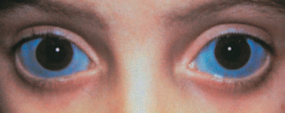
blue s. A hereditary defect in which the sclera has a bluish appearance. The sclera is thinner than normal and is susceptible to rupture if the person engages in contact sports. It is often associated with fragility of the bones and deafness as part of a condition called osteogenesis imperfecta (fragilitas ossium, van der Hoeve’s syndrome), with keratoconus or with acquired scleral thinning (e. g. necrotizing scleritis). Syn. blue sclerotic (Fig. S3).
See syndrome, Ehlers-Danlos; syndrome, Marfan’s.
scleral buckling See retinal detachment, rhegmatogenous.
scleral crescent See crescent, myopic.
scleral contact lens; ectasia; indentation; rigidity; ring See under the nouns.
scleral size, back Maximum internal diameter of the back surface of a scleral contact lens before the outer sharp edge has been rounded. Syn. back haptic size.
scleral spur A ridge of the sclera at the level of the limbus interposed between the posterior portion of Schlemm’s canal and the anterior part of the ciliary body. The scleral spur is the structure to which some of the ciliary muscle fibres are attached.
See gonioscopy; sulcus, internal scleral.
scleral zone The portion of a scleral contact lens designed to lie in front of the sclera. Syn. haptic.
sclerectasia See ectasia, scleral.
sclerectomy Surgical removal of a portion of the sclera performed in glaucoma surgery. A common procedure is to make a conjunctival flap at the limbus, followed by a full thickness scleral opening and iridectomy.
deep s. A type of non-penetrating filtration surgery aimed at lowering intraocular pressure by dissecting a superficial scleral flap and excising a deeper partial thickness scleral flap below leaving a thin membrane consisting of trabeculum and Descemet’s membrane through which the aqueous humour diffuses. It then drains from the anterior chamber to the subconjunctival space or though Schlemm’s canal. A collagen implant is usually inserted under the superficial flap to improve drainage of aqueous humour.
scleritis Inflammation of the sclera, which in its severe necrotizing or in the posterior type may cause sight-threatening complications such as keratitis, uveitis, angle-closure glaucoma or optic neuropathy. It affects females more commonly than males in the fourth to sixth decades of life. Like episcleritis it has a tendency to recur. It is characterized by pain, which can be severe, redness, tearing and some patients may develop nodules (nodular scleritis). It is often associated with a systemic disease (e.g. rheumatoid arthritis, Wegener’s granulomatosis, polyarteritis nodosa, lupus erythematosus, ankylosing spondylitis, syphilis, herpes zoster). It can involve part of the sclera, e.g. anterior scleritis (which is the most common, and it is classified as diffuse non-necrotizing or nodular non-necrotizing) or posterior scleritis. Treatment includes topical and systemic steroids and immunosuppressive drugs for very severe cases.
See keratitis, acute stromal; syndrome, Brown’s superior oblique tendon sheath.
necrotizing s. The most severe form of scleritis, much less common than the other types. About half the patients have one of the following diseases: rheumatoid arthritis, Wegener’s granulomatosis, polyarteritis nodosa, systemic lupus erythematosus, or herpes zoster. It is characterized by pain, and white, avascular areas next to damaged areas through which one can see the brown colour of the underlying uveal tissue, and to congested areas of the sclera. In most cases visual acuity is decreased. The necrosis gradually spreads around the globe. Treatment typically consists of topical steroids, immunosuppressive agents and occasionally surgery to repair scleral or corneal perforation.
See keratolysis; scleromalacia. s.
necroticans See scleromalacia.
posterior s. Inflammation of the sclera involving the posterior segment of the eye. The condition is often associated with a systemic disease (e.g. rheumatoid arthritis). It is characterized by pain and reduced visual acuity. The severity of the visual impairment depends on the involved tissue and its location. Signs include eyelid oedema, proptosis, limitation of ocular movements and, if anterior scleritis is present, redness. The ocular fundus may present disc swelling, choroidal folds, macular oedema and serous retinal detachment. Treatment consists mainly of systemic steroids and immunosuppressive agents.
See choroidal folds.
sclerochoroiditis Inflammation of the sclera and the choroid. It can occur as a result of pathological myopia and it presents with a posterior staphyloma in the region of the optic disc.
scleroconjunctival Pertaining to the sclera and the conjunctiva.
scleroconjunctivitis Inflammation of the sclera and of the conjunctiva.
sclerocornea A rare, congenital condition in which the sclera and cornea are considered as a single layer. The limbus is ill defined and portions of opaque scleral tissue with conjunctival vessels cover the cornea. The condition is usually bilateral and frequently associated with cornea plana. Visual acuity is reduced and often it is merely light perception if the entire cornea is involved. The eye is usually hyperopic. Systemic associations include mental retardation, deafness and craniofacial abnormalities. Treatment includes correction of the refractive error but in cases of central corneal opacification keratoplasty may be indicated.
scleroderma Thickening and hardening of the skin due to new collagen formation. It occurs either in localized or in systemic disease. It may very occasionally produce keratoconjunctivitis sicca. Syn. dermatosclerosis.
scleroiritis Inflammation of the sclera and of the iris.
sclerokeratitis Inflammation of the sclera and of the cornea.
scleromalacia A bilateral and painless degenerative thinning of the sclera occurring in people with rheumatoid arthritis. In this condition rheumatoid nodules may develop in the sclera and cause perforation (scleromalacia perforans). Syn. necrotizing scleritis without inflammation; scleritis necroticans.
sclerosis, central areolar choroidal See dystrophy, central areolar choroidal.
sclerosis, multiple (MS) An autoimmune disease in which there are disseminated patches of demyelination and sclerosis (or hardening) of the brain, spinal cord and peripheral and optic nerves causing paralysis, tremor, disturbance of speech, nystagmus, diplopia due to involvement of the extraocular muscles, and frequently retrobulbar optic neuritis.
See myokymia; oscillopsia; potential, visual evoked cortical; pupil, Marcus Gunn; Uhthoff’s symptom.
sclerotic See sclera.
sclerotic scatter illumination See illumination, sclerotic scatter.
sclerotomy Surgical incision of the sclera. It may be performed to extract an intraocular cyst, or to drain a choroidal haemorrhage.
scopolamine hydrobromide See hyoscine hydrobromide.
scotoma An area of partial or complete blindness surrounded by normal or relatively normal visual field.
See angioscotoma; hemianopia; quadrantanopia.
absolute s . A scotoma in which vision is entirely absent in the affected area.
See retinoschisis; scotoma, relative.
annular s. See scotoma, arcuate; scotoma, ring.
arcuate s . Scotoma running from the blind spot into the nasal visual field and following the course of the retinal nerve fibres. A double arcuate scotoma extending both in the upper and lower part of the field may join to make an annular scotoma or ring scotoma. A common cause is glaucoma. Syn. comet scotoma; scimitar scotoma.
See fibres, arcuate; retinal raphe; scotoma, Bjerrum’s.
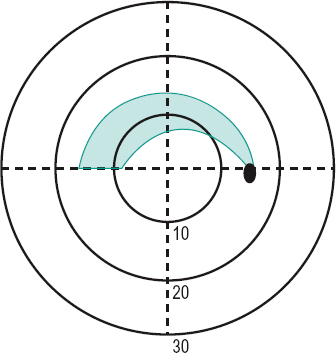
Bjerrum’s s . An arcuate scotoma extending around the fixation point (usually located between the 10° and 20° circles), which occurs in open-angle glaucoma. It often extends from the horizontal midline to the optic disc (Fig. S4). Syn. Bjerrum’s sign.
See Roenne nasal step; scotoma, Seidel’s.
central s. A scotoma involving the fixation area.
comet s. See scotoma, arcuate.
congruous s’. Scotomas in the two visual fields that are identical. They form a single defect in the binocular visual field. Such scotomas are often the result of lesions in the visual cortex.
flittering s. See scotoma, scintillating.
incongruous s’. Scotomas in the two visual fields that differ in one or more ways. Such scotomas are often the result of lesions in the optic tract.
junction s. A visual defect due to a lesion (e.g. a pituitary tumour) at the junction of one optic nerve with the chiasma where it is believed that the inferior nasal fibres of the contralateral optic nerve loop before passing backward to the optic tract. The visual defects typically consist of an upper temporal quadrantanopia in the field of the contralateral eye with, usually, a temporal hemicentral scotoma in the ipsilateral eye. Some authors attribute these visual defects to prechiasmal compression of one optic nerve plus compression of the whole chiasma.
See Wilbrand’s knee.
negative s. A scotoma of which the person is unaware. The physiological blind spot is an example of a negative scotoma but it is usually referred to as a physiological scotoma.
paracentral s. A scotoma involving the area adjacent to the fixation area.
physiological s. See scotoma, negative.
positive s. A scotoma of which the person is aware.
relative s. A scotoma in which there is some vision left or in which there is blindness to some stimuli, but not to others.
See scotoma, absolute.
ring s. 1. An annular scotoma surrounding the fixation point. It may be formed by the development of two arcuate scotomas. Syn. annular scotoma. 2. A circular area in the peripheral field of view at the edge of a strong convex spectacle lens which is not seen (Fig. S5). This scotoma is due to the prismatic effect at the edge of the lens and unlike other scotomas, not from a pathological condition. When the head turns the ring scotoma also turns and it is then called a roving ring scotoma.
See phenomenon, jack-in-the-box.
roving ring s. See phenomenon,jack-inthe-box; scotoma, ring.
scimitar s. See scotoma, arcuate.
scintillating s. The sudden appearance of a transient, shimmering scotoma with a zigzag outline of brightly coloured lights (also called a fortification spectrum or fortification figures). It usually occurs as one of the first symptoms of a migraine attack. Syn. flittering scotoma.

See migraine; teichopsia.
Seidel’s s. An arcuate scotoma extending above and below the blind spot found in glaucoma. Syn. Seidel’s sign.
scotometer An instrument such as a campimeter or a perimeter for detecting and plotting the position and magnitude of a scotoma.
See campimeter; perimeter.
scotopia See vision, scotopic.
scotopic eye See eye, dark-adapted.
scotopic sensitivity syndrome See syndrome, Meares-Irlen.
scotopic vision See vision, scotopic.
screen, Bjerrum’s See screen, tangent.
screen, Hess A black tangent screen for measuring and classifying strabismus. It consists of a chart divided by red lines into small sections of 5° separations. As the screen is flat, the lines are curved in a pincushion pattern (i.e. the middle of the lines is closer to the centre of the screen than the ends of the lines). At the respective positions on the lines in the eight major meridians are small red dots indicating positions 15° and 30° from the fixation point. Two green threads extend from the upper corners of the screen and meet a third green thread, which originates on the end of a pointer. The three threads form a figure Y. The patient, wearing a red lens in front of one eye and a green lens in front of the other, moves the pointer until the common origin of the green threads is superimposed on each of the red dots. The discrepancy between the two on the screen indicates the extent of the deviation in various directions of gaze. It is a particularly useful test to indicate an incomitant strabismus as the field plots obtained with each eye are compared and an estimate of which muscle/s is affected can be made. Moreover, if the inner (15°) and outer (30°) plots are affected differently the incomitancy is of mechanical origin. Note: there exists a computerized version of the Hess screen.
screen, Lees An instrument similar to the Hess screen. It consists of two internally illuminated screens placed at right angles. One eye views one screen directly, the other views the second screen via a mirror; hence, the eyes are dissociated. When one eye fixates a point on one illuminated screen, its position as seen by the second eye is indicated with a wand on the second, non-illuminated screen. When this is illuminated, the position of the wand relative to the screen markings indicates whether a muscle or set of muscles is paretic or not.
screen, tangent A large plane surface for detecting and plotting the central visual field (about 50° in diameter) by moving the position of a stimulus (e.g. a white 1 mm pinhead). It consists of dull black cloth or other material perpendicular to the line of sight and placed usually 1 m away from the subject (2 m gives more accuracy). In the centre of the screen is a white spot that provides a fixation point and a series of radial and circumferential lines are sewn or drawn to facilitate the localization of the stimulus. Syn. Bjerrum’s screen.
See campimeter; perimeter.
screener, vision See vision screener.
SEAL See staining, fluorescein.
sebaceous gland carcinoma See carcinoma, sebaceous gland.
sebum See glands, meibomian.
seclusio pupillae A complete blocking of the anterior chamber from the posterior chamber by a posterior annular synechia.
See glaucoma, inflammatory; pupillary block.
secondary deviation; glaucoma; position See under the nouns.
secretion 1 . The substance produced by a cell or organ (e.g. a gland). 2. Production by a cell or organ of a physiologically active substance. This flow out of a cell is driven by an osmotic pressure gradient across the membrane, which is created by active transport of one or more ion species from one side to the other.
See transport, active; ultrafiltration.
see 1 . To perceive by the eye. 2. To discern. 3. To note: to understand.
see-saw nystagmus See nystagmus.
segment height The vertically measured distance from the lowest point on the lens to the top of the segment of a bifocal (or trifocal) ophthalmic lens. Syn. seg height.
segment of a bifocal lens (seg) An area of a bifocal lens of a power different to that of the main portion. Syn. portion. There is the distance portion, which has the correction for distance vision, and the near or reading portion, which has the correction for near vision.
See portion, intermediate.
segment of the eye, anterior Portion of the eye comprising all the structures situated between the front surface of the cornea and the front surface of the vitreous. The eyelids are sometimes included in this definition.
segment of the eye, posterior Posterior portion of the eye comprising the vitreous humour, the retina, the optic disc, the choroid and most of the sclera.
Seidel aberration See aberration, monochromatic.
Seidel’s scotoma See scotoma, Seidel’s.
semidecussation See hemidecussation.
semi-finished lens See lens, semi-finished.
semilunar fold See plica semilunaris.
senile macular degeneration See macular degeneration, age-related.
senilis, arcus See arcus, corneal.
sensation The conscious response to the effect of a stimulus exciting any sense organ.
See perception.
sensation, visual A sensation produced by the sense of sight.
sense Any faculty (or ability) by which some aspect of the environment is perceived. The five main senses are those of sight, hearing, smell, taste and touch. The sense of sight may be further divided into the colour sense, the form sense, the light sense, the space sense, etc.
sense organ A structure especially adapted for the reception of stimuli and the transmission of the relevant information to the brain. The organ of sight is the eye, in which light is transduced into nerve signals in the photoreceptors of the retina.
sensitive period See period, critical.
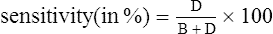
sensitivity 1. The capability of responding to or transmitting a stimulus. 2. The reciprocal of the threshold. 3. The extent to which a test gives results that are free from false negatives (i.e. people found not to have the defect when they actually have it). The fewer the number of false negatives, the greater is the sensitivity of the test. It is usually presented as a percentage of the number of people truly identified as defectives, referred to as true positives, A (or hit), divided by the total number of defective people tested. The total number includes all the true positives, A, plus the false negatives, C (or miss). Hence
< ?xml:namespace prefix = "mml" />
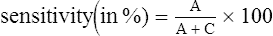
See specificity.
sensitivity, contrast The ability to detect luminance contrast. In psychophysical terms it is the reciprocal of the minimum perceptible contrast. The measurement of the contrast sensitivity of the eye is a more complete assessment of vision than standard visual acuity measurement. It provides an evaluation of the detection of objects (usually sinusoidal gratings) of varying spatial frequencies and of variable contrast and thus obtaining a contrast sensitivity function (CSF). Example: following amblyopia treatment, some cases still have the same visual acuity while the CSF is improved.
See chart, contrast sensitivity; function, contrast sensitivity; resolution, spurious; test, Arden grating; Vistech.
sensitivity, corneal The capability of the cornea to respond to stimulation. Corneal sensitivity to touch is assessed by an aesthesiometer that measures the corneal touch threshold (CTT), which is the reciprocal of corneal sensitivity. Sensitivity varies across the cornea, with the centre being the most sensitive. Diseases and rigid contact lens wear greatly reduce the sensitivity of the cornea.
See aesthesiometer; corneal touch threshold; hyperaesthesia, corneal.
sensitization 1 . A state or condition in which the response to a second or later stimulus (e.g. a drug) is greater than the response to the original stimulus (e.g. first administration of the drug). 2. The process in which exposure to an antigen results in the development of hypersensitivity.
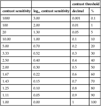
Table S1
Relationship between contrast sensitivity and contrast threshold (contrast sensitivity = 1/contrast threshold). Neither values have units
| contrast threshold | |||
| contrast sensitivity | log10 contrast sensitivity | decimal | % |
| 1000 | 3.00 | 0.001 | 0.1 |
| 100 | 2.00 | 0.01 | 1 |
| 20 | 1.30 | 0.05 | 5 |
| 10.00 | 1.00 | 0.1 | 10 |
| 5.00 | 0.70 | 0.2 | 20 |
| 3.33 | 0.52 | 0.3 | 30 |
| 2.50 | 0.40 | 0.4 | 40 |
| 2.00 | 0.30 | 0.5 | 50 |
| 1.67 | 0.22 | 0.6 | 60 |
| 1.43 | 0.15 | 0.7 | 70 |
| 1.25 | 0.10 | 0.8 | 80 |
| 1.11 | 0.05 | 0.9 | 90 |
| 1.00 | 0.00 | 1 | 100 |
sensory fusion See fusion, sensory.
septum orbitale See orbital septum.
serous chorioretinopathy See retinopathy, central serous.
sessile drop test See test, sessile drop.
sex chromosome See chromosome.
sex-linked recessive inheritance See inheritance.
shadow A darkened area from which rays from a source of light are excluded. The shadow pattern cast by light (e.g. sunlight, ceiling fixtures) is such a common sight that if light shines from the opposite direction (e.g. from the ground upward) the normal shadow pattern will be reversed and so will perception; depressions will appear as mounds or vice versa. Shadows offer a cue to depth perception, as when trying to judge the shape of objects.
See penumbra; perception, depth.
Shaffer classification See gonioscopy.
Shafer’s sign See sign, Shafer’s.
shagreen, crocodile Polygonal greyish-white corneal opacities separated by relatively clear spaces and located in the stroma, either near Bowman’s layer (anterior crocodile shagreen) or occasionally near Descemet’s membrane (posterior crocodile shagreen). They are the result of an irregularity of the stromal collagen lamellae. The condition occurs in old people who are asymptomatic, with little or no effect on vision. Syn. crocodile shagreen of Vogt.
shagreen of the crystalline lens The slightly irregular or granular appearance of the surfaces of the crystalline lens when viewed with the slit-lamp with specular reflection illumination. The posterior lens shagreen has a slightly yellower tint and is less coarse than the anterior. It is believed to represent variations in the refraction of light within the lens capsule.
shape constancy See constancy, shape.
shape factor; magnification See magnification, shape; magnification, spectacle.
Sheard criterion See criterion, Sheard.
sheath syndrome See syndrome, Brown’s superior oblique tendon sheath.
shell See impression, eye.
Sheridan–Gardner test See test, Sheridan–Gardner.
Sherrington’s law of reciprocal innervation See law of reciprocal innervation, Sherrington’s.
shield, eye 1. See occluder.2. A protecting screen against injury or light (Fig.S6).
See goggles; spectacles, industrial.
shield ulcer See ulcer, shield.
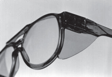
Table S2
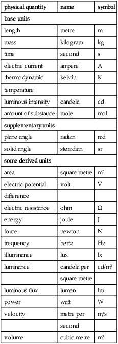
Base and supplementary SI units, and some derived units
| physical quantity | name | symbol |
| base units | ||
| length | metre | m |
| mass | kilogram | kg |
| time | second | s |
| electric current | ampere | A |
| thermodynamic | kelvin | K |
| temperature | ||
| luminous intensity | candela | cd |
| amount of substance | mole | mol |
| supplementary units | ||
| plane angle | radian | rad |
| solid angle | steradian | sr |
| some derived units | ||
| area | square metre | m2 |
| electric potential | volt | V |
| difference | ||
| electric resistance | ohm | Ω |
| energy | joule | J |
| force | newton | N |
| frequency | hertz | Hz |
| illuminance | lux | lx |
| luminance | candela per | cd/m2 |
| square metre | ||
| luminous flux | lumen | lm |
| power | watt | W |
| velocity | metre per | m/s |
| second | ||
| volume | cubic metre | m3 |
shingles See herpes zoster.
short-pointing See pointing, short-.
short sight See myopia.
shutter A device that provides a means (mechanical or electro-optical) for letting a beam of light pass during a given length of time.
SI unit Abbreviation of Système International d’unités (or International System of Units). It is derived from the metric system of physical units. There are seven base units, two supplementary units and many derived units. See candela per square metre; lumen; lux; micrometre (micron); nanometre.
sickle-cell disease See disease, sickle-cell.
side An attachment to the front of a spectacle frame passing towards or over the ear for the purpose of holding the frame in position. Syn. temple.
See spectacle frame markings; spectacles; temple, library.
curl s. The end of the side of a spectacle frame that lies along the greater part of the groove behind the ear. It contributes to the stability of the frame and it is particularly useful to children and to people engaged in sports. Syn. earpiece.
siderosis bulbi Deposit of iron produced by an iron foreign body in the ocular tissues. It is characterized by a reddish-brown discoloration of the surrounding tissue.
See heterochromia; line, iron.
Siegrist’s streaks Line of pigment spots seen along choroidal arteries in the fundus in advanced hypertensive retinopathy. They are indicative of fibrinoid necrosis.
sight The special sense by which the colour, form, position, shape, etc. of objects is perceived when light from these objects impinges upon the retina of the eye.
sight, line of See line of sight.
sight, night See hemeralopia.
sight, old See presbyopia.
sight, partial See vision, low.
sight, second Improvement of near vision in the elderly resulting from increased refraction of the nucleus of the crystalline lens. It often occurs in people with incipient cataract. Syn. gerontopia; senile lenticular myopia.
sight, sense of See sense.
sight-testing See refraction.
sign Objective evidence of a disease as distinguished from symptom, which is a subjective complaint of a patient.
Argyll Robertson s. See pupil, Argyll Robertson.
Bell’s s. Bell’s phenomenon occurring on the affected side in Bell’s palsy.
Bjerrum’s s. See scotoma, Bjerrum’s.
Cogan’s lid twitch s. A twitch of the upper eyelid in an eye with ptosis when the patient is asked to look in the primary position following a downward look. The eyelid then returns to its ptosis position. This condition occurs in myasthenia gravis.
Collier’s s. Unilateral, or more commonly bilateral, eyelid retraction that exposes an unusual amount of the sclera of the eye above and below the iris; it gives the person a frightened or startled expression. It is due to a midbrain lesion.
See syndrome, Parinaud’s.
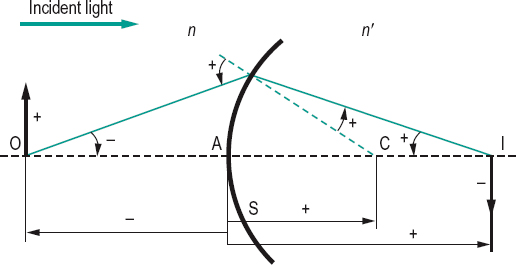
s. convention A set of conventions regulating the direction of distances, lengths, and angles measured in geometrical optics. The most common is the New Cartesian Sign Convention. It stipulates: (1) All distances are measured from the lens, refracting surface or mirror. Those in the same direction as the incident light, which is drawn travelling from left to right, are positive. Those in the opposite direction are negative. (2) All distances are measured from the axis. Those above are positive. Those below are negative. (3) Angles are measured from the incident ray to the axis, with anticlockwise angles positive and clockwise angles negative. (4) The power of a converging lens is positive and that of a diverging lens is negative (Fig. S7).
See focal length; law, Lagrange’s; law of refraction; Newton’s formula; paraxial equation, fundamental.
Dalrymple’s s. Retraction of the eyelids causing an abnormally widened palpebral fissure, in primary gaze. This is a sign of Graves’ disease. The patient appears to stare and to be frightened as some white sclera may be seen above the upper limbus.
doll’s eye s. See phenomenon, doll’s head.
von Graefe’s s . Immobility or lagging of the upper eyelid when looking downward. This is a sign of Graves’ disease.
Gunn’s crossing s. Tapering of veins on either side of the arteriovenous crossings seen in hypertensive retinopathy.
Hutchinson’s s. A triad of signs present in congenital syphilis. They are interstitial keratitis, notched teeth and deafness.
local s. See direction, oculocentric.
Moebius’ s. Convergence weakness occurring in Graves’ disease.
Mizuo’s s. See phenomenon, Mizuo’s.
Munson’s s . A sign observed in keratoconus in which the lower lid is bulging as a cone when the patient looks downward.
pseudo-von Graefe s . See aberrant regeneration.
Rizzuti’s s. An arrowhead pattern near the nasal part of the corneoscleral limbus, sometimes seen in advanced keratoconus.
Salus’ s. Retinal vein deflection from its normal course at arteriovenous crossings seen in hypertensive retinopathy.
Seidel’s s. See scotoma, Seidel’s.
Shafer’s s. The presence of pigment granules of various sizes floating in the anterior vitreous. They usually result from a retinal break/s, which may progress into rhegmatogenous retinal detachment. Then the pigment cells appear as small black dust-like particles (‘tobacco dust’) seen on clinical examination.
Vogt’s s . Loss of the normal shagreen of the front surface of the crystalline lens indicating anterior capsular cataract.
Uhthoff’s s. See Uhthoff’s symptom.
significance In statistics, an indication that the results of an investigation on a population (e.g. patients) differ from those of another population (e.g. general) by an amount that could not happen by chance alone. This is evaluated by establishing a significance level, that is the probability, called p value, which leads us to reject or accept the null hypothesis Ho (there is no significant difference between two populations and the difference is attributed to chance) and accept or reject the alternative hypothesis H1 that there is a statistically significant difference between two populations. A p value p 0.05 is often considered significant, but the lower this figure, the stronger the evidence.
See trial, randomized controlled.
silicone hydrogel lens See lens, silicone hydrogel.
silicone rubber A polymeric elastomeric, transparent material that is used in the manufacture of silicone rubber contact lenses. It has very high oxygen permeability but it is also very hydrophobic (wetting angle greater than 90°). The surfaces must be specially treated to render them wettable. A silicone component is used in silicone hydrogel and gas permeable contact lenses. Syn. poly(dimethyl siloxane).
See angle, contact; index of refraction; lens, silicone hydrogel; modulus of elasticity.
simultanagnosia Inability to comprehend a whole picture or sustain visual attention across simultaneous elements, although its constituent elements may be recognized. Objects may look fragmented or even sometimes disappear hence the patient may have difficulty recognizing a face as only one part is seen, or difficulty reading. Visual acuity and visual fields are normal. This is often a symptom of Balint’s syndrome or the result of a lesion, usually, in both parietal visual cortices, although temporal lobes may also be involved. Syn. simultagnosia; visual disorientation.
SILO response See response, SILO.
silver wire artery See arteriosclerosis.
simple astigmatism; cell See under the nouns.
simultaneous binocular vision See vision, Worth’s classification of binocular.
simultaneous vision See lens, contact.
Simultantest Trade name for an accessory to be inserted in a lens aperture of a phoropter or trial frame used to refine both the sphere and cylindrical components of a refraction. It provides simultaneous viewing of a distant test object through either a plus and minus sphere lens (+0.25 and –0.25 D) or through two +0.25 sphere, –0.50 cylinder lens systems with their cylindrical axes perpendicular to each other. Either mode of viewing is obtained by turning a knob, which actually rotates one of the lenses in the system.
See phoropter; trial frame.
sine condition The elimination of distortion and coma in an image is met when
M = n sin u/n’ sin u’ or nh sin u = n’h’ sin u’
where M is the lateral magnification, n and n’ the refractive indices of the media in the object and image space, u and u’ are the angular apertures on the object and image sides, and h and h’ the sizes of the object and image, respectively. Syn. Abbé’s condition.
See aperture, angular; coma; correction; distortion.
single binocular vision See vision, binocular single.
single-vision lens See lens, single-vision.
sinus A hollow space in bone or other tissue. cavernous s. One of the two venous sinuses in the dura mater of the brain extending on each side of the pituitary body, behind the orbit. It receives blood from the superior and the inferior ophthalmic veins and the central retinal veins. The third, fourth, fifth and sixth nerves pass through as well as the internal carotid artery. The cavernous plexus is located within this sinus.
See fistula, carotid-cavernous. s.
circularis iridis See canal, Schlemm’s.
ethmoidal s. Mucus-lined air cavities within the ethmoid bone, between the nose and the orbit. They drain into the nasal cavity. They contain the anterior and posterior ethmoidal nerves and blood vessels and are filled with air. They are separated from adjacent areas by very thin plates through which infection can pass easily and in particular ethmoiditis, which is the most common cause of orbital cellulitis.
s . of Maier See lacrimal apparatus.
scleral s. See canal, Schlemm’s.
s. venosus sclerae See canal, Schlemm’s.
situs inversus of the disc See crescent, congenital scleral.
sixth cranial nerve See nerve, abducens.
sixth nerve paralysis See paralysis of the sixth nerve.
size, angular The size of an object measured in terms of the angle it subtends. The point of reference for the eye can be either the nodal point or the centre of the entrance pupil.
See object, extended; size, apparent.
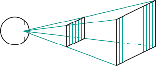
apparent s. 1. The size of an object represented by the angular size. 2. The perceived size of an object as distinguished from the actual size (Fig. S8).
s. constancy See constancy, size.
s. lens See lens, aniseikonic.
Sjögren’s syndrome See syndrome, Sjögren’s.
skiascope See retinoscope.
skiascopy See retinoscopy.
slab-off lens See lens, slab-off.
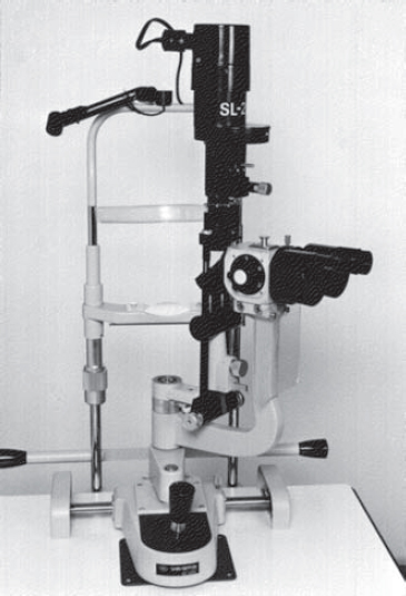
slit-lamp Instrument producing a bright focal source of light with a slit of variable width and height. It may be used to examine the tissues of the anterior segment of the eye in conjunction with a microscope (usually binocular) of variable magnification. To examine the internal structures of the eye including the retina an auxiliary lens is required. Many such lenses exist, some which do not make contact with the eye (e.g. Hruby lens, Volk lens) and others which come in contact with the eye, usually like a scleral type of lens (e.g. Goldmann lens, Thorpe four mirror fundus lens, Wilson three mirror fundus lens). A slit-lamp is essential in contact lens practice. This instrument is also commonly called a biomicroscope (Fig. S9).
See biomicroscopy, fundus; gonioscope; illumination; method, van Herick, Shaffer and Schwartz; method, Smith’s; microscope, slit-lamp; polymegethism, endothelial.
small angle strabismus See microtropia.
small eye artifact See retinoscopic artifact.
smooth pursuit See movement, pursuit.
Smith’s method See method, smith’s.
smoothing Grinding of the surface of a lens to the exact curvature and thickness with a very fine abrasive. This procedure comes after the roughing process and prior to polishing.
See abrasive; surfacing
Snell’s law See law of refraction.
Snellen acuity See acuity, Snellen.
Snellen chart See chart, Snellen.
Snellen equivalent See acuity, near visual.
Snellen fraction A representation of visual acuity in the form of a fraction (e.g. 6/6 (20/20), 6/24 (20/80), etc.) in which the numerator is the testing distance, usually expressed in metres (or in feet), and the denominator is the distance at which the smallest Snellen letter read by the eye has an angular size of 5 minutes.
See acuity, near visual; acuity, visual; acuity, decimal visual.
Snellen test type See chart, Snellen.
Snell–Sterling visual efficiency scale See visual efficiency scale, Snell–Sterling.
snow blindness See keratoconjunctivitis, actinic.
sodium chloride See saline, physiological; tears, artificial.
Table S3
Relationship between Snellen fractions in metres and feet, and for two testing distances (4 and 6 metres)
| (6 m) | (20 ft) | (4 m) |
| 6/3 | 20/10 | 4/2 |
| 6/4.8 | 20/16 | 4/3.2 |
| 6/6 | 20/20 | 4/4 |
| 6/7.5 | 20/25 | 4/5 |
| 6/9.5 | 20/32 | 4/6.3 |
| 6/12 | 20/40 | 4/8 |
| 6/15 | 20/50 | 4/10 |
| 6/18 | 20/60 | 4/12 |
| 6/21 | 20/70 | 4/14 |
| 6/24 | 20/80 | 4/16 |
| 6/30 | 20/100 | 4/20 |
| 6/38 | 20/125 | 4/25 |
| 6/48 | 20/160 | 4/32 |
| 6/60 | 20/200 | 4/40 |
| 6/90 | 20/300 | 4/60 |
| 6/120 | 20/400 | 4/80 |
| 6/150 | 20/500 | 4/100 |
| 6/180 | 20/600 | 4/120 |
| 6/240 | 20/800 | 4/160 |
| 6/300 | 20/1000 | 4/200 |
sodium cromoglicate See conjunctivitis, giant papillary; conjunctivitis, vernal; keratoconjunctivitis, superior limbic; mast cell stabilizers.
sodium fluorescein See fluorescein.
sodium pump See potential, action.
sodium/potassium pump See potential, action; transport, active.
soft drusen; exudate See under the nouns.
solar retinopathy; spectrum See under the nouns.
solution, hypertonic A solution with an osmotic pressure greater than that of an isotonic solution. Hypertonic ophthalmic solutions cause some stinging when instilled. Examples: sodium chloride 5%: when applied to an oedematous cornea this solution reduces oedema by drawing water from it; sulfacetamide sodium 30%; glycerol (or glycerin), at a dose of 1–1.5 g/kg body weight given as a solution with water or other liquid, which draws water from the eye into the blood and thereby reduces the intraocular pressure. Syn. hyperosmotic agent.
See hyperosmotic agent; pressure, osmotic; saline, physiological.
hypotonic s. A solution with an osmotic pressure lower than that of an isotonic solution. Hypotonic ophthalmic solutions generally cause less irritation than hypertonic ones.
isotonic s. A solution with an osmotic pressure equal to that on the other side of a semipermeable membrane. Example: sodium chloride 0.9% is considered to be approximately isotonic with the tears.
See pressure, osmotic; saline, physiological.
source, coherent See coherent sources.
source, extended A source consisting of many point sources separated laterally and such that it has a very low degree of coherence. Thus, an extended source has a given size and subtends a given angle.
See coherent sources; light, beam of; light, diffuse.
source, glare See glare.
source, light See light source .
source, point A source whose dimensions are sufficiently small to cause a radiation with a high degree of coherence. If the point source is situated on the axis of an optical system it gives rise to an axial ray and it is referred to as an axial point source.
See coherent sources; ray, axial.
space 1. An area or a cavity within the body. 2. A limited area, usually three dimensional.
Berger’s postlenticular s. A space between the posterior surface of the crystalline lens and the hyaloid fossa of the vitreous. The space is believed to be filled with aqueous humour. Syn. retrolental space of Berger.
colour sp. A two- or three-dimensional representation of colour stimuli. Example: CIE chromaticity diagram.
gaussian s. See paraxial region.
horopter s. The horopter consisting of all object points in space which stimulate corresponding retinal points as distinguished from the two-dimensional cases such as the apparent frontoparallel plane, longitudinal or nonius horopters.
image s. Region on one side of an optical system in which the image is formed.
intertrabecular s. See meshwork, trabecular.
object s . Region on one side of an optical system or a lens in which the object is situated.
Panum’s fusional s . An area in space corresponding to Panum’s area within which there is fusion and stereopsis of a non-fixated target.
perichoroidal s. See space, suprachoroidal.
suprachoroidal s. A potential space located between the choroid and the sclera. Anteriorly it is continuous with the supraciliary space. It contains thin, pigmented strands of collagen fibres and it is traversed by the long and short posterior ciliary arteries and nerves. Syn. perichoroidal space.
supraciliary s. A potential space located between the ciliary body and the sclera. In this space are thin strands of collagen fibres derived partly from the suprachoroid and partly from layers of the ciliary muscle. This space together with the suprachoroidal space form part of the unconventional route of aqueous humour outflow, the uveoscleral pathway.
See pathway, uveoscleral.
Spaeth classification See gonioscopy.
span of recognition The amount of reading matter that can be correctly identified or perceived during a brief exposure. It is often evaluated in testing and training reading ability.
See reading.
sparing of the macula See macula, sparing of the.
spasm of accommodation; of the near reflex See accommodation, spasm of.
spasm of the lid See blepharospasm.
spasmus nutans A pendular, rapid nystagmus of small amplitude which presents in infancy. It is generally bilateral, although asymmetrical and horizontal in most cases. Rotary and vertical types have been reported. The condition is associated with head nodding and less frequently with torticollis. The cause is unknown and the condition subsides spontaneously in early childhood. It must be differentiated from other disorders, such as congenital nystagmus and intracranial tumours, which also result in head nodding, and nystagmus.
spatial frequency; induction; summation See under the nouns.
spatial vision See perception, depth.
specificity The extent to which a test gives results that are free from false positives (i.e. people found to have the defect when they are actually free of it). The fewer the number of false positives, the greater is the specificity of the test. It is usually presented as the percentage of people truly identified as not defectives, or normal, referred to as true negatives, D (or correct reject), divided by the total number of not defectives or normal people tested. The total number includes all the true negatives, D, plus the false positives, B (or false alarm). Hence
See sensitivity.
spectacle blur See blur, spectacle.
spectacle crown See glass, crown.
spectacle frame See spectacles.
spectacle frame markings Numbers shown on a spectacle frame. They are usually the eyesize, the distance between lenses and the side length. Metal frames may also indicate the amount of gold (if any) found in the frame (e.g. 12 k meaning that the material is one-half gold and one-half of another metal). Example: 52/22 on the front of the frame means that the eyesize is 52 mm and the DBL is 22 mm, and 140 on the side means that the total length of the side is 140 mm.
See distance between lenses; eyesize; side.
spectacle frame, metal A structure in metal for enclosing or supporting ophthalmic lenses. Common materials include nickel/silver, Hi-nickel alloy, bronze, stainless steel, gold, gold plated, gold coating (commonly referred to as gold filled in which the base metal is most often nickel silver and for better quality frames, monel), copper, beryllium, titanium and sometimes aluminium. Metal spectacle frames are usually more rigid and maintain their shape better than plastic spectacle frames. However, the contacts with the skin of the nose and ears are done with plastic (or silicone) nose pads and temple covers.
spectacle frame, plastic A structure in plastic for enclosing or supporting ophthalmic lenses. Common materials include carbon fibre composite materials, which are a mixture of principally carbon and nylon in different percentages, cellulose acetate, cellulose propionate, nylon, epoxy resin (trade name Optyl), and Perspex and Plexiglas (which are both trade names for acrylic plastics containing mainly polymethyl methacrylate). Most of the plastics used are thermoplastic, i.e. a material which softens with heat and can thus be manipulated to fit the patient’s head and to insert and remove the lenses.
See frame heater; polymethyl methacrylate.
spectacle lens See lens, spectacle.
spectacle magnification See magnification, spectacle.
spectacle refraction See refractive error.
spectacles An optical appliance consisting of a pair of ophthalmic lenses mounted in a frame or rimless mount, resting on the nose and held in place by sides extending towards or over the ears. Syn. eyeglass frame; eyeglasses; eyewear (colloquial); glasses; spectacle frame.
See acetone; angle, pantoscopic; angle, retroscopic; angling; bridge; clipover; eczema; endpiece; eyesize; front; hinge; lens washer; lorgnette; mount; pad; plastic; rim; side; spectacle frame markings; sunglasses; temple; tortoiseshell.
aphakic s. Spectacles mounted with aphakic lenses used to compensate the loss of optical power resulting from a cataract extraction when no intraocular lens implant has been inserted. Syn. cataract glasses.
billiards s . Spectacles incorporating joints that enable the wearer to adjust the angle of the sides (British Standard).
folding s. Spectacles that are hinged at the bridge and in the sides, so as to fold with the two lenses in apposition.
half-eye s. A pair of spectacles for near vision, designed so that the lenses cover only half of the field of view, usually the lower half (Fig. S10). Syn. half-eyes.
hemianopic s. Spectacles incorporating a device that provides a lateral displacement of one or both fields of view. The device is usually a prism such as a Fresnel Press-On prism, which is placed over the blind (hemianopic) side of the visual field. A mirror system may also be used. The view within that side of the field is imaged on the seeing side of the visual field of the eye.
industrial s. Spectacles made with plastic or safety glass and solid frame, sometimes with side shields. They are used in industrial occupations where there are possible hazards to the eye.
See Fig. S6; glass, safety; goggles; lens, safety.
library s . A plastic spectacle frame with heavyweight front and sides. Syn. library frame.
magnifying s. Spectacles containing lenses of high convex power (+10 D or higher) used for near vision.
orthopaedic s. Spectacles with attachments designed to relieve certain anatomical deformities such as entropion, ptosis, etc.
pinhole s. Spectacles fitted with opaque discs having one or more small apertures. They are used as an aid in certain types of low vision (e.g. corneal scar).
See spectacles, stenopaeic; vision, low.
recumbent s. Spectacles intended to be used while recumbent. They usually incorporate a prism that deflects a beam of light through approximately 90° while keeping the image erect.
reversible s. Spectacles that are designed to be worn with either lens before either eye.
rimless s. Spectacles without rims, the lenses being fastened to the frame by screws, clamps or similar devices.
See lens groove; rim.
stenopaeic s. Spectacles fitted with opaque discs having a slit. They are used as an aid in certain types of low vision.
See disc, stenopaeic; spectacles, pinhole; vision, low.
supra s . Spectacles in which the lenses are held in position by thin nylon threads attached to the rims.
See lens groove; rim.
telescopic s. See lens, telescopic.
spectral 1 . Relating or belonging to a spectrum. 2. Relating to wavelength.
spectral luminous efficiency See efficiency, spectral luminous.
spectral transmission factor See spectrophotometer.
spectrometer An instrument for making measurements of the angle of a prism and the index of refraction. It consists of a collimator, an astronomical telescope, a table for carrying a prism and a graduated circle.
spectrophotometer An instrument for measuring the relative intensities of the spectrum, wavelength by wavelength. Specifically it measures the spectral transmission factor (i.e. the ratio of radiant flux transmitted through a medium to that incident on it) of a medium for a given wavelength. This factor is usually plotted as a graph against many wavelengths providing a spectral transmission curve. Ophthalmic lenses, contact lenses and any other substances (e.g. tear lipids) through which light may be passed can thus be analysed.
See density.
spectroscope An instrument for producing and observing spectra. It consists of a slit, a diffraction grating or a prism to disperse the radiations, achromatic lenses, and an eyepiece to observe them.
See monochromator; spectrum.
spectrum 1 . Spatial display of a complex radiation produced by separation of its monochromatic components. 2. Composition of a complex radiation, e.g. continuous spectrum, line spectrum (CIE). Plural: spectra.
See light.
absorption s. The curve representing the relative absorption of a pigment or chemical substance as a function of the wavelength of light. Example: the absorption spectrum of rhodopsin. Syn. absorbance spectrum.
action s. A graphical representation of the relative energy necessary to produce a constant biological effect. Example: frequency of action potentials in a ganglion cell as a function of wavelength.
continuous s. A spectrum in which, over a considerable range, all wavelengths exist without any abrupt variation in intensity. Example: the spectrum of hot solids.
electromagnetic s. The total range of all electromagnetic waves. It extends from the longest radio waves of some thousands of metres in wavelength through radar, microwave, infrared rays, visible rays (between wavelengths 780 nm and 380 nm) to ultraviolet rays, X-rays, gamma rays and cosmic rays with wavelengths as short as 8 × 10–12 mm. All these electromagnetic waves differ only in frequency (and wavelength) but have the same speed as light in a vacuum.
equal energy s. Spectrum in which all wavelengths have about the same amount of energy.
See achromatic; light, white.
fortification s. See scotoma, scintillating.
invisible s. The portions of the entire electromagnetic spectrum that are made up of radiations other than those of the visible spectrum.
line s. Spectrum consisting of a series of discrete monochromatic lines (or narrow bands of monochromatic light) with large intensity differences and separated by intervals without radiations. Example: the spectrum emitted by an electric discharge through a gas or vapour under low pressure.
s. locus The representation of the spectral colour stimuli on a chromaticity diagram.
solar s. The spectrum formed by sunlight. It is crossed at intervals by Fraunhofer’s lines.
visible s. The portion of the electromagnetic spectrum that can be perceived by the visual system. It is composed of radiations of wavelengths in the range between 380 nm and 780 nm in younger eyes. This range decreases with age especially due to lens absorption of short wavelengths becoming closer to 420 nm than 380 nm.
See light.
specular microscope; reflection See under the nouns.
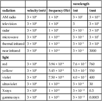
Table S4
Approximate values of the velocity, frequency and wavelength of electromagnetic radiations in a vacuum (the values represent a point within a range of radiations)
| wavelength | ||||
| radiation | velocity (m/s) | frequency (Hz) | (m) | (nm) |
| AM radio | 3 × 10⁸ | 1 × 10⁶ | 3 × 10² | 3 × 10¹¹ |
| television | 3 × 10⁸ | 1 × 10⁸ | 3 | 3 × 10⁹ |
| radar | 3 × 10⁸ | 1 × 10⁹ | 3 × 10⁻¹ | 3 × 10⁸ |
| microwave | 3 × 10⁸ | 1 × 10¹⁰ | 3 × 10⁻² | 3 × 10⁷ |
| thermal infrared | 3 × 10⁸ | 1 × 10¹³ | 3 × 10⁻⁵ | 3 × 10⁴ |
| near infrared | 3 × 10⁸ | 1 × 10¹⁴ | 3 × 10⁻⁶ | 3000 |
| light | ||||
| red | 3 × 10⁸ | 3.94 × 10¹⁴ | 7.6 × 10⁻⁷ | 760 |
| yellow | 3 × 10⁸ | 5.45 × 10¹⁴ | 5.5 × 10⁻⁷ | 550 |
| violet | 3 × 10⁸ | 7.50 × 10¹⁴ | 4.0 × 10⁻⁷ | 400 |
| ultraviolet | 3 × 10⁸ | 1 × 10¹⁶ | 3 × 10⁻⁸ | 30 |
| X-rays | 3 × 10⁸ | 1 × 10¹⁸ | 3 × 10⁻¹⁰ | 0.3 |
| gamma rays | 3 × 10⁸ | 1 × 10²¹ | 3 × 10–13 | 0.0003 |
speculum, eye See eye speculum.
speed of light See light, speed of.
sphenoid bone See orbit.
sphere A term commonly used to denote the spherical component of a prescription or of the power of a lens, or even a spherical lens.
See lens, spherical; prescription.
far point s . The imaginary spherical surface on which lie the far points of accommodation for all directions of gaze.
See accommodation, far point of.
near point s. The imaginary spherical surface on which lie the near points of accommodation for all directions of gaze.
See accommodation, near point of.
spherical aberration; equivalent See under the nouns.
spherical error See prescription.
spherical lens See lens, spherical.
spherometer See lens measure.
sphero-cylindrical lens See lens, sphero-cylindrical.
spherophakia A congenital, bilateral defect in which the crystalline lens is smaller and more spherical than normal. The zonule of Zinn may also be defective or absent.
See syndrome, Weill–Marchesani.
spherule, rod See rod spherule.
sphincter oculi muscle See muscle, orbicularis.
sphincter pupillae muscle See muscle, sphincter pupillae.
sphingomyelin lipidosis See disease, Niemann–Pick.
sphygmomanometer An instrument for measuring the arterial blood pressure. There are various types, the most common consisting of an inflatable cuff that is placed around the upper arm (usually the left) and air pressure within the cuff is balanced against the pressure of the blood in the brachial artery. The pressure is estimated by means of a mercury or an aneroid manometer. A stethoscope is normally used in conjunction with the instrument to listen to the blood pressure sounds (a stethoscope is not needed with an electronic sphygmomanometer). Normal systolic and diastolic blood pressures in a young adult are about 120/80, respectively. The difference between the two pressures is called the pulse pressure. Blood pressure varies with age, gender, altitude, disease, stress, fear, excitement, exercise, etc. A normal range for systolic pressure is usually considered to be 100–140 mmHg and for diastolic pressure below 90 mmHg.
See arteriosclerosis; hypertension; retinopathy, hypertensive.
Spielmeyer–Stock disease See disease, Batten–Mayou.
spin-cast contact lens See lens, spin-cast contact.
spindle, Krukenberg’s See Krukenberg’s spindle.
spindle, muscle See muscles, extraocular.
spiral, Plateau’s See Plateau’s spiral.
spot A small, circumscribed area visibly different in colour or texture from the surrounding tissue.
baring of the blind s. A visual field defect in which there is such a marked contraction of the peripheral temporal visual field that it lies on, or nasal to, the blind spot. Although it may occur in open-angle glaucoma, it is not indicative of the disease as it also occurs in other conditions (e.g. miosis).
Bitot’s s . Foamy patch found on the bulbar conjunctiva near the limbus in xerophthalmia and due to vitamin A deficiency. Syn. Bitot’s patch.
blind s . Physiological negative scotoma in the visual field corresponding to the head of the optic nerve. It is not seen in binocular vision, as the two blind spots do not correspond in the field. In monocular vision it is usually not noticed. It has the shape of an ellipse with its long axis vertical and measuring approximately 7.5°, whereas its shorter axis along the horizontal measures approximately 5.5°. Its centre is located 15.5° to the temporal side of the centre of the visual field and 1.5° below the horizontal meridian. Syn. blind spot of Mariotte; physiological blind spot; punctum caecum (Fig. S11).
See fibres, myelinated nerve; image, retinal.
blind s. enlargement A visual field defect in which the blind spot appears larger than normal. One of the common causes is papilloedema.
blind s. esotropia; syndrome See syndrome, Swann’s.
cherry-red s . Bright red appearance of the macular area in an eye with occlusion of the central retinal artery, Tay–Sachs disease or Niemann–Pick disease. In the case of central retinal artery occlusion the surrounding area is white due to ischaemia but the reddish reflex from the intact choroidal vessels beneath the fovea shows at that spot since the retina is thinnest there. There is a very marked, if not complete, loss of vision which appears suddenly. In cases of storage disease (i.e. Niemann–Pick or Tay–Sachs), the area surrounding the fovea is artificially whitened and opaque, offsetting the normal pinkish colour of the fovea (Fig. S12).

See disease, Niemann–Pick; disease, Sandhoff’s; disease, Tay–Sachs; retinal arterial occlusion.
cotton-wool s’s. See bodies, cytoid; exudate.
Elschnig’s s’s . Small, yellowish spots found in the fundus in advanced hypertensive retinopathy. They are choroidal infarcts caused by insufficient blood supply.
Fuchs’s . A round or elliptical, pigmented spot, usually located in the macular or paramacular area. It occurs in patients who have pathological myopia. It is due to breaks in Bruch’s membrane (called lacquer cracks) and to the development of a choroidal neovascular membrane followed by subretinal haemorrhage which has changed colour and has become pigmented. The patient may notice photopsia when the membrane breaks but eventually it causes a loss of vision with a central scotoma. Syn. Forster–Fuchs spot.
Maxwell’s s. Entopic phenomenon in which the subject can observe a dark or greyish spot in the visual field corresponding to the fovea. This is accomplished by viewing a


