Rotaviruses, Caliciviruses, and Astroviruses

Published on 22/03/2015 by admin

Filed under Pediatrics

Last modified 22/04/2025

Print this page

rate 1 star rate 2 star rate 3 star rate 4 star rate 5 star
Your rating: none, Average: 0 (0 votes)

This article have been viewed 1596 times

Chapter 257 Rotaviruses, Caliciviruses, and Astroviruses

Diarrhea is a leading cause of childhood mortality in the world, accounting for 5-10 million deaths/yr. In early childhood, the single most important cause of severe dehydrating diarrhea is rotavirus infection. Rotavirus and other gastroenteritis viruses not only are major causes of pediatric mortality but also lead to significant morbidity. Children in the USA have been estimated to have a risk of hospitalization for rotavirus diarrhea of 1:43, corresponding to 80,000 hospitalizations annually.

Etiology

Rotavirus, astrovirus, caliciviruses such as the Norwalk agent, and enteric adenovirus are the medically important pathogens of human viral gastroenteritis (Chapter 332).

Rotaviruses are in the Reoviridae family and cause disease in virtually all mammals and birds. These viruses are a wheel-like, triple-shelled icosahedron containing 11 segments of double-stranded RNA. The diameter of the particles on electron microscopy is approximately 80 nm. Rotaviruses are classified by serogroup (A, B, C, D, E, F, and G) and subgroup (I or II). Rotavirus strains are species specific and do not cause disease in heterologous hosts. Group A includes the common human pathogens as well as a variety of animal viruses. Group B rotavirus is reported as a cause of severe disease in infants and adults in China only. Occasional human outbreaks of group C rotavirus are reported. The other serogroups infect only nonhumans.

Subgrouping of rotaviruses is determined by the antigenic structure of the inner capsid protein, VP6. Serotyping of rotaviruses, described for group A only, is determined by classic cross-neutralization testing and depends on the outer capsid glycoproteins, VP7 and VP4. The VP7 serotype is referred to as the G type (for glycoprotein). There are 10 G serotypes, of which 4 cause most illness and vary in occurrence from year to year and region to region. The VP4 serotype is referred to as the P type. There are 11 P serotypes. Although both VP4 and VP7 elicit neutralizing immunoglobulin (Ig) G antibodies, the relative role of these systemic antibodies compared with that of mucosal IgA antibodies and cellular responses in protective immunity remains unclear.

Caliciviruses, which constitute the Caliciviridae family, are small 27- to 35-nm viruses that are the most common cause of gastroenteritis outbreaks in older children and adults. Caliciviruses also cause a rotavirus-like illness in young infants. They are positive-sense, single-stranded RNA viruses with a single structural protein. Human caliciviruses are divided into 2 genera, the noroviruses and sapoviruses. Caliciviruses have been named for locations of initial outbreaks: Norwalk, Snow Mountain, Montgomery County, Sapporo, and others. Caliciviruses and astroviruses are sometimes referred to as small, round viruses on the basis of appearance on electron microscopy.

Astroviruses, which constitute the Astroviridae family, are important agents of viral gastroenteritis in young children, with a high incidence in both the developing and developed worlds. Astroviruses are positive-sense, single-stranded RNA viruses. They are small particles, approximately 30 nm in diameter, with a characteristic central 5- or 6-pointed star when viewed on electron microscopy. The capsid consists of 3 structural proteins. There are 8 known human serotypes.

Enteric adenoviruses are a common cause of viral gastroenteritis in infants and children. Although many adenovirus serotypes exist and are found in human stool, especially during and after typical upper respiratory tract infections (Chapter 254), only serotypes 40 and 41 cause gastroenteritis. These strains are very difficult to grow in tissue culture. The virus consists of an 80-nm-diameter icosahedral particle with a relatively complex double-stranded DNA genome.

Aichi virus is a picornavirus that has been associated with gastroenteritis and was initially described in Asia. Several other viruses that may cause diarrheal disease in animals have been postulated but not well established as human gastroenteritis viruses. These include coronaviruses, toroviruses, and pestiviruses. The picobirnaviruses are an unclassified group of small (30 nm), single-stranded RNA viruses that have been found in 10% of patients with HIV-associated diarrhea.

Epidemiology

Worldwide, rotavirus is estimated to cause >111 million cases of diarrhea annually in children younger than 5 yr. Of these, 18 million cases are considered at least moderately severe, with approximately 500,000 deaths per year. Rotavirus causes 3 million cases of diarrhea, 80,000 hospitalizations, and 20-40 deaths annually in the USA.

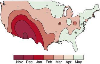
Rotavirus infection is most common in winter months in temperate climates. In the USA, the annual winter peak spreads from west to east (Fig. 257-1). Unlike the spread of other winter viruses, such as influenza, this wave of increased incidence is not due to a single prevalent strain or serotype. Typically, several serotypes predominate in a given community for 1 or 2 seasons, while nearby locations may harbor unrelated strains. Disease tends to be most severe in patients 3-24 mo of age, although 25% of the cases of severe disease occur in children >2 yr of age, with serologic evidence of infection developing in virtually all children by 4-5 yr of age. Infants younger than 3 mo are relatively protected by transplacental antibody and possibly breast-feeding. Infections in neonates and in adults in close contact with infected children are generally asymptomatic. Some rotavirus strains have stably colonized newborn nurseries for years, infecting virtually all newborns without causing any overt illness.

image

Figure 257-1 Peak rotavirus activity by month in the USA for July 1996–June 1997. This pattern is typical of the annual rotavirus activity each year.

(From the Centers for Disease Control and Prevention: Laboratory-based surveillance for rotavirus—United States, July 1996-June 1997, MMWR Morb Mortal Wkly Rep 46:1092–1094, 1997.)

Rotavirus and the other gastrointestinal viruses spread efficiently via a fecal-oral route, and outbreaks are common in children’s hospitals and child-care centers. The virus is shed in stool at very high concentration before and for days after the clinical illness. Very few infectious virions are needed to cause disease in a susceptible host.

The epidemiology of astroviruses is not as thoroughly studied as that of rotavirus, but they are a common cause of mild to moderate watery winter diarrhea in children and infants and an uncommon pathogen in adults. Hospital outbreaks are common. Enteric adenovirus gastroenteritis occurs year-round, mostly in children younger than 2 yr. Nosocomial outbreaks occur but are less common than with rotavirus and astrovirus. Calicivirus is best known for causing large, explosive outbreaks among older children and adults, particularly in settings such as schools, cruise ships, and hospitals. Often a single food, such as shellfish or water used in food preparation, is identified as a source. Like astrovirus and rotavirus, caliciviruses are also commonly found in winter infantile gastroenteritis.

Pathogenesis

Viruses that cause human diarrhea selectively infect and destroy villus tip cells in the small intestine. Biopsies of the small intestines show variable degrees of villus blunting and round cell infiltrate in the lamina propria. Pathologic changes may not correlate with the severity of clinical symptoms and usually resolve before the clinical resolution of diarrhea. The gastric mucosa is not affected despite the commonly used term gastroenteritis, although delayed gastric emptying has been documented during Norwalk virus infection.

In the small intestine, the upper villus enterocytes are differentiated cells, which have both digestive functions, such as hydrolysis of disaccharides, and absorptive functions, such as the transport of water and electrolytes via glucose and amino acid co-transporters. Crypt enterocytes are undifferentiated cells that lack the brush border hydrolytic enzymes and are net secretors of water and electrolytes. Selective viral infection of intestinal villus tip cells thus leads to (1) decreased absorption of salt and water and an imbalance in the ratio of intestinal fluid absorption to secretion and (2) diminished disaccharidase activity and malabsorption of complex carbohydrates, particularly lactose. Most evidence supports altered absorption as the more important factor in the genesis of viral diarrhea. It has been proposed that a rotavirus nonstructural protein (NSP4) functions as an enterotoxin.

Viremia may occur often in severe, primary infections, but symptomatic extraintestinal infection is extremely rare in immunocompetent persons—although immunocompromised patients may rarely experience hepatic and renal involvement. The increased vulnerability of infants (compared with older children and adults) to severe morbidity and mortality from gastroenteritis viruses may relate to a number of factors, including decreased intestinal reserve function, lack of specific immunity, and decreased nonspecific host defense mechanisms such as gastric acid and mucus. Viral enteritis greatly enhances intestinal permeability to luminal macromolecules and has been postulated to increase the risk for food allergies.

Clinical Manifestations

Rotavirus infection typically begins after an incubation period of <48 hr (range 1-7 days) with mild to moderate fever as well as vomiting, followed by the onset of frequent, watery stools. All 3 symptoms are present in about 50-60% of cases. Vomiting and fever typically abate during the 2nd day of illness, but diarrhea often continues for 5-7 days. The stool is without gross blood or white blood cells (WBCs). Dehydration may develop and progress rapidly, particularly in infants. The most severe disease typically occurs among children 4-36 mo of age. Malnourished children and children with underlying intestinal disease such as short-bowel syndrome are particularly likely to acquire severe rotavirus diarrhea. Rarely, immunodeficient children experience severe and prolonged illness. Although most newborns infected with rotavirus are asymptomatic, some outbreaks of necrotizing enterocolitis have been associated with the appearance of a new rotavirus strain in the affected nurseries.

The clinical course of astrovirus appears to be similar to that of rotavirus, with the notable exception that the disease tends to be milder, with less significant dehydration. Adenovirus enteritis tends to cause diarrhea of longer duration, often 10-14 days. The Norwalk virus has a short (12-hr) incubation period. Vomiting and nausea tend to predominate in illness associated with the Norwalk virus, and the duration is brief, usually consisting of 1-3 days of symptoms. The clinical and epidemiologic picture of Norwalk virus often closely resembles so-called food poisoning from preformed toxins such as Staphylococcus aureus and Bacillus cereus.

Treatment

Avoiding and treating dehydration are the main goals in treatment of viral enteritis. A secondary goal is maintenance of the nutritional status of the patient (Chapters 55 and 332).

There is no routine role for antiviral drug treatment of viral gastroenteritis. Controlled studies have shown no benefit from antiemetics or antidiarrheal drugs, and there is a significant risk for serious side effects with both types of agents. Antibiotics are similarly of no benefit. Immunoglobulins have been administered orally to both normal and immunodeficient patients with severe rotavirus gastroenteritis, but this treatment is currently considered experimental. Therapy with probiotic organisms such as Lactobacillus species has been shown to be helpful only in mild cases and not in dehydrating disease.

Supportive Treatment

Rehydration via the oral route can be accomplished in most patients with mild to moderate dehydration (Chapters 55 and 332). Severe rehydration requires immediate intravenous therapy followed by oral rehydration. Modern oral rehydration solutions containing appropriate quantities of sodium and glucose promote optimum absorption of fluid from the intestine. There is no evidence that a particular carbohydrate source (rice) or addition of amino acids improves the efficacy of these solutions for children with viral enteritis. Other clear liquids, such as flat soda, fruit juice, and sports drinks, are inappropriate for rehydration of young children with significant stool loss. Rehydration via the oral (or nasogastric) route should be done over 6-8 hr, and feedings begun immediately thereafter. Providing the rehydration fluid at a slow, steady rate, typically 5 mL/min, reduces vomiting and improves the success of oral therapy. Rehydration solution should be continued as a supplement to make up for ongoing excessive stool loss. Initial intravenous fluids are required for the infant in shock or the occasional child with intractable vomiting.

After rehydration has been achieved, resumption of a normal diet for age has been shown to result in a more rapid recovery from viral gastroenteritis. Prolonged (>12 hr) administration of exclusive clear liquids or dilute formula is without clinical benefit and actually prolongs the duration of diarrhea. Breast-feeding should be continued even during rehydration. Selected infants may benefit from lactose-free feedings (such as soy formula and lactose-free cow’s milk) for several days, although this step is not necessary for most children. Hypocaloric diets low in protein and fat such as BRAT (bananas, rice, cereal, applesauce, and toast) have not been shown to be superior to a regular diet.

Prevention

Good hygiene reduces the transmission of viral gastroenteritis, but even in the most hygienic societies, virtually all children become infected as a result of the efficiency of infection of the gastroenteritis viruses. Good handwashing and isolation procedures can help control nosocomial outbreaks. The role of breast-feeding in prevention or amelioration of rotavirus infection may be small, given the variable protection observed in a number of studies. Vaccines offer the best hope for control of these ubiquitous infections.

Vaccines

A trivalent rotavirus vaccine was licensed in the USA in 1998 and was subsequently linked to an increased risk for intussusception, especially during the 3- to 14-day period after the 1st dose and the 3- to 7-day period after the 2nd dose. The vaccine was withdrawn from the market in 1999. Subsequently 2 new live, oral rotavirus vaccines have been approved in the USA after extensive safety and efficacy testing.

A live, oral, pentavalent rotavirus vaccine was approved in 2006 for use in the USA. The vaccine contains 5 reassortant rotaviruses isolated from human and bovine hosts. Four of the reassortant rotaviruses express 1 serotype of the outer protein VP7 (G1, G2, G3, or G4), and the 5th expresses the protein P1A (genotype P[8]) from the human rotavirus parent strain. The pentavalent vaccine protects against rotavirus gastroenteritis when administered as a 3-dose series at 2, 4, and 6 mo of age. The 1st dose should be administered between 6 and 12 wk of age, with all 3 doses completed by 32 wk of age. The vaccine provides substantial protection against rotavirus gastroenteritis, with primary efficacy of 98% against severe rotavirus gastroenteritis caused by G1-G4 serotypes and 74% efficacy against rotavirus gastroenteritis of any severity through the 1st rotavirus season after vaccination. It provides a 96% reduction in hospitalizations for rotavirus gastroenteritis through the 1st 2yr after the 3rd dose. In a study of >70,000 infants, the pentavalent vaccine did not increase the risk for intussusception.

Another new monovalent rotavirus vaccine was licensed in the USA and also appears to be safe and effective. It is an attenuated monovalent human rotavirus and is administered as 2 oral doses at 2 and 4 mo of age. The vaccine has 85% efficacy against severe gastroenteritis and was found to reduce hospital admissions for all diarrhea by 42%. Despite being monovalent, the vaccine is effective in prevention of all 4 common serotypes of human rotavirus.

Preliminary surveillance data on rotavirus incidence from the U.S. Centers for Disease Control and Prevention (CDC) suggest that rotavirus vaccination greatly reduced the disease burden in the USA during the 2007-2008 rotavirus season. Given the incomplete vaccine coverage during this period, the results suggest a degree of “herd immunity” from rotavirus immunization. Vaccine-associated disease has been reported in vaccine recipients who have severe combined immunodeficiency disease (a contraindication). In addition, vaccine-derived virus may undergo reassortment and become more virulent, producing diarrhea in unvaccinated siblings.

Bibliography

Bresee JS, Parashar UD, Widdowson MA, et al. Update on rotavirus vaccines. Pediatr Infect Dis J. 2005;24:947-952.

Centers for Disease Control and Prevention. Addition of severe combined immunodeficiency as a contraindication for administration of rotavirus vaccine. MMWR. 2010;59(22):687-688.

Centers for Disease Control and Prevention (CDC). Delayed onset and diminished magnitude of rotavirus activity-United States, Nov 2007–May 2008. MMWR Morb Mortal Wkly Rep. 2008;57:697-700.

Chandron A, Heinzen RR, Santosham M, et al. Nosocomial rotavirus infections: a systematic review. J Pediatr. 2006;149:441-447.

Clark B, McKendrick M. A review of viral gastroenteritis. Curr Opin Infect Dis. 2004;17:461-469.

Clark HF, Offit PA, Plotkin SA, et al. The new pentavalent rotavirus vaccine composed of bovine (strain WC3)–human rotavirus reassortants. Pediatr Infect Dis J. 2006;25:577-582.

Coffin SE, Elser J, Marchant C, et al. Impact of acute rotavirus gastroenteritis on pediatric outpatient practices in the United States. Pediatr Infect Dis J. 2006;25:584-589.

Costa-Ribeiro H, Ribeiro TC, Mattos AP, et al. Limitations of probiotic therapy in acute, severe dehydrating diarrhea. J Pediatr Gastroenterol Nutr. 2003;36:112-115.

Glass RI, Bresee JS, Parashar UD, et al. The future of rotavirus vaccines: a major setback leads to new opportunities. Lancet. 2004;363:1547-1550.

Grimwood K, Kirkwood CD. Human rotavirus vaccines: too early for the strain to tell. Lancet. 2008;371:1144-1145.

Guandalini S, Pensabene L, Zikri MA, et al. Lactobacillus GG administered in oral rehydration solution to children with acute diarrhea: a multicenter European trial. J Pediatr Gastroenterol Nutr. 2000;30:54-60.

Guerrero ML, Noel JS, Mitchell DK, et al. A prospective study of astrovirus diarrhea of infancy in Mexico City. Pediatr Infect Dis J. 1998;17:723-727.

Jiang B, Gentsch JR, Glass RI. The role of serum antibodies in the protection against rotavirus disease: an overview. Clin Infect Dis. 2002;34:1051-1061.

Lynch M, Shieh WJ, Tatti K, et al. The pathology of rotavirus-associated deaths, using new molecular diagnostics. Clin Infect Dis. 2003;37:1027-1033.

O’Ryan M, Diaz J, Mamani N, et al. Impact of rotavirus infections on outpatient clinic visits in Chile. Pediatr Infect Dis J. 2007;26:41-45.

O’Ryan M, Matson DO. New rotavirus vaccines: renewed optimism. J Pediatr. 2006;149:448-451.

Parashar UD, Holman RC, Clarke MJ, et al. Hospitalizations associated with rotavirus diarrhea in the United States, 1993 through 1995: surveillance based on the new ICD-9-CM rotavirus-specific diagnostic code. J Infect Dis. 1998;77:13-17.

Parashar UD, Hummelman EG, Bresee JS, et al. Global illness and deaths caused by rotavirus disease in children. Emerg Infect Dis. 2003;9:565-572.

Payne DC, Edwards KM, Bowen MD, et al. Sibling transmission of vaccine-derived rotavirus (RotaTeq) associated with rotavirus gastroenteritis. Pediatrics. 2010;125:e438-e441.

Plotkin SA. New rotavirus vaccines. Pediatr Infect Dis J. 2006;25:575-576.

Ruiz-Palacios GM, Perez-Schael I, Velazquez FR, et al. Safety and efficacy of an attenuated vaccine against severe rotavirus gastroenteritis. N Engl J Med. 2006;354:11-22.

Soares-Weiser K, Goldberg E, Tamimi G, et al: Rotavirus vaccine for preventing diarrhoea, Cochrane Database Syst Rev (1):CD002848, 2004.

Staat MA, Azimi PH, Berke T, et al. Clinical presentations of rotavirus infection among hospitalized children. Pediatr Infect Dis J. 2002;21:221-227.

Tucker AW, Haddix AC, Bresee JS, et al. Cost-effectiveness analysis of a rotavirus immunization program for the United States. JAMA. 1998;279:1371-1376.

Vesikari T, Matson DO, Dennehy P, et al. Safety and efficacy of a pentavalent human-bovine (WC3) reassortant rotavirus vaccine. N Engl J Med. 2006;354:23-33.