Roseola (Human Herpes Viruses 6 and 7)

Published on 22/03/2015 by admin

Filed under Pediatrics

Last modified 22/04/2025

Print this page

rate 1 star rate 2 star rate 3 star rate 4 star rate 5 star
Your rating: none, Average: 0 (0 votes)

This article have been viewed 2929 times

Chapter 248 Roseola (Human Herpes Viruses 6 and 7)

Human herpesvirus 6 (HHV-6) and human herpesvirus 7 (HHV-7) cause ubiquitous infection in infancy and early childhood. HHV-6 is responsible for the majority of cases of roseola infantum (exanthema subitum or sixth disease) and has been associated with other diseases, including encephalitis, especially in immunocompromised hosts. A small percentage of children with roseola have primary infection with HHV-7.

Etiology

HHV-6 and HHV-7 are the sole members of the Roseolovirus genus in the Betaherpesvirinae subfamily of human herpesviruses. Human cytomegalovirus (CMV), the only other β-herpesvirus, shares limited sequence homology with HHV-6 and HHV-7. Morphologically all human herpesviruses are composed of an icosahedral nucleocapsid, protein-dense tegument, and lipid envelope. Within the nucleocapsid, HHV-6 and HHV-7 each contain large, linear, double-stranded DNA genomes that encode >80 unique proteins.

Two strain groups of HHV-6 have been recognized, HHV-6 variant A and variant B. The genomes of HHV-6 A and B are highly conserved, with approximately 90% sequence identity. However, they can be distinguished by restriction fragment length polymorphisms, reactivity with monoclonal antibodies, differential growth in tissue culture cell lines, and epidemiology. Accordingly, some researchers propose that they should be separate viruses. Although the frequency of detection of HHV-6 variant A DNA differs among studies, variant B is the overwhelmingly predominant strain found in both normal and immunocompromised hosts by both culture and polymerase chain reaction (PCR). Primary infection with HHV-6 variant A has been detected in children in Africa. It is not clear whether the differences in the detection of HHV-6 variant A DNA and variant B DNA relate to different tissue tropism, differences in mode or age of acquisition, differences in the ability to cause human disease, or the geographical location of the population studied.

Epidemiology

Primary infection with HHV-6 is acquired rapidly by essentially all children following the loss of maternal antibodies in the first few months of infancy, 95% of children being infected with HHV-6 by 2 yr of age. The peak age of primary HHV-6 infection is 6-9 mo of life, with infections occurring sporadically and without seasonal predilection. Infection with HHV-7 is also widespread but occurs later in childhood and at a slower rate; only 50% of children have evidence of prior infection with HHV-7 by 3 yr of age. Seroprevalence reaches 75% at 3-6 yr of age. In a small study of children with primary HHV-7 infection, the mean age of the patients was 26 mo, significantly older than that of children with acute HHV-6 infection.

Although it is presumed that children acquire primary infection with HHV-6 and HHV-7 from the saliva of asymptomatic adults, congenital infection with HHV-6 occurs in 1% of newborns. Two mechanisms of vertical transmission of HHV-6 have been identified, transplacental infection and chromosomal integration (CI-HHV6). HHV-6 is unique among the human herpesviruses in that it is integrated at the telomere end of human chromosomes at a frequency of 0.2-2.2% of the population and is passed from parent to child via the germline. Chromosomal integration has been identified as the major mechanism by which HHV-6 is vertically transmitted, accounting for 86% of congenital infections, with one third due to HHV-6 variant A. The clinical consequences of chromosomal integration or transplacental infection with HHV-6 have yet to be determined. In one series of infants identified with HHV-6 congenital infection, no evidence of disease was present in the early neonatal period. Congenital infection with HHV-7 has not been demonstrated. DNA of both HHV-6 and HHV-7 has been identified in the cervical secretions of pregnant women, suggesting an additional role for sexual or perinatal transmission of these viruses. Breast milk does not appear to play a role in transmission of either HHV-6 or HHV-7.

Pathology/Pathogenesis

Primary HHV-6 infection causes a viremia that can be demonstrated by co-culture of the patient’s peripheral blood mononuclear cells (PBMCs) with mitogen-stimulated cord blood mononuclear cells. HHV-6 has a recognizable cytopathic effect, consisting of the appearance of large refractile mononucleated or multinucleated cells with intracytoplasmic and/or intranuclear inclusions. Infected cells exhibit a slightly prolonged life span in cultures; however, lytic infection predominates. HHV-6 infection also induces apoptosis of T cells and may lead to cell expiration via loss of mitochondrial membrane potential as well as alteration of interferon and retinoic acid-induced cell death signals. In vitro, HHV-6 can infect a broad range of cell types, including primary T cells, monocytes, natural killer (NK) cells, dendritic cells, and astrocytes. HHV-6 has also been documented to infect B-cell, megakaryocytic, endothelial, and epithelial cell lines. Human astrocytes, oligodendrocytes, and microglia have been infected with HHV-6 ex-vivo. The broad tropism of HHV-6 is consistent with the recognition that CD46, a complement regulatory protein present on the surface of all nucleated cells, is a cellular receptor for HHV-6. The CD4 molecule has been identified as a receptor for HHV-7. HHV-7 has been demonstrated to reactivate HHV-6 from latency in vitro. Whether this phenomenon occurs in vivo remains unknown.

Primary infection with HHV-6 and HHV-7 is followed by lifelong latency or persistence of virus at multiple sites. HHV-6 exists in a true state of viral latency in monocytes and macrophages. The detection of replicating HHV-6 in cultures of primary CD34+ hematopoietic stem cells has also been described, suggesting that cellular differentiation is a trigger of viral reactivation. This observation may be clinically significant because of the possibility that HHV-6 may cause either primary or reactivated infection during hematopoietic stem cell transplantation. Additionally, HHV-6 and HHV-7 infection may be persistent in salivary glands, and DNA for both HHV-6 and HHV-7 can be routinely detected in the saliva of both adults and children. HHV-7 can also be isolated in tissue culture from saliva, but HHV-6 cannot. HHV-6 DNA has been identified in the cerebrospinal fluid (CSF) of children both during and subsequent to primary infection as well as in brain tissue from immunocompetent adults at autopsy, implicating the central nervous system (CNS) as an additional important site of either viral latency or persistence. HHV-7 DNA has also been found in adult brain tissue but at a significantly lower frequency.

Clinical Manifestations

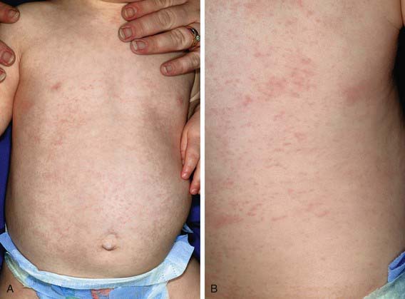
Roseola infantum (exanthem subitum, or sixth disease) is an acute, self-limited disease of infancy and early childhood. It is characterized by the abrupt onset of high fever, which may be accompanied by fussiness. The fever usually resolves acutely after 72 hr (“crisis”) but may gradually fade over a day (“lysis”) coincident with the appearance of a faint pink or rose-colored, nonpruritic, 2- to 3-mm morbilliform rash on the trunk (Fig. 248-1). The rash usually lasts 1-3 days but is often described as evanescent and may be visible only for hours, spreading from the trunk to the face and extremities. Because the rash is variable in appearance, location, and duration, it is not distinctive. Associated signs are sparse but can include mild injection of the pharynx, palpebral conjunctivae, or tympanic membranes and enlarged suboccipital nodes. In Asian countries, ulcers at the uvulopalatoglassal junction (Nagayama spots) are commonly reported in infants with roseola.

High fever (mean 39.7°C) is the most consistent finding associated with primary HHV-6 infection. Rash detected either during the illness or following defervescence has been reported in approximately 20% of infected children in the USA. Additional symptoms and signs include irritability, inflamed tympanic membranes, rhinorrhea and congestion, gastrointestinal complaints, and encephalopathy. Symptoms of lower respiratory tract involvement such as cough are identified significantly less frequently in children with primary HHV-6 infection than in children with other febrile illnesses. The mean duration of illness due to primary HHV-6 infection is 6 days, with 15% of children having fever for 6 or more days. Primary infection with HHV-6 accounts for a significant burden of illness on the health care system; one study found that 24% of visits to emergency departments by infants between 6 and 9 mo of age were due to primary HHV-6 infection. A population-based study of primary HHV-6 infection has confirmed that 93% of infants had symptoms and were more likely to visit a physician than non-infected infants. Fever was less likely to be present with HHV-6 infection in children <6 mo of age but was significantly more common in older infants and children.

Much less is known about the clinical manifestations of HHV-7 infection. Primary infection with HHV-7 has been identified in a small number of children with roseola in whom the illness is indistinguishable from that due to HHV-6. Secondary cases of roseola due to infection with HHV-7 have also been reported. Additionally, primary infection with HHV-7 may be asymptomatic or may cause a nonspecific febrile illness lasting approximately 3 days.

Diagnosis

Although roseola is generally a benign self-limited disease, its diagnosis can exclude other, more serious disorders that cause fever and rash. A history of 3 days of high fever in an otherwise nontoxic 10 mo old infant with a blanching maculopapular rash on the trunk suggests a diagnosis of roseola. Likewise, a specific diagnosis of HHV-6 is not usually necessary except in situations in which the manifestations of the infection are severe or unusual and might benefit from antiviral therapy.

The diagnosis of primary infection with either HHV-6 or HHV-7 is confirmed by demonstrating the presence of actively replicating virus in the patient’s blood sample coupled with seroconversion. Viral culture is the gold standard method to document active viral replication. Unfortunately, culture is expensive, time consuming, and available only in research laboratories. Two other methods used to identify active HHV-6 replication are the detection of viral DNA by PCR on acellular fluids such as plasma and reverse transcriptase PCR (RT-PCR) on PBMC samples designed to detect viral transcription and protein production. Quantitative PCR for HHV-6 genome copy numbers on various specimens is also frequently reported and is commercially available. However, the role of this methodology is not clear, as a specific value of DNA that can discriminate between patients with viremia and those who are culture negative has not been determined. Complicating the use of molecular assays for the detection of active replication of HHV-6 is the recognition that individuals with chromosomally integrated HHV-6 have persistently high viral loads of HHV-6 DNA in plasma, PBMCs, and CSF in the absence of disease and viremia.

Serologic methods including indirect immunofluorescence assays, enzyme-linked immunosorbent assay (ELISA), neutralization assays, and immunoblot have been described for the measurement of concentrations of antibodies to HHV-6 and HHV-7 in serum or plasma and are commercially available. Although IgM antibody is produced early in infection with HHV-6, assays designed to measure this response have not proved useful in the diagnosis of primary or reactivated infection. The absence of IgG antibody in an infant >6 mo of age combined with the presence of replicating virus is strong evidence of primary infection with either HHV-6 or HHV-7. Alternatively, the demonstration of seroconversion between acute and convalescent samples also confirms primary infection but is not clinically useful in the acute care setting. Unfortunately, serologic assays have not been found reliable in the detection of HHV-6 reactivation and cannot be used to differentiate between infection with HHV-6 variants A and B. Additionally, limited antibody cross reactivity has been demonstrated between HHV-6 and HHV-7, complicating the interpretation of serologic assays, especially if low titers are reported.

Differential Diagnosis

Primary infection with either HHV-6 or HHV-7 usually causes an undifferentiated febrile illness that may be very difficult to distinguish from other common viral infections of childhood. This difficulty also applies to the early stages of roseola, before the development of rash. Once the rash is present, roseola may be confused with other exanthematous diseases of childhood, especially measles and rubella. Children with rubella often have a prodrome characterized by mild illness with low-grade fever, sore throat, arthralgia, and gastrointestinal complaints, unlike those with roseola. On physical examination, suboccipital and posterior auricular lymph nodes are prominent up to 1 wk before the rash of rubella is evident and persist during the exanthematous phase. Additionally, the rash of rubella usually begins on the face and spreads to the chest, like that in measles. The associated symptoms of measles virus infection include cough, coryza, and conjunctivitis, with high fever coincident with the development of rash, unlike in roseola. Roseola may also be confused with scarlet fever, though the latter is rare in children <2 yr of age and causes a characteristic sandpaper-like rash concurrent with fever.

Roseola may be confused with illness due to enterovirus infections, especially in the summer and fall months. Drug hypersensitivity reactions may also be difficult to distinguish from roseola. Antibiotics are frequently prescribed for children with fever due to roseola before the appearance of rash. A child who then demonstrates rash after the resolution of fever may erroneously be labeled as being drug allergic.

Complications

Convulsions are the most common complication of roseola and are recognized in up to one third of patients. Seizures are also the most common complication of children with primary HHV-6 infection, occurring in approximately 15%, with a peak age at 12-15 mo. Children with primary HHV-6 infection have also been reported to have a higher frequency of partial seizures, prolonged seizures, post-ictal paralysis, and repeated seizures than children with febrile seizures not associated with HHV-6. In a study limited to children with primary HHV-6 infection and seizures, 30% of patients had prolonged seizures, 29% had focal seizures, and 38% had repeated seizures.

A prospective study of children 2-35 mo of age with suspected encephalitis or severe illness with convulsions and fever found that 17% had primary infection with either HHV-6 or HHV-7, and status epilepticus was the most common presentation. Despite the reported severity of seizures during primary infection with HHV-6 and HHV-7, limited data suggest that there may be a decreased risk of recurrent seizures after primary infection with HHV-6 than of febrile seizures due to other causes.

Case reports and small patient series have described additional complications in children with primary HHV-6 infection, including encephalitis, acute disseminated demyelination, acute cerebellitis, hepatitis, and myocarditis. Late-developing long-term sequelae, including developmental disabilities and autistic-like features, have been reported rarely in children who have CNS symptoms during primary HHV-6 infection.

An association between recurrent seizures and reactivated or persistent infection of the CNS by HHV-6 has been suggested. Studies evaluating brain tissue specimens have implicated HHV-6 in as many as 35% of patients with temporal lobe epilepsy, high viral loads being found in the hippocampus or lateral temporal lobe regions. HHV-6 protein production has also been identified in a small number of resected tissue specimens. Primary astrocytes obtained from these samples had undetectable levels of a glutamate transporter, suggesting the loss of ability to control glutamate levels as a possible mechanism for the development of recurrent seizures.

Reactivation of HHV-6 has been reported in several different populations with and without disease with the use of various methods of detection. The best documentation of HHV-6 reactivation has been in immunocompromised hosts, especially those patients who have undergone hematopoietic stem cell transplantation (HSCT). Such reactivation occurs in approximately 35-50% of patients, typically at 2-4 weeks after transplantation. Many of the clinical complications seen following HSCT have been associated with HHV-6 reactivation, including fever, rash, delayed engraftment of platelets or monocytes, and graft versus host disease with variable degrees of support in the literature for each.

HHV-6 reactivation has also been reported as a cause of encephalitis in both normal and immunocompromised hosts. A distinct syndrome of post-HSCT limbic encephalitis has been described; it is characterized by short-term memory dysfunction, confusion, and insomnia with seizures noted either clinically or on prolonged electroencephalography (EEG) monitoring. HHV-6 DNA has been identified in the CSF in the majority of these patients. Additionally, HHV-6 proteins were identified in astrocytes of the hippocampus in one post mortem specimen, suggesting active HHV-6 infection at the time of death. Because of the variability in sensitivity of PCR methods and the high prevalence of HHV-6 DNA in multiple body sites following primary infection, it is very difficult to evaluate the validity of these reports.

Bibliography

Caserta MT, Hall CB, Schnabel K, et al. Primary human herpesvirus 7 infection: a comparison of human herpesvirus 7 and human herpesvirus 6 infections in children. J Pediatr. 1998;133:386-389.

De Bolle LL, Naesens De Clercq E. Update on human herpesvirus 6 biology, clinical features, and therapy. Clin Microbiol Rev. 2005;18:217-245.

Fotheringham J, Donati D, Akhyani N, et al. Association of human herpesvirus-6B with mesial temporal lobe epilepsy. PLoS Med. 2007;4:e180.

Hall CB, Caserta MT, Schnabel K, et al. Chromosomal integration of human herpesvirus 6 is the major mode of congenital human herpesvirus 6 infection. Pediatrics. 2008;122:513-520.

Hall CB, Long CE, Schnabel KC, et al. Human herpesvirus-6 infection in children: a prospective study of complications and reactivation. N Engl J Med. 1994;331:432-438.

Seeley WW, Marty FM, Holmes TM, et al. Post-transplant acute limbic encephalitis: clinical features and relationship to HHV6. Neurology. 2007;69:156-165.

Ward KN, Andrews NJ, Verity CM, et al. Human herpesviruses-6 and -7 each cause significant neurological morbidity in Britain and Ireland. Arch Dis Child. 2005;90:619-623.

Ward KN. Human herpesviruses-6 and -7 infections. Curr Opin Infect Dis. 2005;18:247-252.

Ward KN. The natural history and laboratory diagnosis of human herpesviruses-6 and -7 infections in the immunocompetent. J Clin Virol. 2005;32:183-193.

Yao K, Honarmand S, Espinosa A, et al. Detection of human herpesvirus-6 in cerebrospinal fluid of patients with encephalitis. Ann Neurol. 2009;65:257-267.

Zerr DM. Human herpesvirus 6 and central nervous system disease in hematopoietic cell transplantation. J Clin Virol. 2006;37:S52-S56.

Zerr DM, Meier AS, Selke SS, et al. A population-based study of primary human herpesvirus 6 infection. N Engl J Med. 2005;352:768-776.