CHAPTER 9 Right Middle Lobectomy—video 9
Approach to Video-Assisted Right Middle Lobectomy
Order of Operative Steps
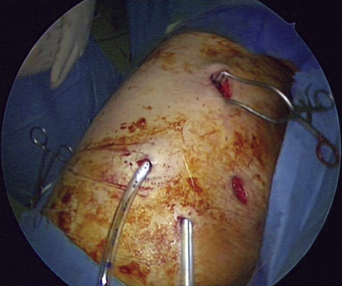
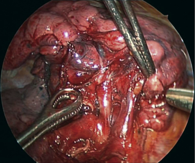
The order of steps for the operation is as follows: RML vein, major fissure between the RML and the right lower lobe (RLL), RML bronchus, RML artery, and the minor fissure. The incisions are described in Chapter 1 (see Figure 1-2). For a right middle lobectomy, place the utility incision (i.e., incision 3) one intercostal space below the superior pulmonary vein.
Video-Assisted Right Middle Lobectomy
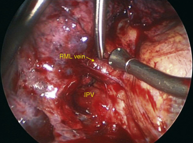
Step 1. Right Middle Lobe Vein
♦ Use the standard incisions, although the auscultatory incision is often not necessary (Figure 9-1). The utility incision is made directly up from the superior pulmonary vein, as for an upper lobectomy.
♦ Dissect between the inferior and superior pulmonary veins to define the inferior border of the RML vein, and determine whether there is drainage from the middle lobe to the inferior pulmonary vein.
♦ With DeBakey pickups and Metzenbaum scissors, define the superior and inferior aspect of the RML vein (Figure 9-2).
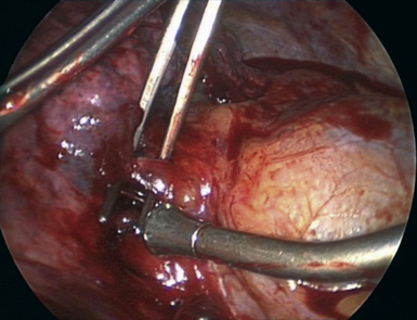
Step 2. Major Fissure
♦ Exposure: To line up the major fissure for stapling, retract the RLL inferiorly and slightly anteriorly while the RML is retracted inferiorly and medially to line up the major fissure with incision 1.
♦ Aim the thoracoscope anteriorly with the 30-degree lens pointed posteriorly and superiorly, directly down the fissure.
♦ Through incision 1, the anvil of the stapler is placed between the superior pulmonary vein and the inferior pulmonary vein. Hold the stapler in place as the lung parenchyma is pulled into the stapler. The cartridge of the stapler is lined up with the fissure, and the stapler is fired (Figure 9-4).
♦ Define the pulmonary artery with the Metzenbaum scissors through incision 1 by spreading on the surface of the artery or with the Yankauer suction bluntly dissecting along the surface of the pulmonary artery.
Step 3. Accessory Right Middle Lobe Artery
♦ A few patients have an accessory right middle lobe artery that is located inferior and posterior to the RML bronchus (Figure 9-5).
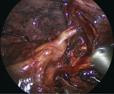
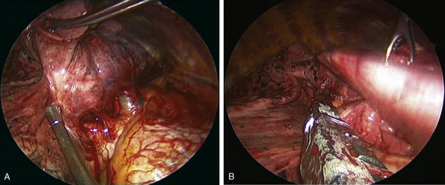
Step 4. Right Middle Lobe Bronchus
♦ Aim the thoracoscope anteriorly with the 30-degree lens pointed posteriorly but pulled back into the trocar.
♦ Remove the lobar nodes around the bronchus. Watch for the right middle lobe artery, which lies behind and parallel to the RML bronchus (Figure 9-6).
♦ With DeBakey pickups and Metzenbaum scissors introduced through the utility incision, dissect the bronchus away from the artery.
♦ Pass a right-angle clamp between the bronchus and the artery. Spread widely to create a tunnel for the stapler (Figure 9-7).
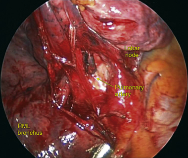
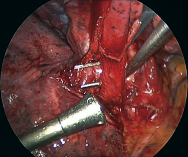
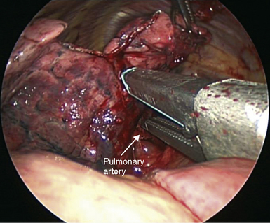
Step 5. Right Middle Lobe Artery
♦ Exposure: Retract the RLL posteriorly and inferiorly as the RML is retracted superiorly and anteriorly toward the apex of the chest.
♦ Aim the thoracoscope anteriorly with the 30-degree lens pointed posteriorly. The camera is rotated slightly clockwise.
♦ With the vein and bronchus transected, the RML artery is exposed as shown in Figure 9-9. Handling for the artery parallels that for the bronchus.
♦ With DeBakey pickups and Metzenbaum scissors introduced through the utility incision, dissect the artery. A right-angle clamp mobilizes the artery to create a tunnel through which the stapler will pass to transect the artery.
Step 6. Minor Fissure
♦ Exposure: Retract the RLL posteriorly and inferiorly. A ring forceps through the mid-clavicular incision retracts the RML inferiorly.
♦ Place the anvil just inferior to the right upper lobe vein and on the surface of the artery (Figure 9-10).
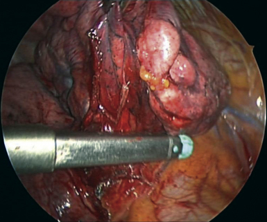
Step 7. Removal of the Lobe
♦ Exposure: Retract the RML low in the chest near the diaphragm through the posterior auscultatory triangle incision.
♦ Aim the thoracoscope anteriorly with the 30-degree lens pointed toward the apex of the chest for a panoramic view of the chest.
♦ With a ring forceps through the incision in the auscultatory triangle, hold the RML posteriorly and inferiorly by the diaphragm to keep it out of view while the Lapsac bag is passed through the utility incision to the apex of the chest.