Rhesus Isoimmunization

Published on 10/03/2015 by admin

Filed under Obstetrics & Gynecology

Last modified 22/04/2025

Print this page

rate 1 star rate 2 star rate 3 star rate 4 star rate 5 star
Your rating: none, Average: 0 (0 votes)

This article have been viewed 9340 times

Chapter 15 Rhesus Isoimmunization

Rhesus (Rh) isoimmunization is an immunologic disorder that occurs in a pregnant, Rh-negative patient carrying an Rh-positive fetus. The immunologic system in the mother is stimulated to produce antibodies to the Rh antigen, which then cross the placenta and destroy fetal red blood cells.

image Pathophysiology

The Rh complex is made up of a number of antigens, including C, D, E, c, e, and other variants, such as Du antigen. More than 90% of cases of Rh isoimmunization are due to antibodies to D antigens. Therefore, this chapter is mainly limited to a discussion of the D antigen, although the same principles apply to any other antigen-antibody combination. A person who lacks the D antigen on the surface of the red blood cells is called “Rh negative,” and an individual with the D antigen is considered “Rh positive.”

Among African Americans, about 8% are Rh negative, whereas among white Americans, about 15% are Rh negative. Only 1% to 2% of Asians and Native Americans are Rh negative. When Rh-negative patients are exposed to the Rh antigen, they may become sensitized. Two mechanisms are proposed for this sensitization. The most likely mechanism is the occurrence of an undetected placental leak of fetal red blood cells into the maternal circulation during pregnancy. The other proposal is the “grandmother” theory. This theory suggests that an Rh-negative woman may have been sensitized from birth by receiving enough Rh-positive cells from her mother during her own delivery to produce an antibody response.

In general, two exposures to the Rh antigen are required to produce any significant sensitization, unless the first exposure is massive. The first exposure leads to primary sensitization, whereas the second causes an anamnestic response leading to the rapid production of immunoglobulins, which can cause a “transfusion reaction” or hemolytic disease of the fetus during pregnancy.

The initial response to exposure to Rh antigen is the production of immunoglobulin M (IgM) antibodies for a short period of time, followed by the production of IgG antibodies that are capable of crossing the placenta. If the fetus has the Rh antigen, these antibodies will coat the fetal red blood cells and cause hemolysis. If the hemolysis is mild, the fetus can compensate by increasing the rate of erythropoiesis. If the hemolysis is severe, it can lead to profound anemia, resulting in hydrops fetalis from congestive cardiac failure and intrauterine fetal death. High bilirubin levels can damage the central nervous system and lead to neonatal kernicterus. Before the widespread use of Rh immune globulin for prevention of Rhesus isoimmunization, neonatal kernicterus was one of the leading causes of cerebral palsy.

The fetal and maternal circulations are normally separated by the placental “barrier.” Small hemorrhages occur in either direction across the intact placenta throughout pregnancy. With advancing gestational age, the incidence and size of these transplacental hemorrhages increase, with the largest hemorrhages usually occurring at delivery. Most immunizations occur at the time of delivery, and antibodies appear either during the postpartum period or following exposure to the antigen in the next pregnancy.

If a pattern of mild, moderate, or severe disease has been established with two or more previous pregnancies, the disease tends either to be of the same severity or to become progressively more severe with subsequent pregnancies. If a woman has a history of fetal hydrops with a previous pregnancy, the risk for hydrops with a subsequent pregnancy is about 90%. Hydrops usually develops at the same time as, or earlier than, in the previous pregnancy.

image Incidence

Although transplacental hemorrhage is very common, the incidence of Rh immunization within 6 months of the delivery of the first Rh-positive, ABO-compatible infant is only about 8%. In addition, the incidence of sensitization with the development of a secondary immune response before the next Rh-positive pregnancy is 8%. Therefore, the overall risk of immunization for the second full-term, Rh-positive, ABO-compatible pregnancy is about 1 in 6 pregnancies. The risk for Rh sensitization following an ABO-incompatible, Rh-positive pregnancy is only about 2%. The protection against immunization in ABO-incompatible pregnancies is due to the destruction of the ABO-incompatible cells in the maternal circulation and the removal of the red blood cell debris by the liver.

Transplacental hemorrhage may also occur before delivery. Establishment of the fetal circulation occurs at about 4 weeks’ gestation, and the presence of the Rh D antigen has been demonstrated as early as 38 days after conception. Consequently, Rh isoimmunization can occur at any time during pregnancy, from the early first trimester on. In the first trimester, the most common causes of transplacental hemorrhage are spontaneous or induced abortions. The incidence of immunization following spontaneous abortion is 3.5%, whereas that following induced abortion is 5.5%. The risk is low in the first 8 weeks, but it rises to significant levels by 12 weeks’ gestation. The risk for immunization following ectopic pregnancy is about 1%. Transplacental hemorrhage can also occur in the setting of second- or third-trimester vaginal bleeding, after invasive procedures such as amniocentesis or chorionic villus sampling, after abdominal trauma, or after external cephalic version. If necessary the amount of fetal blood entering the maternal circulation after an episode associated with transplacental hemorrhage can be estimated using the Kleihauer-Betke test (described later). All pregnant Rh-negative women who are not sensitized to the D antigen should routinely receive prophylactic Rh immune globulin (RhOGAM) at 28 weeks of gestation, within 72 hours of delivery of an Rh-positive fetus, and at the time of recognition of any of the problems cited previously that are associated with transplacental hemorrhage.

image 
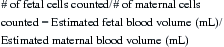
Recognition of the Pregnancy at Risk

A blood sample from every pregnant woman should be sent at the first prenatal visit for determination of the blood group and Rh type and for antibody screening. In Rh-negative patients, whose anti-D antibody titers are positive (i.e., those who are Rh sensitized), the blood group and Rh status of the father of the baby should be determined. If the father is Rh negative, the fetus will be Rh negative, and hemolytic disease will not occur. If the father is Rh positive, his Rh genotype and ABO status should be determined. This may be done by testing the father’s red blood cells with the reagents available for the antigens D, E, C, e, and c. Newer molecular techniques are now available to assess fetal Rh genotype. If he is homozygous for the D antigen, every fetus he fathers will be Rh positive and could potentially be affected. If he is heterozygous, only half of his children will be affected. Information regarding the zygosity of the father is of value in absolutely predicting the presence or absence of the Rh antigen in the fetus if the father is homozygous and in signaling the potential need for fetal antigen testing if the father is heterozygous. About 56% of Rh-positive whites are heterozygous for the Rh D antigen. If it is not possible to test the antigen status and zygosity of the father, it must be assumed that he is antigen positive.

ULTRASONIC DETECTION OF RH SENSITIZATION

Serial ultrasonic examinations of a woman with a fetus at risk for hemolytic disease can be a useful adjunct to amniocentesis in confirming fetal well-being and determining the advent of fetal hydrops. The examination should include a routine fetal assessment plus a determination of placental size and thickness and hepatic size. Both the placenta and the fetal liver are enlarged with hydrops. Fetal hydrops is easily diagnosed by the characteristic appearance of one or more of the following: ascites, pleural effusion, pericardial effusion, or skin edema. Appearance of any of these factors during an ultrasonic examination eliminates the need for diagnostic amniocentesis and necessitates therapeutic intervention based on fetal gestational age.

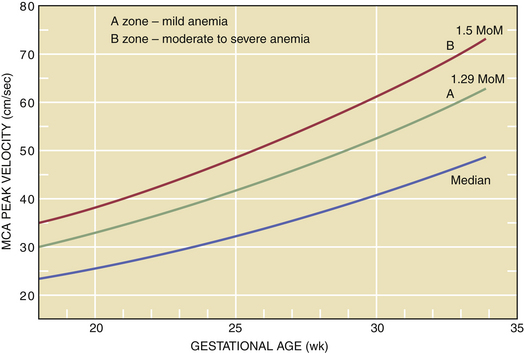
Doppler assessment of peak velocity in the fetal middle cerebral artery (MCA) in cm/sec has proved to be the most valuable tool for detecting fetal anemia. At-risk pregnancies should have this test performed every 1 to 2 weeks from 18 to 35 weeks of gestation. A fetal MCA peak systolic velocity (PSV) value above 1.5 multiples of the median for gestational age is predictive of moderate to severe fetal anemia and is an indication for percutaneous umbilical blood sampling for precise determination of fetal hemoglobin concentration. Intrauterine fetal transfusion should follow if indicated. After 35 weeks’ gestation, this test may produce a higher false-positive rate (Figure 15-1), and amniotic fluid spectrophotometry may be indicated.

image

FIGURE 15-1 Middle cerebral artery (MCA) Doppler peak velocities based on gestational age. MoM, multiples of the median.

(Data from Moise KJ Jr: Management of rhesus alloisoimmunization. Obstet Gynecol 100:600-611, 2002.)

AMNIOTIC FLUID SPECTROPHOTOMETRY

Before widespread use of MCA Doppler studies, analysis of amniotic fluid was the most frequently used method of gauging the severity of fetal hemolysis. It is still used in settings in which expertise in the performance of an MCA Doppler is not available, or occasionally, after 35 weeks of gestation. There is an excellent correlation between the amount of biliary pigment in the amniotic fluid and the fetal hematocrit, beginning at 27 weeks’ gestation.

The most likely source of bilirubin in the amniotic fluid is tracheal and pulmonary efflux with some transudate from the umbilical and placental vessels. Because of the small concentrations found in the amniotic fluid, spectrophotometric analysis is the most widely used technique for estimating amniotic fluid bilirubin concentration.

The optical density deviation (ΔOD) at 450 μ from a baseline drawn between the optical density values at 365 and 550 μ forms a peak that can be used to calculate the change in ΔOD in nanomoles (nm) at 450 μ measuring the amniotic fluid unconjugated bilirubin, which correlates with the cord blood hemoglobin of the newborn at birth.

Bilirubin is oxidized to colorless pigments when it is exposed to light; therefore, the fluid should be protected from light. Heme pigments and meconium may cause falsely high spectrophotometric values.

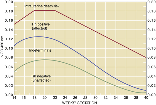
Bilirubin is normally found in amniotic fluid in a concentration that gradually diminishes toward term. For predictive interpretation, Liley devised a spectrophotometric graph based on the correlation of cord blood hemoglobin concentrations at birth and the amniotic fluid change in optical density at 450 μ. Using this method, he was able to establish predictive zones for mild, moderate, and severe disease. The Liley chart (Figure 15-2) can be used to determine, with accuracy, the severity of the disease and the appropriate management, beginning at 27 weeks’ gestation. The Queenan curve, a modified Liley curve with four zones instead of three, is used as a predictive tool in some centers from 14 to 40 weeks’ gestation (Figure 15-3). Because single ΔOD 450 values are helpful only if they are very high (zone III) or very low (zone I), serial sampling of amniotic fluid is generally indicated.

image

FIGURE 15-3 Queenan curve for optical density at a wavelength of 450-nm (ΔOD 450-nm) values for the management of the patient with isoimmunization. Rh, rhesus.

(Adapted from Queenan JT, Tomai TP, Ural SH, et al: Deviations in amniotic fluid optical density at a wavelength of 450-nm in Rh-immunized pregnancies from 14 to 40 weeks’ gestation: A proposal for clinical management. Am J Obstet Gynecol 168:1370-1376, 1993.)

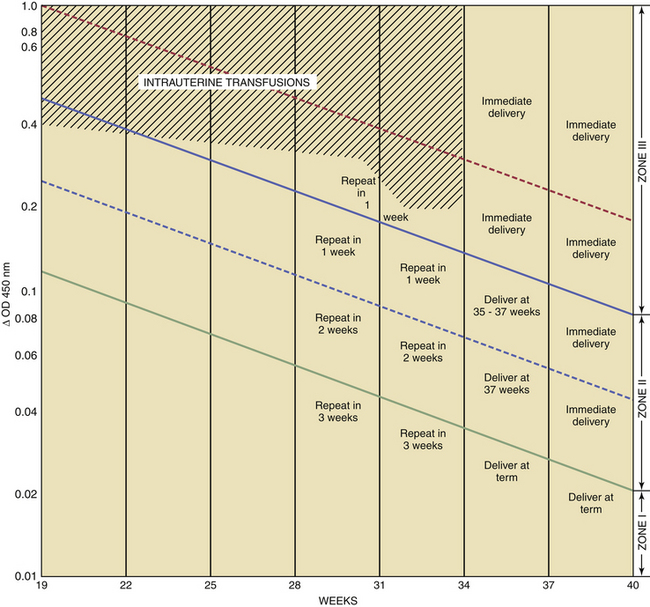
The severity of hemolytic disease in the prior pregnancy provides an approximate index for the timing of the first amniocentesis. This may range from 22 to 30 weeks with prior severe disease indicating the initial procedure as early as 22 weeks. With repeat sampling one of three trends will emerge: (1) Falling ΔOD 450 values are indicative of a fetus that is either unaffected (e.g., Rh negative) or very mildly affected. No intervention is indicated in these patients (see Figures 15-1 and 15-2). (2) If the ΔOD 450 is either stable or rising, frequent ΔOD 450 determinations are necessary to determine the timing of delivery. (3) If the ΔOD 450 enters zone III (refer to zone levels on the right side of Figure 15-2) before 34 weeks, percutaneous umbilical blood sampling is performed for determination of fetal hemoglobin followed by intrauterine transfusion if indicated.

INTRAUTERINE TRANSFUSION

Intrauterine transfusion, initially introduced in 1963 as an intraperitoneal transfusion and presently usually administered as an intravascular transfusion has markedly changed the prognosis for severely affected fetuses. The goal is to transfuse fresh group O, Rh-negative packed red blood cells. In addition to routine blood screening, the blood for transfusion is irradiated, washed, processed through a leukocyte-poor filter, and screened for cytomegalovirus. Curare is usually injected directly into the fetal thigh with a 22-gauge spinal needle before transfusion, regardless of method, to immobilize the fetus during the procedure. Repeat transfusions are generally scheduled at 1- to 3-week intervals. The final transfusion is typically performed at 32 to 34 weeks’ gestation. In general, the fetus is delivered when the lungs are mature.

The overall survival rate following intrauterine transfusion is about 85%. In fetuses with no evidence of hydrops, the survival rate is about 90%, and for fetuses with hydrops before the transfusion, the survival rate is about 75%.

image Prevention of Rhesus Isoimmunization

Because Rh isoimmunization occurs in response to exposure of an Rh-negative mother to the Rh antigen, the mainstay for prevention is the avoidance of maternal exposure to the antigen. Rh immune globulin (RhO-GAM) diminishes the availability of the Rh antigen to the maternal immune system, although the exact mechanism by which it prevents Rh isoimmunization is not well understood.

RhO-GAM is prepared from fractionated human plasma obtained from hyperreactive sensitized donors. The plasma is screened for hepatitis B surface antigen and anti-HIV-1. The globulin is available in several dosages for intramuscular injection. Since the advent of its use in 1967, Rh immune globulin has dramatically reduced the incidence of Rh isoimmunization.

Because the greatest risk for fetal-to-maternal hemorrhage occurs during labor and delivery, Rh immune globulin was initially administered only during the immediate postpartum period. This resulted in a 1% to 2% failure rate, thought to be due to exposure of the mother to fetal red blood cells during the antepartum period. The indications for the use of Rh immune globulin have therefore been broadened to include any antepartum event (such as amniocentesis) that may increase the risk for transplacental hemorrhage. The routine prophylactic administration of Rh immune globulin at 28 weeks’ gestation is now the standard of care. Despite adherence to this suggested Rh immune globulin protocol, 0.27% of primiparous Rh-negative patients still become sensitized.

INDICATIONS FOR ADMINISTRATION OF RhO-GAM

It is the responsibility of every health care practitioner who is involved in the care of pregnant women to prevent Rh isoimmunization. This is done by the timely administration of RhO-GAM to pregnant Rh-negative women, whose anti D titers are negative, at about the time of events associated with fetal-maternal hemorrhage. The following provides a practical approach to the administration of Rh immune globulin to an Rh-negative unsensitized patient.

All pregnant women should have a blood type and antibody screen on their first prenatal visit. During an uncomplicated pregnancy, the Rh-negative woman whose initial antibody screen is negative should have a repeat antibody titer at 28 weeks’ gestation. If the antibody screen is still negative, she should routinely receive an intramuscular injection of 300 μg of RhO-GAM prophylactically. A positive antibody screen does not necessarily mean the woman is not a candidate for RhO-GAM. In this case, an antibody identification and titer must be requested. If the antibody is not an anti-D antibody, then she is still a candidate for RhO-GAM.

RhO-GAM should also be administered during the antepartum period at any gestational age to an Rh-negative unsensitized (anti-D–negative) woman at the time of spontaneous or induced abortion, treatment of an ectopic pregnancy, significant vaginal bleeding, performance of an amniocentesis, abdominal trauma, or external cephalic version. Before 12 weeks of gestation, 50 to 100 μg of RhO-GAM should be sufficient to prevent isoimmunization.

Because chorionic villi in gestational trophoblastic disease are avascular and are devoid of fetal erythrocytes, RhO-GAM is probably not necessary following termination of a “complete” molar pregnancy. A “partial” molar pregnancy may have fetal tissue, and theoretically fetal cells could enter the maternal circulation. At least one case of sensitization following a molar pregnancy has been reported.

The risk for transplacental hemorrhage increases at the time of delivery, especially with cesarean birth or manual removal of the placenta. At delivery, cord blood must be sent for determination of the fetal blood group, Rh type, and for a direct Coombs’ test. RhO-GAM (300 μg) is routinely given to all Rh-negative, anti-D–negative women who deliver an Rh-positive infant within 72 hours of delivery. If a transplacental hemorrhage of greater than 30 mL of fetal blood is suspected (as might occur in the setting of an abruption, manual removal of the placenta, or severe maternal abdominal trauma), a Kleihauer-Betke test is helpful in determining the volume of the hemorrhage. Additional RhO-GAM may then be given at a dose of 10 μg of RhO-GAM per 1 ml of fetal blood that entered the maternal circulation.