Retroperitoneal Sarcoma

Published on 16/04/2015 by admin

Filed under Surgery

Last modified 22/04/2025

Print this page

rate 1 star rate 2 star rate 3 star rate 4 star rate 5 star
Your rating: none, Average: 0 (0 votes)

This article have been viewed 1721 times

Chapter 50

Retroperitoneal Sarcoma

image

Introduction

Retroperitoneal sarcomas account for 10% to 15% of adult soft tissue sarcomas, or about 1000 new cases each year. Patients have relatively poor survival rates because these tumors often do not produce symptoms until late in the disease. Retroperitoneal sarcomas can become quite large before symptoms begin; about half will exceed 20 cm at diagnosis.

Nonsurgically treated lymphoma should be ruled out in patients with retroperitoneal masses. Computed tomography (CT) scans should be performed to determine the extent of the tumor, show its relationship to surrounding structures, and identify possible distant metastases. CT-guided biopsy of the mass is the technique of choice for definitive diagnosis.

Complete margin-negative resection is the standard of care for retroperitoneal sarcoma. These tumors can invade surrounding organs, with kidney, colon, pancreas, and spleen most often involved. Also, these tumors create intense reactions in surrounding tissues; these reactions make it difficult to assess whether the tumors are invading, rather than just pushing on, surrounding structures.

Preoperative Imaging and Incision

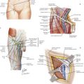
The retroperitoneum has many vital structures, including the kidneys, pancreas, inferior vena cava (IVC), and the aorta (Fig. 50-1). These structures are close to each other and can become greatly distorted by a large retroperitoneal tumor. Preoperative imaging is particularly useful; the CT scan in Figure 50-1 shows a large, right retroperitoneal tumor. This tumor encroaches on the IVC and clearly involves the right kidney. Availability of this information before surgery can allow for preoperative assessment of kidney function in the event of nephrectomy and planning for possible vascular resection (consulting with vascular surgeon).

Incisions for this operation are varied. A standard midline laparotomy incision works well for most cases. In other situations, placement of the patient in the lateral decubitus position and performing a curvilinear flank incision allows the best exposure. Most cases can be performed through a midline incision, and this chapter focuses on the procedure through this incision.

Abdominal Exposure for Tumor Removal

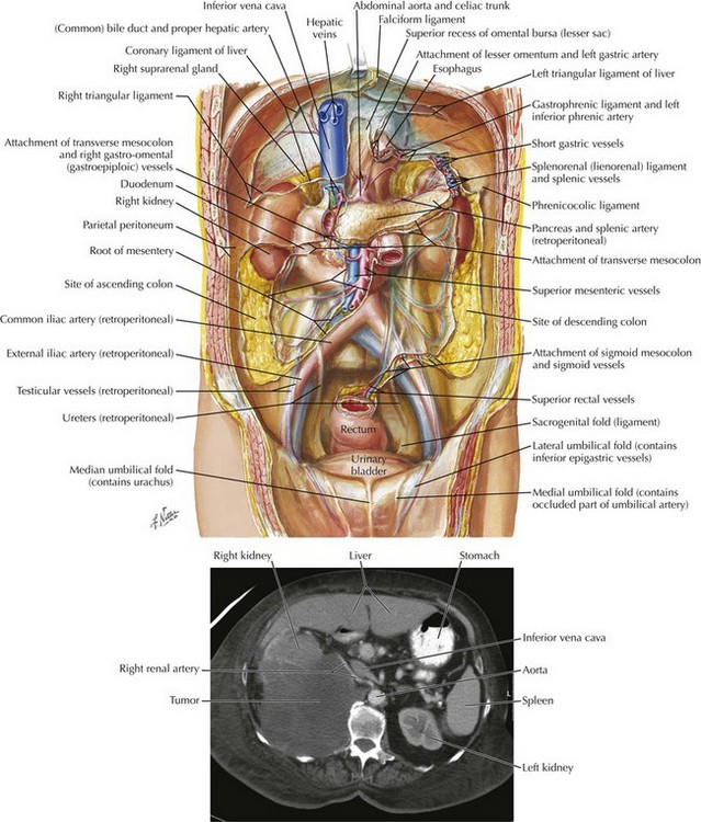
After the surgeon enters the abdomen, the retroperitoneal tumor is often the first structure to be seen and distorts the normal anatomy; a common site is shown in Figure 50-2. It is in these situations that correctly recognizing structures and being in the correct plane are vital.

Typically, the first step to approaching a retroperitoneal tumor, or gaining access to the retroperitoneum for any procedure, is to retract the colon (either ascending or descending) medially and find the reflection of peritoneum, the white line of Toldt. This line will guide the dissection and provide exposure into the right or left retroperitoneum, where tumors typically develop. (Toldt’s fascia, or membrane, is part of the renal fascia anterior to the kidney.)

If more superior exposure is required in the right retroperitoneum, a Kocher maneuver can be performed. This maneuver involves freeing the lateral border of the duodenum from its retroperitoneal attachments and retracting it medially.

Once full exposure of the tumor is obtained, a more detailed assessment can be made of the surrounding structures that are involved. Margin status has a major impact on survival rate, so every attempt should be made for a complete en bloc, margin-negative resection. This often means that the kidney, portions of the colon, vascular structures, or portions of the duodenum are resected.

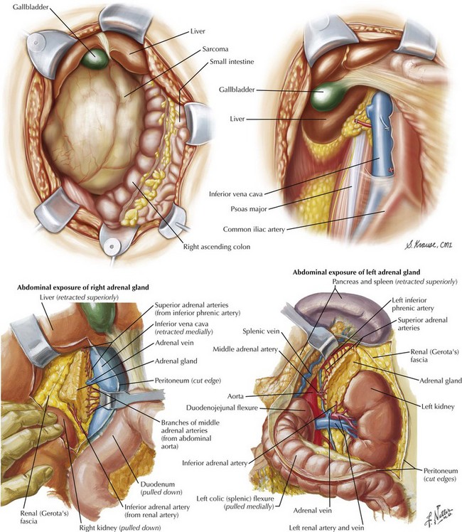
Figure 50-2 shows the right retroperitoneum after removal of a large retroperitoneal tumor. Notice that the right kidney was resected as well. In this case the right colon and the duodenum have been mobilized and retracted medially.