Retinoblastoma
Clinical Features:
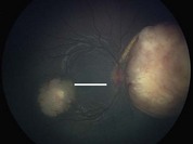
The most common presenting feature is leukocoria, but others include strabismus, decreased vision, and a painful eye. Characteristic clinical features include a white or yellow-white elevated, fungating, singular or multifocal retinal tumor (Fig. 22.2.1). Associated vitreous and subretinal seeding may also be present.
OCT Features:
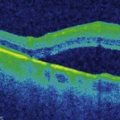
OCT shows involvement of the neurosensory retina, particularly the photoreceptors and outer retina. Early in tumor growth or along the leading edge of the tumor, these features can be more clearly seen (Fig. 22.2.2). In larger or more advanced tumors, the entire neurosensory retina can be obscured but the underlying retinal pigment epithelial layer is preserved (Figs 22.2.3 and 22.2.4).
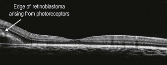
Figure 22.2.2 OCT (corresponding to line mark section of the scan in Figure 22.2.1) shows the edge of the tumor located above the RPE and involving the outer retina. The tumor appears to be arising from the photoreceptor layer. The central fovea is uninvolved. (Courtesy of Carol Shields, MD.)
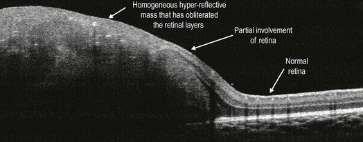
Figure 22.2.3 OCT (corresponding to Figure 22.2.1, horizontal scan through macular lesion) shows a fairly homogeneous hyper-reflective mass that has obliterated the retinal layers completely. The transition to partial involvement of the retina and normal retina can also be seen. (Courtesy of Carol Shields, MD.)
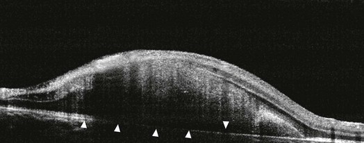
Figure 22.2.4 OCT (corresponding to Figure 22.2.1, vertical scan through macular lesion) illustrates that the underlying RPE layer is intact and not involved (arrowheads). (Courtesy of Carol Shields, MD.)