207 Retinitis pigmentosa
Salient features
Examination
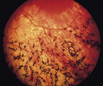
• Peripheral retina shows perivascular ‘bone spicule pigmentation’ and arteriolar narrowing (Fig. 207.1). The retinal veins (never the arteries) often have a sheath of pigmentation for part of their course. The pigment spots that lie near the retinal veins are seen to be anterior to them, and so they hide the course of the vessel. (In this respect, they differ from the pigment around the spots of choroidal atrophy in which the retinal vessels can be traced over the spots.)
Questions
Advanced-level questions
Mention a few systemic disorders associated with retinitis pigmentosa
• Laurence–Moon–Biedl–Bardet syndrome, which is a recessively inherited disorder characterized by mental disability, polydactyly, syndactyly, hypogonadism, obesity and renal disease (structural abnormalities such as calyceal cysts or calyceal clubbing and blunting).
• Bassen–Konzweig syndrome (abetalipoproteinaemia), characterized by fat malabsorption, abetalipoproteinaemia, acanthocytosis and spinocerebellar ataxia (Ophthalmology 1984;91:991).
• Refsum’s disease (phytanic acid storage disease), an autosomal recessive disorder characterized by hypertrophic peripheral neuropathy, deafness, ichthyosis, cerebellar ataxia, raised CSF protein levels in the absence of pleocytosis.
• Kearns–Sayre syndrome, a triad of retinitis pigmentosa, progressive external ophthalmoplegia and heart block (Br J Ophthalmol 1985;69:63).
• Usher’s disease, a recessively inherited disorder characterized by congenital, non-progressive, sensorineural deafness (Arch Ophthalmol 1983;101:1367).
What do you know about the genetics of retinitis pigmentosa?
What is retinitis pigmentosa sine pigmento?
A variety of retinitis pigmentosa but without visible pigmentation of the retina.
What is inverse retinitis pigmentosa?
Bone corpuscles are visible in the perifoveal area, whereas the retinal periphery is normal.
How would you manage this patient?
• Refer for genetic counselling
• Impaired vision training and aids for daily living
• Regular ophthalmology follow-up including visual fields, electroretinography.


