212 Retinal changes in AIDS
Instruction
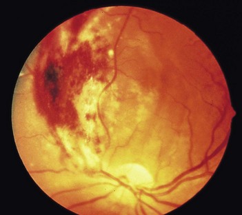
At a distance from the patient, the examiner will say that this patient has HIV, examine his fundus.
Salient features
Questions
How would you treat these cotton-wool spots?
In a patient with known HIV infection, such infarcts require no specific therapy.
Advanced-level questions
How would you treat retinitis?
• Therapy with intravenous ganciclovir or foscarnet is given lifelong to protect unaffected areas but does not restore functional areas already affected. The optimal treatment for patients with AIDS, normal renal function and cytomegalovirus (CMV) retinitis is foscarnet plus an antiretroviral nucleoside such as zidovudine. For similar patients with impaired renal function, ganciclovir would be the drug of choice, perhaps with an antiretroviral agent such as didanosine as it has few overlapping side effects. Late retinal detachment occurs despite therapy in 20% of these patients
• Sustained-release ganciclovir implant in the pars plana of the eye. Oral ganciclovir in conjunction with an implant reduces the incidence of a new CMV disease and delays the progression of retinitis (N Engl J Med 1999;340:1063–70)
What are the other retinal manifestations in patients with AIDS?
• Toxoplasmosis retinochoroiditis: usually nasal to the disc, characterized by extensive whitish infarction and inflammation, with minimal associated intraretinal haemorrhage (outer retinal involvement may show ‘brush-fire’ advancement of infection) (see Fig. 208.1A)
• Pneumocystis jiroveci (was carinii) choroiditis: characterized by scattered nodular yellowish-orange lesions deep in the retina, usually throughout the posterior pole
• Acute retinal necrosis syndrome: with severe peripheral retinal infarction and retinal vasculitis. Atrophic retinal holes appear later in these areas
• Progressive outer retinal necrosis syndrome: characterized by diffuse whitish opacification of the retina beneath the retinal vessels and a cherry-red spot in the fovea. It is associated with herpes zoster, and the prognosis for visual acuity is dismal
• Ocular presentations of syphilis include anterior and posterior uveitis, retinitis, retinal vasculitis and papillitis
• Acute syphilitic posterior placoid chorioretinitis: large placoid yellowish lesions at the level of the retinal pigment epithelium in the posterior retina with inflammation of the vitreous and loss of vision.
What diagnostic tests would you perform to detect retinal manifestations of AIDS?
• Careful clinical observations
• Serology: CD4 cell count (usually <50 × 106 cells/l in presentation of untreated CMV retinitis)
• Retinal biopsy by pars plana vitrectomy
• Fluorescein angiography: helpful in differentiating retinal lesions
• Sequential fundus photographs: for diagnosis and monitoring of lesions


