Restenosis and Drug-Eluting Stents

Published on 21/06/2015 by admin

Filed under Cardiovascular

Last modified 22/04/2025

Print this page

rate 1 star rate 2 star rate 3 star rate 4 star rate 5 star
Your rating: none, Average: 0 (0 votes)

This article have been viewed 2619 times

7 Restenosis and Drug-Eluting Stentsimage

Restenosis

Restenosis is defined as vessel narrowing of a previous angioplasty site. After balloon expansion, this narrowing occurs as a result of two mechanisms, negative remodeling (vessel constriction after stretching) and reaccumulation of material within the lumen (or stent). Reaccumulation of cellular material (also known as endothelial proliferation) is initiated by vessel injury with a release of thrombogenic, vasoactive, and mitogenic factors. Endothelial and deeper injury leads to platelet aggregation, thrombus formation, inflammation, and activation of smooth muscle cells and macrophages. The production and release of growth factors and cytokines promotes further synthesis of such factors and release from the cells involved. These factors result in the migration of new smooth muscle cells from their location within the arterial media to the endovascular lumen. These cells become a synthetic type of cell that produces extracellular matrix, leading to cellular proliferation and mechanical obstruction of the vessel lumen.

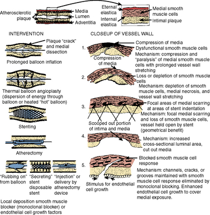
The second component of restenosis, recoil and negative remodeling of the arterial wall is inhibited by stents. Compared to balloon angioplasty, stents have reduced restenosis from 40% to 50% after percutaneous transluminal coronary angioplasty (PTCA) to 20% after bare-metal stenting. Drug-eluting stents (DESs) have brought restenosis rates to <10% in most patient subgroups. Restenosis still occurs inside stents (called in-stent restenosis [ISR]) mostly, if not exclusively, as a result of endothelial cell proliferation. Rarely does vascular recoil make a contribution, but it must be considered in treating the ISR lesion. Restenosis is not device-specific but rather a function of the anatomic substrate and the type of injury produced (Fig. 7-1).

image

Figure 7-1 Interventional devices and presumed mechanisms of action of arterial plaque in vessel wall lead to restenosis. The indication of immediate outcome and restenosis rates depend on both the device and the arterial substrate encountered.

(From Waller BF, Pinkerton CA, Orr CM. Restenosis 1 to 24 months after clinically successful coronary balloon angioplasty: A necropsy study of 20 patients. J Am Coll Cardiol 1991;17:58–70.)

Definitions of Restenosis

There are two types of restenosis recognized in patients, angiographic and clinical, which are not mutually exclusive.

Angiographic Restenosis

Angiographically measured luminal renarrowing after PCI has been the “gold standard” for restenosis. Angiographic restenosis is a continuous phenomenon, with no obvious threshold separating “restenosers” from “nonrestenosers.” Studies have shown that the percentage of stenosis or minimal lumen diameter has a near Gaussian (normal) distribution on follow-up angiograms after balloon angioplasty. Thus, restenosis is best measured as a continuous variable. Nevertheless, because of practicality, the most commonly used definition of restenosis employs a dichotomous value (e.g., 50% diameter narrowing).

Several different angiographic definitions of restenosis have been published with overlapping differences in some patients. Most studies define angiographic restenosis as either a greater than 50% loss of initial gain after intervention or an absolute lesion stenosis of greater that or equal to 50% at follow-up angiogram. The late loss of the acute luminal enlargement, or net gain in millimeters at the lesion site 6 months after treatment by quantitative angiography, should be around 0.7 mm for balloon angioplasty.

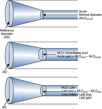
The late loss index is the loss at the lesion site divided by the amount of acute gain (Fig. 7-2). The loss index is accepted as the most sensitive measure of the effectiveness of the technique and should range from 0.4 to 0.6 mm for balloon angioplasty. The lower the loss index is, the more effective the antirestenosis treatment will be.

Restenosis is both a lumen-related and a vessel wall–related phenomenon. It appears that 40% to 60% of the acute luminal gain is lost during follow-up in all patients treated, independent of the devices. A similar degree of intimal thickening (restenosis by wall measurements) may or may not cause a significant luminal narrowing (restenosis by lumen measurement). As expected, the vessel size itself exerts a significant positive influence on minimal lumen diameter at follow-up and an equally negative effect on late loss. A larger artery will have a larger lumen at follow-up and vice versa for a smaller artery. Using percentage stenosis rather than absolute lumen diameter will neutralize this effect by correcting automatically for artery size.

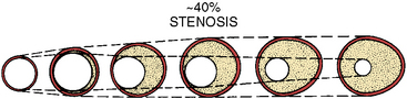
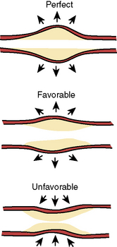
Intravascular ultrasound (IVUS) imaging is superior to angiography for anatomic and morphologic restenosis definitions. Recent IVUS studies have shown that an important component of restenosis is vessel recoil, a feature prevented by stenting. Normal vessel modeling maintains the coronary lumen. Late negative remodeling of the injured vessel is also prevented by stenting (Figs. 7-3 and 7-4).

image

Figure 7-4 Adequacy of arterial remodeling with respect to changes in vessel size.

(From Schwartz RS. Pathophysiology of restenosis: interaction of thrombosis, hyperplasia, and/or remodeling. Am J Cardiol 1998;81:16E.)

Time Course

Stents prevent very early (<24 hr) restenosis due to elimination of acute elastic recoil. The incidence of restenosis increases, peaking around 6 months. Late restenosis occurs uncommonly after 12 months. Restenosis after angioplasty using non-balloon, non-stent devices alone was highly variable with a reported incidence of restenosis between 15% and 55% (see Suggested Readings).

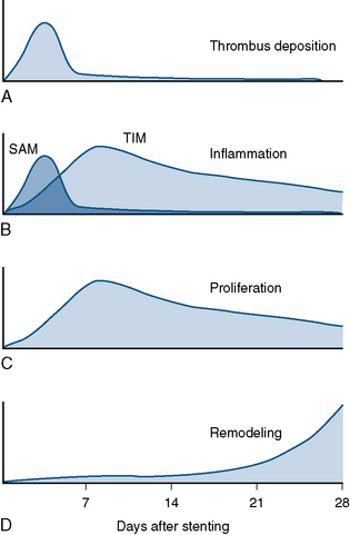
Different mechanisms produce restenosis in a time-dependent manner. Early restenosis is due to thrombus, whereas late restenosis is related more to remodeling (Fig. 7-5).

Patient Subsets at Higher Risk of Restenosis

In-Stent Restenosis

In-stent restenosis (ISR) is primarily due to neointimal hyperplasia produced by vessel injury of the balloon and/or stent struts. The injured segments promote activation of platelets, mural thrombus, and inflammatory cells. Vascular injury, mural thrombus, and a metallic foreign body activate circulating neutrophils and tissue macrophages. These elements release cytokines and growth factors, activating smooth muscle as well as stimulating upregulation and expression of genes promoting cell division, such as c-myc, leading to further cell proliferation. Metalloproteinases are produced, leading to increased matrix material and remodeling of the extracellular support matrix, initiating smooth muscle cell migration. Uncontrolled proliferation of vascular smooth muscles into the vessel intima and the deposition of extracellular matrix lead to significant in-stent luminal narrowing 3 to 6 months after PCI.

There are two major categories of in-stent restenosis—focal and diffuse (Fig. 7-6)—and within each category several subtypes of responses relative to the proliferation within and/or adjacent to the stent are noted. Figure 7-7 suggests a scheme for the treatment of ISR. Note that brachytherapy is now rarely used.

image

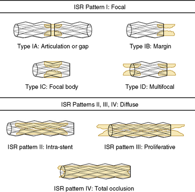
Figure 7-6 Classification system proposed for in-stent restenosis (ISR).

(From Mehran R, Dangas G, Abizaid AS, et al. Angiographic patterns of in-stent restenosis classification and implications for long-term outcome. Circulation 1999;100:1872–1878.)

image

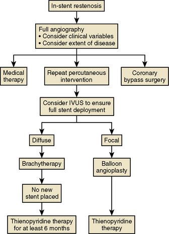
Figure 7-7 Algorithm for treatment of in-stent restenosis.

(Modified from Topol EJ. Textbook of interventional cardiology, 4th ed. Philadelphia: Saunders, 2003: 468.)

Stent fracture is associated with in-stent restenosis in Drug-Eluting Stents.

Lee MS et al. reviewed angiograms on 530 of 2728 patients who underwent drug-eluting stenting from 2003 to 2005. The incidence of DES fracture–related adverse events was rare and identified in 10 patients. None of these fractures was detectable at the time of stent placement. The median time from implant to fracture detection was 226 days (range, 7–620 days). Adverse stent fracture events occurred in seven patients (six patients had binary restenosis and one patient had stent thrombosis), all necessitating repeat intervention. Factors predisposing to stent fracture included excessive tortuosity in the proximal segment, and overlapping stents. In this small patient registry, all stents were sirolimus DESs.

Management of Restenosis

IVUS, Balloon Angioplasty, and Restenting

The ISR lesion may be due to underexpansion of the stent for the size of the vessel on the initial implantation or exuberant neointimal hyperplasia or a combination of these mechanisms. The treatment of ISR should begin with IVUS assessment of vessel/stent relationship. Then an appropriately sized balloon can be used to expand the stent and compress the hyperplastic tissue. A repeat IVUS is often helpful to gauge success of this maneuver. Recently, use of optical coherence tomography in patients undergoing balloon angioplasty of ISR shows tissue disruption, leading the operator to select another stent to seal this material. Although appealing to the operator, there are few data to support this routine approach at this time.

The success and complication rates are lower than for the initial procedure because the restenosis lesion is primarily fibroproliferative rather than an atherosclerotic plaque. Stenting of restenotic lesions, although easily performed, may not resolve the problem long term since outcomes after multiple overlapping stents may predisposed to higher subacute thrombosis rates.

Surveillance angiography is not recommended because of the potential for false positive restenosis, that is, narrowing without significant clinical importance. Intermediately severe ISR lesions (40%–70%) should be associated with evidence of ischemia. It is appropriate to use stress testing or fractional flow reserve measurement (FFR) before treatment.

Use of intravenous ultrasound is critical to decide whether to reexpand an underdeployed stent relative to the vessel size or attack the endothelial proliferation and consider an additional stent.

Use of a cutting balloon should be considered because blades provide excellent balloon stability inside stent during inflations.

Brachytherapy

Vascular brachytherapy, using beta and gamma emitters for treating PCI-related restenosis, and especially in-stent restenosis, had demonstrated safety and marginal efficacy. Brachytherapy is not currently used for routine treatment of in-stent restenosis because of logistical complexity and complications such as late thrombosis and edge effects after radiation therapy. A full discussion of this topic can be found elsewhere.

The adverse effects of brachytherapy included the following:

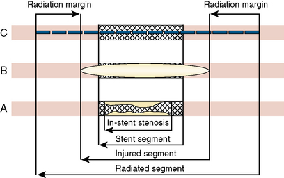
Of all the adverse effects, late thrombosis occurring more than 30 days after radiation therapy was one of the most feared major complications of vascular brachytherapy. Late thrombosis in early clinical trials was reported in up to 14% of patients. Late thrombosis also occurs with other vascular brachytherapy and relates to the healing arrest and lack of stent re-endothelialization. An effective strategy to prevent late rethrombosis is limiting restenting at the time of radiation treatment. It is essential to administer at least 12 months of antiplatelet therapy, preferably clopidogrel in addition to aspirin, for all radiation cases, both beta and gamma emitters. Definitions of artery and stent segments address by brachytherapy are shown on Figure 7-8.

Drug-Eluting Stents

Compared to BMSs, DESs reduce the rate of TLR and in-stent restenosis. The components of a DES system are (1) the specific metal stent design, (2) the use and type of polymer for drug absorption, and (3) the type of antiproliferative agent and its elution kinetics.

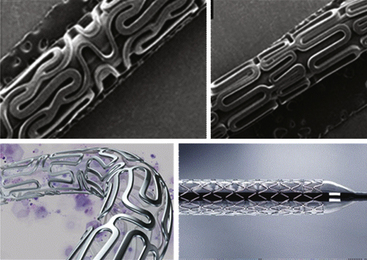
Current DESs available in the United States include drug compounds that contain antiproliferative agents such as paclitaxel, sirolimus, everolimus, or zotarolimus (Fig. 7-9).

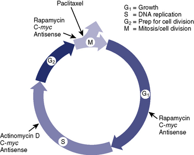
Drugs for Coated Stents (Fig. 7-10)

Drugs used for stent coatings to reduce neointimal proliferation should have a large therapeutic window and a low inflammatory potential, inhibit multiple mechanisms of the complex restenotic biology, and reduce smooth muscle cell proliferation without unacceptable toxicity to the medial and adventitial cell layers. Unlike radiation, local drug elution should not inhibit stent re-endothelialization. Drugs for coated stents should have favorable local pharmacokinetics and distribution properties. Hydrophilic drugs, such as heparin, permeate into tissue but are rapidly cleared. Hydrophobic agents, such as paclitaxel or sirolimus, are insoluble in the aqueous phase and bind to hydrophobic sites on the arterial wall. Both hydrophilic and hydrophobic drugs have large spatial concentration gradients across the arterial wall, with hydrophobic drugs distributing better and more homogeneously than hydrophilic agents.

Sirolimus is rapamycin, a naturally occurring macrocyclic lactone discovered in the soil of Easter Island (Rapa Nui) in the 1960s. Rapamycin is a product of fermentation of Streptomyces hygroscopicus and was used as an antifungal antibiotic. Sirolimus blocks the cell cycle of proliferating cells binding to the high-affinity cytosolic receptor protein FK506, leading to the inhibition of mammalian target of rapamycin (mTOR), which prevents downregulation of tumor suppressive cell P27. The gene p27 inhibits cell-dependent kinase activity and blocks G1- to S-phase cell cycle progression. Sirolimus is lipophilic and easily crosses the cell membrane. The inhibition of mTOR suppresses T-cell proliferation and is a powerful antiproliferative and antimigratory agent acting on smooth muscle cells. Systemic sirolimus reduces neointimal proliferation after balloon injury in porcine coronary arteries. Local sirolimus administration inhibits neointimal proliferation.

Paclitaxel is a powerful antineoplastic drug found in the Pacific yew tree (Taxus brevifolia) and is used in the treatment of malignant ovarian and breast cancer. Paclitaxel stabilizes polymerized microtubules and enhances microtubular assembly, forming unorganized and decentralized microtubules in the cytoplasm. Cell replication is inhibited predominately in the G0/G1 and G2/M phase of the cell cycle. Paclitaxel is highly lipophilic, promoting rapid uptake through hydrophobic cell membranes and minimizing systemic loss. Paclitaxel is suitable for polymer-based delivery. It has long-lasting antiproliferative effects after a single administration and can be directly applied to metal as a durable simple coating.

Everolimus is a novel semisynthetic highly lipophilic macrolide with immunosuppressant and antiproliferative properties. The chemical name is 40-O-(2-hydroxyethyl)-rapamycin and it is created by modifying rapamycin. Similar to rapamycin, inhibiting mTOR is the likely mechanism for suppression of cell proliferation. At the cellular level, it blocks growth factor–driven transduction signals in the T-cell response to alloantigen and proliferation of both hematopoietic and non-hematopoietic cells. Following stimulation of the IL-2 receptor on the activated cell, it inhibits p70 S6 kinase, thereby arresting the cell cycle in the late G1 phase. Systemic everolimus suppresses in-stent neointimal growth in the rabbit iliac artery following stenting.

Zotarolimus is also a novel semisynthetic derivative of rapamycin, which was designed to have a shorter in vivo half-life. It is a highly lipophilic immunosuppressive and antiproliferative agent. As with rapamycin and everolimus, zotarolimus inhibits mTOR, blocks growth factor–driven cell proliferation, which ultimately results in cell cycle arrest in the G1 phase.

Drug-Eluting Stent Systems

The Cypher (Cordis Corp., Bridgewater, NJ) sirolimus-eluting coronary stent system is comprised of two components: a BX Velocity coronary stent system (Cordis Corp., Bridgewater, NJ) and the sirolimus drug product. It was approved for clinical use by the U.S. Food and Drug Administration (FDA) in April 2003. It comes premounted on either an over-the-wire (OTW) or rapid exchange (RX) delivery system. The Cypher stent is comprised of 316 L stainless steel with six to seven circumferential cells. The inactive ingredients are parylene C and two non-erodible polymers: polyethylene-co-vinyl acetate (PEVA) and poly n-butyl methacrylate (PBMA). The base coat is made of a combination of the two polymers and sirolimus (67%/33%). This is then applied to the stent, which has been pretreated with parylene C. A solution of PBMA polymer is applied as a topcoat to the stent. Lastly, the drug/polymer combination solution is applied to the entire surface. The drug delivery kinetics allows for 80% of the sirolimus to be released over 30 days. The stent is available in 2.25 mm to 3.5 mm diameter sizes and 8 mm to 33 mm in length. It currently has the FDA-approved indications for improving coronary luminal diameter in patients with symptomatic ischemic heart disease due to discrete de novo lesions of 30 mm in native coronary arteries with a reference vessel diameter of 2.25 mm to 3.50 mm.

This stent is contraindicated in patients with known hypersensitivity to sirolimus, polymethacrylate, or polyolefin copolymers. It is also contraindicated in patients who are unable to take recommended antiplatelet and anticoagulant therapy.

The benefit of the Cypher stent in reducing in-stent restenosis was documented in the large, pivotal, randomized controlled trial SIRIUS and the smaller randomized supportive trial RAVEL.

The SIRIUS trial compared the Cypher stent with the control BX Velocity bare-metal stent in de novo coronary artery lesions in 1058 patients. These were considered somewhat complex patients because many were diabetics and had, on average, long lesions (14.4 mm) and small vessels (2.8 mm). Overall, 92% of patients had AHA/ACC type B1, B2, or C lesions. It examined target vessel failure at 9 months as the primary end point, which was defined as cardiac death, MI, or TVR. The Cypher stent was found to significantly lower the primary end point to 8.6% from 21% in the BMS group. This was largely driven by reduction in TLR in the Cypher arm (4% vs. 17%). Overall improvements were seen in rates of in-stent restenosis (3% vs. 35%) and late lumen loss (0.17 mm vs. 1.0 mm), which were assessed by angiography and IVUS. The lower rates of TLR persisted at 2 years (5.8% vs. 21.3%). Follow-up data reveal no significant difference in late stent thrombosis compared with BMS at 4 to 5 years (0.8% vs. 0.6%).

The RAVEL trial compared the Cypher stent with the control BX Velocity bare-metal stent in de novo coronary artery lesions in 238 patients. There was a significant reduction in the primary end point, which was late lumen loss assessed by angiography (−0.01 mm vs. + 0.80 mm in BMS) at 6 months. There was also a significant reduction in neointimal hyperplasia (2.5 mm3 vs. 37 mm3 in BMS) and the frequency of restenosis of more than 50% of the lumen diameter (0 vs. 27%). TLR at 1 year was 5.8% in the sirolimus group and 28.8% in the control group. Neointimal hyperplasia was also markedly inhibited in the sirolimus group by IVUS substudy.

The Taxus (Boston Scientific Corp., Natick, MA) paclitaxel-eluting coronary stent system is comprised of a balloon-expandable Liberté stent coated with an 8.8% slow-release formulation of paclitaxel. It is available in either the Taxus Express or Taxus Liberté versions. The Taxus Express was first approved for clinical use by the FDA in March 2004, and the Liberté was approved in October 2008. Liberté is newer, has thinner struts, and has more flexible cell geometry. Express and Liberté have similar polymers, drug delivery, and release kinetics but different stent geometry and different strut size. Both are available premounted on either an RX or OTW delivery system.

The stent itself is made of 316 L stainless steel. The Taxus system uses the inactive compound Translute, a tri-block copolymer that is made of SIBS [poly(styrene-b-isobutylene-b-styrene)]. The polymer is mixed with paclitaxel and then applied to the entire surface of the metal stent without primer or top coat. The stent is currently available in 2.25 mm to 4.0 mm diameter sizes and 8.0 mm to 32 mm (Express) or 38 mm (Liberté) in length. It currently has FDA-approved indications for improving the luminal diameter for de novo native coronary artery lesions of 2.25 mm to 4.00 mm in diameter in lesions of 28 mm in length (Express) or 34 mm (Liberté). Taxus Express has the additional indication in bare-metal stent restenotic lesions of 2.5 to 3.75 mm in diameter and 28 mm in length. It is contraindicated in patients with known hypersensitivity to paclitaxel or Translute. It is also contraindicated in patients who are unable to take recommended antiplatelet and anticoagulant therapy.

The benefit of the Taxus stent systems in reducing in-stent restenosis was evaluated in the TAXUS trials.

The TAXUS II randomized controlled trial randomly assigned 536 patients to the Taxus stent or a BMS in patients with a single primary lesion in native coronary arteries. Primary end points were percentage of in-stent net volume obstruction at 6 months as measured by IVUS. Taxus use was associated with significantly lower rates of in-stent restenosis (3.5% vs. 19.1%) and lower rates of TLR (3.9% vs. 13.3%) at 6 months. The incidence of major adverse cardiac events at 12 months was significantly reduced at 10.9% versus 22%. There was no difference in the slow- versus moderate-release formulations in the Taxus stent. Cordis currently markets only a slow-release formulation.

The TAXUS IV trial was a large randomized trial that assigned 1314 patients with a single previously untreated coronary artery stenosis to the Taxus stent or a BMS. The primary end point was the rate of ischemia-driven TVR at 9 months. Taxus use was associated with significant reductions in angiographic restenosis (8% vs. 27%), TLR (3% vs. 11%), and late lumen loss (0.39 mm vs. 0.92 mm) at 9 months. These significant benefits persisted to 1 year, with additional reductions in major adverse cardiac events (10.8% vs. 20%). The incidence of stent thrombosis at 4 years was not significantly different as compared to BMS (1.6% vs. 1.1%).

Xience V (Abbott Vascular, Santa Clara, CA) everolimus-eluting stent system is comprised of the inactive non-erodible polymer PBMA, which adheres to the stent and drug coating. It was first approved for clinical use by the FDA in July 2008. It also contains PVDF-HFP (vinylidene fluoride and hexafluoropropylene monomers) as the drug matrix layer containing everolimus. The drug matrix copolymer is mixed with everolimus (83%/17%) and applied to the entire PBMA coated stent surface without a topcoat. The stent itself is L-605 cobalt chromium alloy. The system is available premounted on either RX or OTW delivery systems. The stent is available in 2.5 mm to 4.0 mm diameter sizes and 8 mm to 28 mm in length. It is indicated in patients with symptomatic heart disease for improving coronary luminal diameter in de novo coronary artery lesions of 28 mm in length and with reference diameters of 2.5 mm to 4.2 mm.

This stent is contraindicated in patients who cannot receive recommended antiplatelet and anticoagulant therapy; in lesions where proper placement and complete balloon angioplasty is not possible; and in patients with hypersensitivity to everolimus, cobalt, chromium, nickel, tungsten, acrylic, or fluoropolymers.

The Xience stent system was studied in SPIRIT clinical trials. SPIRIT III was a randomized clinical trial involving 1002 lower risk patients which demonstrated the non-inferiority of Xience compared to Taxus Express. The primary end point was in-segment late loss at 240 days and the co-primary end point was ischemic-driven TVF (composite of cardiac death, MI, clinically driven TVR) at 270 days. Xience was found to be significantly superior to Taxus with reference to in-segment late loss (0.14 mm vs. 0.28 mm). Xience was also found to be non-inferior in TVF at 9 months (7.6% vs. 9.7%). Two-year follow-up data reveal that stent thrombosis occurred in 1% of patients in the Xience arm at 2 years, with 0.3% occurring very late (>1–2 years). Of note, Boston Scientific also markets the Xience stent under its trade name Promus.

Endeavor (Medtronic Vascular, Santa Rosa, CA) zotarolimus-eluting stent system is comprised of a thin strut, low profile, cobalt chromium alloy Driver (Medtronic Vascular, Santa Rosa, CA) stent, phosphorylcholine polymer, and zotarolimus. It was first approved for clinical use by the FDA in July 2008. The polymer used is a hydrophilic biomimetic polymer that is similar to erythrocyte membranes. The stent contains a dose of 10 μg of zotarolimus per 1 mm of stent length. This allows for 98% of the drug to be eluted within 2 weeks and treatment level doses for approximately 28 days after implantation. The stent is available in 2.5 mm to 3.5 mm diameter sizes and 8 mm to 30 mm in length. It is indicated in patients with ischemic heart disease to improve coronary luminal diameter in de novo coronary artery lesions of 27 mm in length and with reference vessel diameter of 2.5 mm to 3.5 mm.

This stent is contraindicated in patients with known hypersensitivity to zotarolimus, cobalt-based alloy (cobalt, chromium, nickel, tungsten), or phosphorylcholine polymer. As with all coronary stents, it is contraindicated in patients who are unable to receive recommended antiplatelet or anticoagulant therapy, as well as in those that have a lesion that prevents complete balloon angioplasty or proper stent placement.

The Endeavor stent system was studied in the large ENDEAVOR II clinical trial, which compared the stent to the Driver BMS in 1197 patients with single coronary artery stenosis. The primary end point was TVF (composite of TVR, MI, cardiac death) at 9 months. Endeavor was found to significantly reduce TVF (7.9% vs. 15.1%). There was no difference in stent thrombosis between the two arms.

The ENDEAVOR III randomized trial compared the Endeavor stent versus Taxus in 436 patients. The follow-up data at 3 years revealed no significant difference in TVF or major adverse cardiac events between the two arms.

The Future—Bioabsorbable Stents

Bioabsorbable stents are currently being evaluated. Just as the need to deliver antiproliferative drugs is temporary, so is the need to scaffold the vessel with a metal stent. The goal with bioabsorbable DESs is to limit the shortcomings of metal DESs, which include the requirement of prolonged dual antiplatelet therapy, possible permanent side branch occlusion, difficulty of subsequent surgical revascularization, elimination of reactive vasomotion, and the risks of late stent thrombosis. An absorbable stent would need to be in place long enough to protect against vessel recoil and subacute closure, which typically leads to restenosis.

One particular stent system, the bioabsorbable everolimus-eluting stent system by Abbott Vascular (BVS) has been studied in the ABSORB open-label study to assess its safety and efficacy. The stent is made from a bioabsorbable polylactic acid that is coated with a more rapidly absorbed polylactic acid that contains everolimus and controls its release. Elution kinetics allowed for 80% of the everolimus to be eluted within 28 days. A total of 30 patients with de novo single coronary artery lesions were enrolled. The composite end point was cardiac death, MI, and ischemia-driven TLR, measured at 6 and 12 months. The study reported only one MI at 12 months, with no stent thromboses or TLR. At 6 months, there was angiographic late loss of 0.44 mm, mainly due to a mild reduction in stent area as measured by IVUS (−11.8%). This degree of late lumen loss is similar to data reported from Taxus DES trials. Recently, 2-year follow-up data were reported. A multi-imaging approach was taken, which revealed that by echogenicity, virtual histology, and optical coherence tomography, the stent was incorporated into the vessel wall and absorbed. No stent thrombosis occurred despite the discontinuation of thienopyridine drugs. In-stent late loss was 0.48 mm, which did not differ significantly from 6 months. Vasomotion was also noted to occur at stented segments in response to administration of vasoactive agents. There is no FDA-approved bioabsorbable DES available currently, but it may prove to be an option for the treatment of coronary artery disease in the near future.

Suggested Readings

Azar R.R., Prpic R., Ho K.K., et al. Impact of end-stage renal disease on clinical and angiographic outcomes after coronary stenting. Am J Cardiol. 2000;86(5):485–489.

Colombo A., Drzewiecki J., Banning A. Randomized study to assess the effectiveness of slow- and moderate-release polymer-based paclitaxel-eluting stents for coronary artery lesions. Circulation. 2003;108(7):788–794.

Cypher Family of Stents Instructions for Use. Cordis Corporation, a Johnson & Johnson Company, September 2009.

Dangas G., Claessen B.E., Caixeta A., et al. In-stent restenosis in the drug-eluting stent era. J Am Coll Cardiol. 2010;56:1897–1907.

Endeavor Zotarolimus-Eluting Coronary Stent System Instructions for Use. Santa Rosa, CA: Medtronic Vascular, 2009.

Fajadet J., Wijns W., Laarman G.J., et al. Randomized, double-blind, multicenter study of the Endeavor zotarolimus-eluting phosphorylcholine-encapsulated stent for the treatment of native coronary artery lesions: clinical and angiographic results of the ENDEAVOR II Trial. Circulation. 2006;114:798–806.

Garg S., Serruys P.W. Coronary stents: current status. J Am Coll Cardiol. 2010;56:S1–S42.

Garg S., Serruys P.W. Coronary stents: looking forward. J Am Coll Cardiol. 2010;56:S43–S78.

Gupta A., Mancini D., Kirtane A.J., et al. Value of drug-eluting stents in cardiac transplant recipients. Am J Cardiol. 2009;103(5):659–662.

Holmes D.R.Jr, Kereiakes D., Garg S., et al. Stent thrombosis. J Am Coll Cardiol. 2010;56:1357–1365.

Kandzari D., Leon M., Popma J., et al. Comparison of zotarolimus-eluting and sirolimus-eluting stents in patients with native coronary artery disease: a randomized controlled trial. JACC Cardiovasc Interv. 2006 De;48(12):2440–2447.

Kandzari D.E., Rao S.V., Moses J.W., et al. Clinical and angiographic outcomes with sirolimus-eluting stents in total coronary occlusions: the ACROSS/TOSCA-4 trial. JACC Cardiovasc Interv. 2009;2(2):97–106.

Kastrati A., Pache J., Dirschinger J., et al. Primary intracoronary stenting in acute myocardial infarction: long-term clinical and angiographic follow-up and risk factor analysis. Am Heart J. 2000;139:208–216.

Keeley E.C., Boura J.A., Grines C.L. Primary angioplasty versus intravenous thrombolytic therapy for acute myocardial infarction: a quantitative review of 23 randomized trials. Lancet. 2003;361:13–20.

Khot U.N., Friedman D.T., Pettersson G., et al. Radial artery bypass grafts have an increased occurrence of angiographically severe stenosis and occlusion compared with left LIMA and SVGs. Circulation. 2004;109:2086–2089.

Lee M.S., Jurewitz D., Aragon J., et al. Stent fracture associated with drug-eluting stents: clinical characteristics and implications. Catheter Cardiovasc Interv. 2007;69:387–394.

Lee M.S., Kobashigawa J., Tobis J. Comparison of percutaneous coronary intervention with bare-metal and drug-eluting stents for cardiac allograft vasculopathy. JACC Cardiovasc Interv. 2008;1:710–715.

Lee T.T., Feinberg L., Baim D.S., et al. Effect of diabetes mellitus on five-year clinical outcomes after single-vessel coronary stenting. Am J Cardiol. 2006;98(6):718–721.

Mehran R., Dangas G., Abizaid A., et al. Treatment of focal in-stent restenosis with balloon angioplasty alone versus stenting: short- and long-term results. Am Heart J. 2001;141:610–614.

Morice M.C., Serruys P.W., Sousa J.E., et al. A randomized comparison of a sirolimus-eluting stent with a standard stent for coronary revascularization. N Engl J Med. 2002;346:1773–1780.

Moses J.W., Leon M.B., Popma J.J., et al. Sirolimus-eluting stents versus standard stents in patients with stenosis in a native coronary artery. N Engl J Med. 2003;34:1315–1323.

Ormiston J.A., Serruys P.W., Regar E., et al. A bioabsorbable everolimus-eluting coronary stent system for patients with single de-novo coronary artery lesions (ABSORB): a prospective open-label trial. Lancet. 2008;371(9616):899–907.

Sabik J.F.III, Lytle B.W., Blackstone E.H., et al. Comparison of saphenous vein and internal thoracic artery graft patency by coronary system. Ann Thorac Surg. 2005;79:544–551.

Sadeghi H.M., Stone G.W., Grines C.L., et al. Impact of renal insufficiency in patients undergoing primary angioplasty for acute myocardial infarction. Circulation. 2003;108(22):2769–2775.

Serruys P.W., Ormiston J.A., Onuma Y., et al. A bioabsorbable everolimus-eluting coronary stent system (ABSORB): 2-year outcomes and results from multiple imaging methods. Lancet. 2009;373(9667):897–910.

Sousa J.E., Costa M.A., Abizaid A.C., et al. Sustained suppression of neointimal proliferation by sirolimus-eluting stents: one-year angiographic and intravascular ultrasound follow-up. Circulation. 2001;104:2007–2011.

Stone G.W., Ellis S.G., Cox D.A. A polymer-based, paclitaxel-eluting stent in patients with coronary artery disease. N Engl J Med. 2004 Jan 15;350(3):221–231.

Stone G.W., Midei M., Newman W., et al. Comparison of an everolimus-eluting stent and a paclitaxel-eluting stent in patients with coronary artery disease: a randomized trial. JAMA. 2008;299(16):1903–1913.

Stone G.W., Midei M., Newman W., et al. Randomized comparison of everolimus-eluting and paclitaxel-eluting stents: Two-year clinical follow-up from the clinical evaluation of the Xience V Everolimus Eluting Coronary Stent System in the Treatment of Patients with de novo Native Coronary Artery Lesions (SPIRIT) III trial. Circulation. 2009;119:680–686.

Taxus Liberté Paclitaxel-eluting Coronary Stent System Instructions for Use. Natick, MA: Boston Scientific Corporation, August 2009.

West N.E., Ruygrok P.N., Disco C.M., et al. Clinical and angiographic predictors of restenosis after stent deployment in diabetic patients. Circulation. 2004;109(7):867–873.

Xience Everolimus Coronary Stent System Instructions for Use. Santa Clara, CA: Abbott Vascular; July 2008.