[level-membership-for-pathology-category]
Chapter 8 Respiratory System
Upper Respiratory Tract
Acute Inflammation
Infections of the nose, nasal sinuses, pharynx and larynx are common. They are usually mild and self-limiting. Most cases are due to viral infection, but this is often followed by bacterial superinfection.


The wide variety of different viruses involved prevents protective immunity.
Rhinitis
Common Cold (Acute Coryza)
This common respiratory inflammation usually involves the nose and adjacent structures.

The 2 phases, viral and bacterial, are typically seen in this disease.
The infection is acquired by droplet spread of viruses by sneezing.
Allergic Rhinitis ‘Hay Fever’
An allergic (immediate type hyper-sensitivity) type of inflammation (2. p.101) is often seen. Patients develop immediate symptoms of sneezing, itching and watery rhinorrhoea.

The repeated attacks frequently lead to chronic changes in the mucosa with polyp formation.
Nasal Polyps
These form particularly on the middle turbinate bones and within the maxillary sinuses.


Acute Pharyngitis, Tracheitis and Laryngitis
Acute Pharyngitis and Tracheitis
Most sore throats are caused by viruses – including adenovirus and Epstein–Barr virus.
Bacteria include Streptococcus pyogenes, Haemophilus parainfluenzae and Corynbacterium diphtheriae.

Diphtheria, now uncommon in countries where vaccination is widespread, is a serious infection.
The formation of a pseudomembrane is striking.

This pseudomembrane may spread to block the larynx, causing respiratory obstruction.
The exotoxin of diphtheria is encoded by a bacteriophage (a virus which infects the bacterium). It can cause myocarditis (p.212) and neuropathy (p.569).
Acute laryngitis is often due to parainfluenza viruses. Acute oedema of the glottis is seen in some cases of anaphylaxis (p.101) and angioneurotic oedema.
Other Disorders of the Larynx


Tumours of the Upper Respiratory Tract


Lungs – Anatomy
Acinus
This is the functional unit of the lung, where gas transfer takes place. It consists of the respiratory bronchiole and associated alveolar ducts and sacs supplied by one terminal bronchiole. There are approximately 25 000 acini in the normal adult male lung.


Respiration
The normal intake of air is around 7 litres per minute; of this, after allowing for nonfunctioning dead space (trachea, bronchi, etc.), approximately 5 litres per minute are available for alveolar ventilation. A definite flow of air is maintained as far as the terminal bronchiole. Beyond this point the actual flow ceases and gas exchange is effected by diffusion.

Three factors are involved in the maintenance of adequate respiration:

Impaired Diffusion of Gases
Three mechanisms may interfere with diffusion:

Altered Pulmonary Perfusion
Interference with the pulmonary circulation may occur in 4 main ways:
Note: In addition to hypoxaemia, inadequate perfusion tends to cause retention of carbon dioxide.

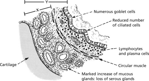
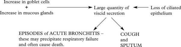
Acute Bronchitis
This is an inflammation of the large and medium bronchi. The condition may be serious if associated with pre-existing respiratory disease. Mucous and serous glands in the walls of the bronchi provide abundant mucoid secretion during the inflammation. Ciliated epithelia lining the bronchi aid passage of the exudate upward and help prevent spread down to the bronchioles.

Bronchial Asthma
In asthma, there are spasmodic attacks of reversible bronchial obstruction with wheezing and dyspnoea and often a dry cough. The prevalence has markedly increased in recent years, although has now plateaued. There are 2 main patterns:
Other varieties include occupational asthma and aspirin-induced asthma.
The basic mechanism is as follows:


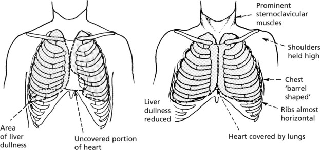
Chronic Obstructive Pulmonary Disease (COPD)
This term describes three entities that show considerable clinical overlap:
COPD is common and is the fourth leading cause of death worldwide.
Aetiology The main factors are:
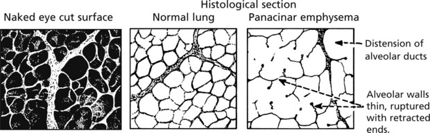
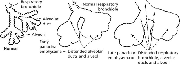
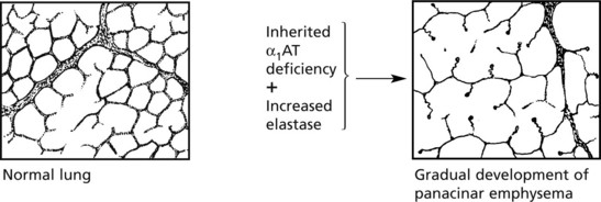
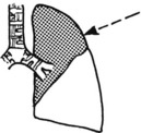
Emphysema
This is defined as a permanent dilatation of air spaces distal to the terminal bronchiole due to destruction of their walls without fibrosis. It is an important component of COPD (p.258) and has the same aetiological factors.
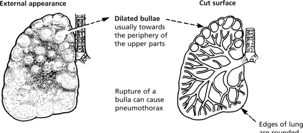
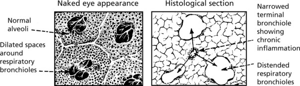
Centrilobular Emphysema
In this form the dilated air spaces immediately surround and involve the respiratory bronchioles.
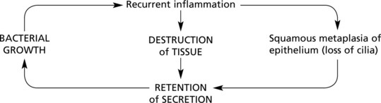
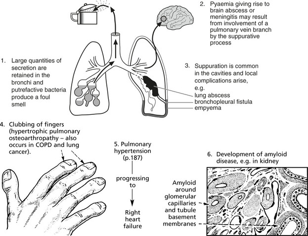
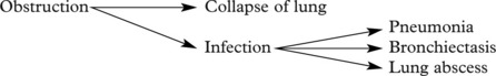
Bronchiectasis
Bronchiectasis means a permanent dilatation of one or more bronchi. There are 2 main subdivisions
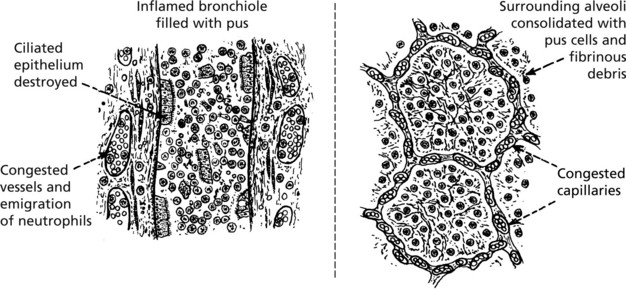
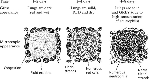
Pneumonia – Bronchopneumonia
Pneumonia is an inflammatory process involving the alveolar tissue of the lungs. It is discussed under 3 main headings:
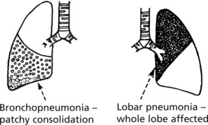
The lesions are initially focal, involving one or more lobules.
First red, then grey, they show a central bronchiole containing pus.
Legionnaire’s Disease
Previously confused with pneumococcal pneumonia, Legionnaire’s disease occurs in small epidemics, is more common in males and is due to a tiny Gram-negative bacillus – Legionella pneumophila. The death rate can be high – up to 20%. Infection is associated with inhalation of aerosol from contaminated water storage systems.
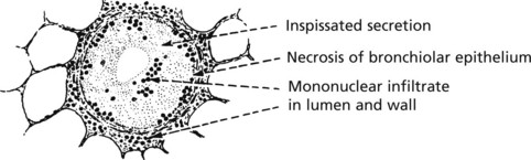
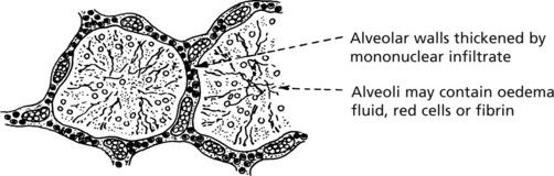
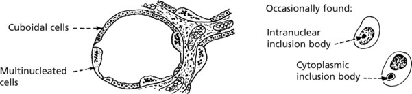
Viral Pneumonias
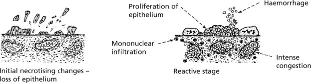
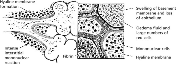
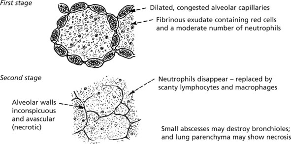
Atypical Pneumonias
There are a number of pneumonic conditions due to viruses and bacteria in which a common pattern of pathological change is observed.
Pneumonia – Special Types
Aspiration Pneumonia
This is due to inhalation of food or infected material from the mouth or pharynx. It may occur during anaesthesia or coma or may complicate conditions associated with frequent regurgitation, e.g. oesophageal obstruction, pyloric stenosis or poor swallowing due to stroke or motor neurone disease.
Possible progression is as follows:

Lipid Pneumonia
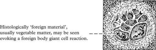
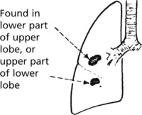
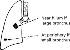
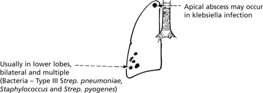
Lung Abscess
This is now an uncommon condition due to prompt treatment of preceding infection. The usual causes are:
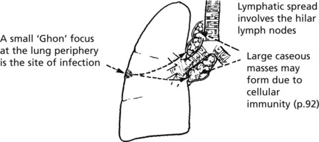
Tuberculosis (TB)
The lungs are more commonly affected by TB than any other organ. The incidence of TB in developed countries is rising, partly due to AIDS. Drug resistant strains of Mycobacteria are an increasing problem.
There are 2 patterns of pulmonary tuberculosis — primary and secondary.
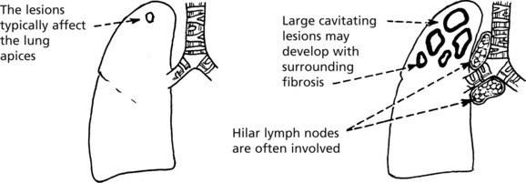
Spread of Tuberculosis
Spread of Tuberculosis
This is typically seen in secondary TB, but may also complicate primary infection.
The infection can spread by several pathways.
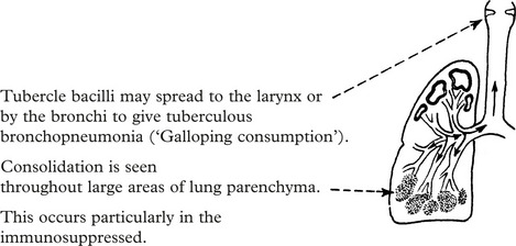
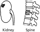
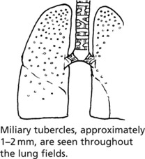
This may lead to isolated blood borne infection e.g. tuberculous meningitis (p.551), renal TB and bone TB, e.g. Pott’s disease of the spine: or to miliary spread throughout the body.
In this, miliary spread occurs throughout the lung fields alone.
The histological features are as described on page 72.
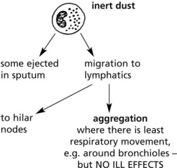
Pneumoconioses (Dust Diseases)
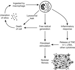
The reaction to inhaled dust varies very considerably. Some dusts, e.g. pure carbon, are inert: others, e.g. silicates, cause severe lung disease.
Basic Pathology
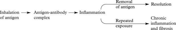
The basic pathology is a Type III hypersensitivity reaction (p. 102) and the term hypersensitivity pneumonitis (Extrinsic allergic alveolitis) is used.
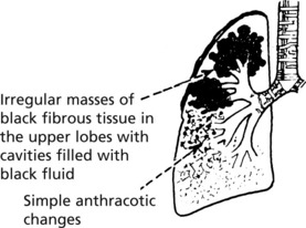
Anthracosis
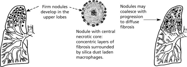
Coal-Miner’s Pneumoconiosis
The severe diseases caused by inorganic dust inhalation, usually occupationally, are described.

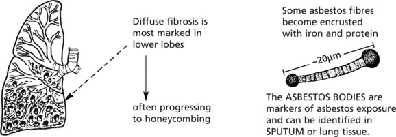
Asbestosis
Asbestos is the generic name for a group of fibrous silicates widely used in heavy industry for insulation (particularly in shipbuilding).
Asbestosis results from chronic exposure.
Other important complications are:
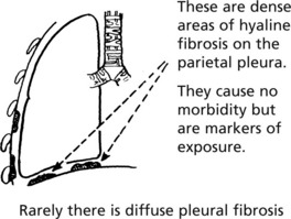
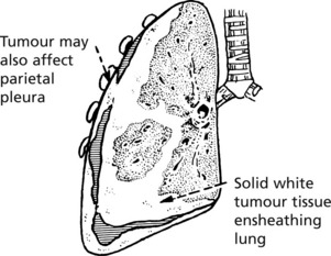
Note: Mesothelioma may also affect the pericardium and peritoneum.
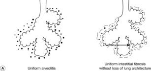
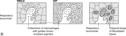
Diffuse Parenchymal Lung Disease
Diffuse Parenchymal Lung Disease
This encompasses a group of conditions in which the lung is altered by a combination of interstitial inflammation and fibrosis. Several histological patterns are recognised but it is thought that regardless of the type the earliest manifestation is alveolitis and that this accumulation of leucocytes results in release of mediators that can injure parenchymal cells and stimulate fibrosis.
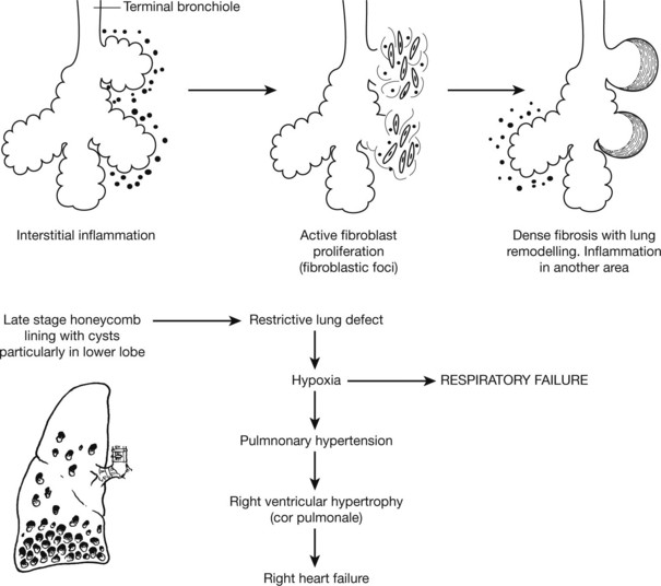
Infection is a common terminal event, and there is an increased risk of lung cancer.
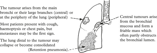
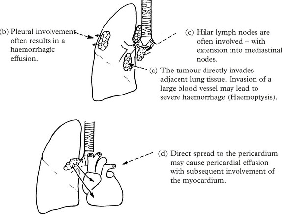
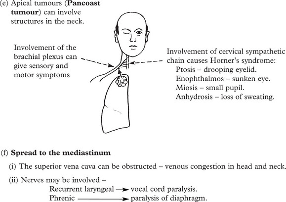
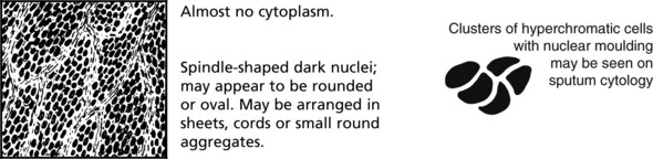
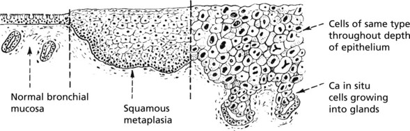
Lung Cancer
Lung cancer (carcinoma of the bronchus) is the commonest form of cancer and one which is largely preventable. Approximately 35000 patients die of lung cancer in the United Kingdom each year.
Histological Types
| 1. Small cell … … … … … … … … … … | 20%. | The proportions vary between series. |
| 2. Squamous carcinoma … … … … … … … | 30%. | |
| 3. Adenocarcinoma … … … … … … … … | 40%. | |
| 4. Large cell carcinoma … … … … … … … | 10%. |
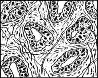
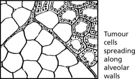
Adenocarcinoma
This tumour is a common tumour in women and is seen in non-smokers, but is also associated with smoking. At least two-thirds arise in the periphery of the lung, sometimes in relation to scarring. In non-smoking women, particularly of East Asian origin, there is a high incidence of epidermal growth factor receptor mutations that confer responsiveness to certain chemotherapeutic agents.
Aetiology of Lung Cancer
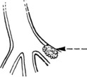
Other Tumours of the Bronchi and Lungs
Benign tumours of the bronchi and lung are unusual. There are 2 main types.
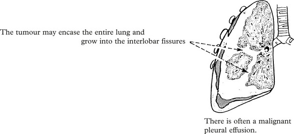
Diseases of the Pleura
Inflammation (Pleurisy)
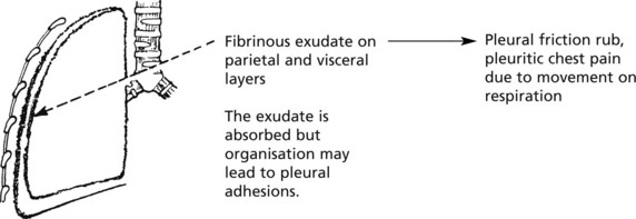
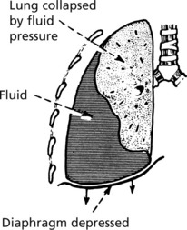
Pleural Effusion
The accumulation of fluid within the pleura can be explained as a form of local oedema.
The composition of the fluid is related to the underlying cause.
| TRANSUDATE | EXUDATE |
|---|---|
| e.g. in heart failure – low protein content; – few inflammatory cells. |
e.g. in fibrinous pleurisy – high protein content; – numerous inflammatory cells. |
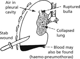
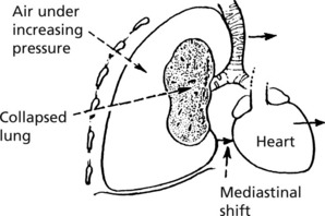
Pneumothorax
This is the accumulation of air in the pleural space. It may follow blunt trauma or penetrating wound, e.g. stabbing. Older patients with emphysema may develop spontaneous pneumothorax. In young people this is usually due to rupture of small bullae or excessive smoking of cannabis.
[/level-membership-for-pathology-category][not-level-membership-for-pathology-category]
Chapter 8 Respiratory System
Upper Respiratory Tract
Acute Inflammation
Infections of the nose, nasal sinuses, pharynx and larynx are common. They are usually mild and self-limiting. Most cases are due to viral infection, but this is often followed by bacterial superinfection.
The wide variety of different viruses involved prevents protective immunity.
Rhinitis
Common Cold (Acute Coryza)
This common respiratory inflammation usually involves the nose and adjacent structures.
The 2 phases, viral and bacterial, are typically seen in this disease.
The infection is acquired by droplet spread of viruses by sneezing.
Allergic Rhinitis ‘Hay Fever’
An allergic (immediate type hyper-sensitivity) type of inflammation (2. p.101) is often seen. Patients develop immediate symptoms of sneezing, itching and watery rhinorrhoea.
The repeated attacks frequently lead to chronic changes in the mucosa with polyp formation.
Nasal Polyps
These form particularly on the middle turbinate bones and within the maxillary sinuses.
Acute Pharyngitis, Tracheitis and Laryngitis
Acute Pharyngitis and Tracheitis
Most sore throats are caused by viruses – including adenovirus and Epstein–Barr virus.
Bacteria include Streptococcus pyogenes, Haemophilus parainfluenzae and Corynbacterium diphtheriae.
Diphtheria, now uncommon in countries where vaccination is widespread, is a serious infection.
The formation of a pseudomembrane is striking.
This pseudomembrane may spread to block the larynx, causing respiratory obstruction.
The exotoxin of diphtheria is encoded by a bacteriophage (a virus which infects the bacterium). It can cause myocarditis (p.212) and neuropathy (p.569).
Acute laryngitis is often due to parainfluenza viruses. Acute oedema of the glottis is seen in some cases of anaphylaxis (p.101) and angioneurotic oedema.
Other Disorders of the Larynx
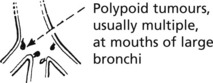
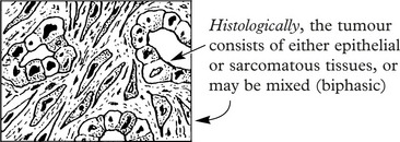
Tumours of the Upper Respiratory Tract
Lungs – Anatomy
Acinus
This is the functional unit of the lung, where gas transfer takes place. It consists of the respiratory bronchiole and associated alveolar ducts and sacs supplied by one terminal bronchiole. There are approximately 25 000 acini in the normal adult male lung.
Respiration
The normal intake of air is around 7 litres per minute; of this, after allowing for nonfunctioning dead space (trachea, bronchi, etc.), approximately 5 litres per minute are available for alveolar ventilation. A definite flow of air is maintained as far as the terminal bronchiole. Beyond this point the actual flow ceases and gas exchange is effected by diffusion.
Three factors are involved in the maintenance of adequate respiration:
Impaired Diffusion of Gases
Three mechanisms may interfere with diffusion:
Altered Pulmonary Perfusion
Interference with the pulmonary circulation may occur in 4 main ways:
Note: In addition to hypoxaemia, inadequate perfusion tends to cause retention of carbon dioxide.
Acute Bronchitis
This is an inflammation of the large and medium bronchi. The condition may be serious if associated with pre-existing respiratory disease. Mucous and serous glands in the walls of the bronchi provide abundant mucoid secretion during the inflammation. Ciliated epithelia lining the bronchi aid passage of the exudate upward and help prevent spread down to the bronchioles.
Bronchial Asthma
In asthma, there are spasmodic attacks of reversible bronchial obstruction with wheezing and dyspnoea and often a dry cough. The prevalence has markedly increased in recent years, although has now plateaued. There are 2 main patterns:
Other varieties include occupational asthma and aspirin-induced asthma.
The basic mechanism is as follows:
Chronic Obstructive Pulmonary Disease (COPD)
This term describes three entities that show considerable clinical overlap:
COPD is common and is the fourth leading cause of death worldwide.
