4 Reflection
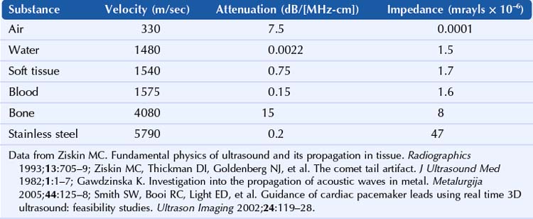
Ultrasonography measures the amplitude of the return echo as a function of time.1 Sound waves are reflected at the interface of tissues with different acoustic impedances. The acoustic impedance (kg/[m2-sec]) is the product of the density (kg/m3) and velocity (m/sec). The extent of reflection is governed by the reflection coefficient: R = (Z1 − Z2)/(Z1 + Z2). If Z1 = Z2, there is no reflected wave.2 Ultrasound characteristics of biologic tissue and interventional materials are summarized in Table 4-1.
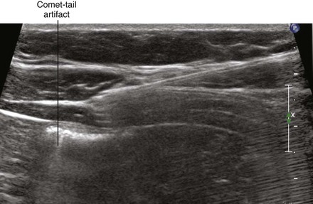
Reflections off a smooth surface are called specular. If two specular reflectors are close to each other, reverberation within the sound field can result, displayed as parallel, equally spaced lines deep to the reflectors. Comet-tail artifact, which is a form of reverberation artifact, is caused by multiple internal reflections from a small, highly reflective interface.3,4
Clinical Pearls
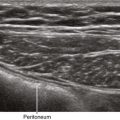
• The normal pleural line is thin and smooth, which generates a few comet-tail artifacts (between one and six artifacts per intercostal space scan). In the presence of parenchymal lung disease, the pleural line is irregular and thickened, generating many more comet-tail artifacts.5
• No comet-tail artifact is observed from the lung when pneumothorax is present.
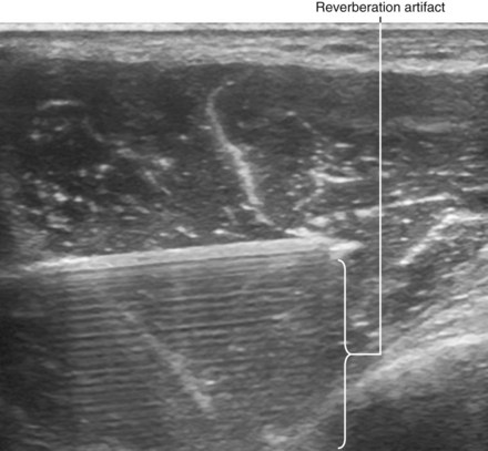
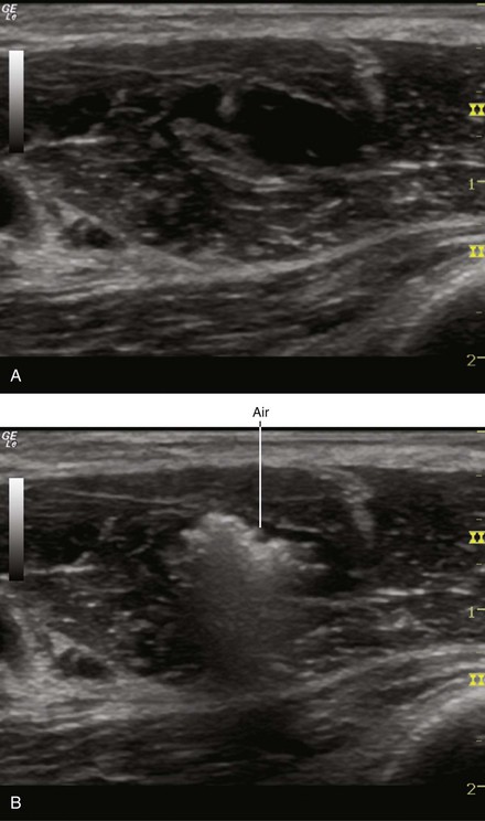
• Hyperechoic reverberation artifacts are seen with metallic foreign bodies such as block needles.
1 Ziskin MC. Fundamental physics of ultrasound and its propagation in tissue. Radiographics. 1993;13:705–709.
2 Ziskin MC. Equation governing the transmission of ultrasound. J Clin Ultrasound. 1982;10:A21.
3 Ziskin MC, Thickman DI, Goldenberg NJ, et al. The comet tail artifact. J Ultrasound Med. 1982;1:1–7.
4 Thickman DI, Ziskin MC, Goldenberg NJ, et al. Clinical manifestations of the comet tail artifact. J Ultrasound Med. 1983;2:225–230.
5 Reissig A, Kroegel C. Transthoracic sonography of diffuse parenchymal lung disease: the role of comet tail artifacts. J Ultrasound Med. 2003;22:173–180.