Recurrent collapse in a 56-year-old truck driver

Published on 10/04/2015 by admin

Filed under Surgery

Last modified 22/04/2025

Print this page

rate 1 star rate 2 star rate 3 star rate 4 star rate 5 star
Your rating: none, Average: 0 (0 votes)

This article have been viewed 1083 times

Problem 30 Recurrent collapse in a 56-year-old truck driver

On further questioning you find out that the patient’s episodes are unprovoked and come on at any time without warning. There is no postural element to the symptoms and they are not exertional. He denies any urinary or faecal incontinence during the attacks and makes a prompt recovery following events. His wife has witnessed the attacks and tells you that there are no associated seizure-like movements. Until now he has not injured himself as a result of the syncopal attacks. In between attacks he has been systemically well. He has not had any recent headaches or head injuries and denies any abdominal symptoms or unplanned weight loss. He denies any chest pain or shortness of breath and there is no history of sudden cardiac death within the family. He seldom drinks alcohol, does not smoke and denies any history of illicit drug use. He is not taking any regular medications.

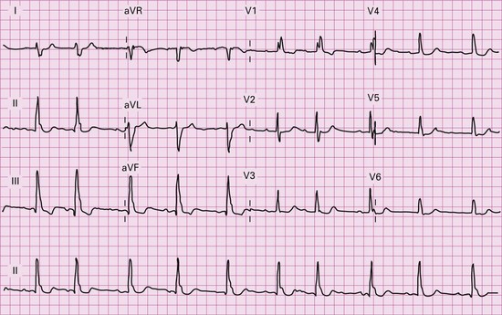
Physical examination is essentially unremarkable with no abnormal cardiovascular findings and a normal neurological exam. The only positive finding is that of centripetal obesity consistent with the patient’s elevated body mass index. You request an ECG in light of the normal physical examination (Figure 30.1).

Following a period of overnight cardiac monitoring that was essentially unremarkable our patient was discharged home for further outpatient evaluation.

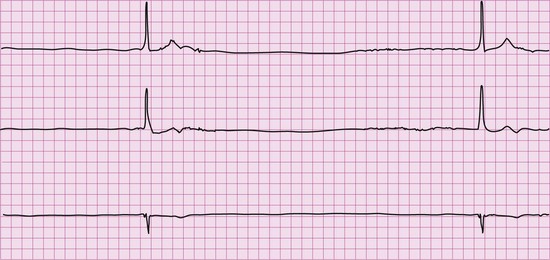
The patient went on to have a 24-hour ambulatory cardiac monitor. He had several episodes of syncope during this time. Figure 30.2 shows the electrocardiogram during the events.

Based on this information, your patient is admitted to hospital and has a dual-chamber pacemaker inserted without complication and recovers well at home. Dual-chamber pacing is preferred over single-chamber left ventricular pacing as it is more physiological and associated with fewer long-term adverse sequelae. Another option, as the patient had sinus node disease, would have been to implant a single-chamber atrial lead. However, given the conduction system disease on his resting ECG it was felt that a ventricular lead should be inserted because of the risk of developing bradycardia due to atrioventricular node/His–Purkinje dysfunction in the future.

An important consideration in any patient presenting with syncope is their suitability to drive a vehicle following the attack. These will vary depending on state or country and will also depend on the type of licence that the patient holds. Generally, patients with a commercial vehicle licence have more stringent regulations placed upon them than patients with a private licence.

Your patient is discharged on the day after pacemaker insertion, with advice to observe the pocket site for infection, and follow-up pacemaker check in 3 months’ time.

Answers

A.1 When approaching a patient presenting with loss of consciousness it is important to use the history to attempt to derive the most likely diagnosis. It may be appropriate to speak to a family member in order to obtain as much collateral history as possible since the patient may have difficulty recalling the situation.

Firstly, the patient’s demographic provides an important clue in the diagnosis. Vasovagal syncope is more common in young patients although syncope in the elderly has a greater risk of adverse outcomes. Also, knowing the situation in which the syncopal event occurred is important. For example, if the patient has been standing for a long time in a hot environment prior to losing consciousness or has a syncopal event following (not during) exercise, again this is suspicious for a vasovagal episode.

Does the patient have a characteristic warning prior to the syncope? Patients with vasovagal syncope classically have a prodrome that consists of a lightheaded sensation, feeling nauseous and diaphoretic prior to the attack. An absence of a warning prior to the episode of loss of consciousness often indicates a more sinister aetiology such as a cardiac arrhythmia.

Syncope occurring soon after standing suggests orthostatic hypotension that may occur on its own or be a part of a systemic process such as autonomic neuropathy. It is worthwhile enquiring what medications the patient is taking as new medications such as diuretics and anti-hypertensives can augment the postural hypotensive response.

A history of headache prior to syncope should make you think of a subarachnoid haemorrhage as a potential cause. Diplopia and vertigo may suggest brainstem ischaemia from cerebrovascular disease; however, syncope is uncommon in anterior circulatory cerebral ischaemia.

It is also important to determine the recovery period following the syncopal episode. Prolonged recovery suggests a possible seizure whereas a more rapid recovery occurs more commonly in vasovagal syndromes and cardiac arrythmias.

A history of exertional syncope suggests hypertrophic cardiomyopathy or aortic stenosis. A family history of sudden cardiac death is a very important component of the history and may make you more suspicious of possible structural heart disease or an acquired genetic disorder such as long QT syndrome. Shortness of breath and chest pain associated with syncope suggests a pulmonary embolism.

Causes of Syncope

A.2 The physical examination is really aimed at trying to identify specific causes of syncope and so it is essentially a system-by-system exam considering possible diagnoses.

Abdominal Examination

A mass in the abdomen could suggest an underlying malignancy that might lead to anaemia. Equally, the presence of an abdominal aortic aneurysm in the context of syncope should alert you to the possibility of a leak or rupture.

A.3 The ECG shows first-degree AV block, left posterior hemiblock and right bundle branch block that together constitute trifasicular block. Alternatively trifasicular block can occur with left axis deviation, right bundle branch block and first degree heart block.

Other specifics to look for on the ECG would be evidence of pre-excitation such as a delta wave and widened QRS that may predispose the patient to developing SVT; prolongation of the QT interval that would again predispose the patient to ventricular arrythmias; ischaemic ECG changes such as ST depression and T wave inversion may alert you to the fact that the patient may have underlying ischaemic heart disease; Evidence of prior AMI with pathological Q waves is also an important finding. ST elevation of aVR is particularly concerning for a proximal main vessel stenosis that may precipitate syncope. Features of right heart strain (RBBB, RAD, T wave inversion V1-V3, S1Q3 T3 is not specific) is also important to exclude.

Further investigations will be guided on your findings from history and examination but as a general rule any patient presenting with syncope should initially have a systematic work-up that includes an ECG and the following:

Blood tests: full blood count – the presence of anaemia should alert you to the possibility of an underlying haemorrhage; urea and electrolytes – might indicate underlying dehydration or suggest adrenal insufficiency. It is not necessary to perform cardiac enzymes on all patients presenting with syncope unless there are ECG changes consistent with ischaemia or the patient is experiencing chest pain. A blood sugar should be a routine part of a syncope evaluation.

Chest X-ray: increased cardiothoracic ratio may indicate an underlying cardiomyopathy or the presence of a pericardial effusion.

In our particular patient the history and ECG suggest that a cardiac cause is a likely diagnosis and in such patients a period of inpatient cardiac monitoring may be necessary. An assessment of left ventricular function with an echocardiogram is also an important test. Patients with systolic left ventricular dysfunction are at increased risk of developing ventricular arrhythmias. The echocardiogram can also determine the presence of any structural heart defects or valvular pathologies such as hypertrophic cardiomyopathy. It can also provide you with an assessment of right heart function, which may be abnormal in patients who have had a pulmonary embolism and in those rare patients with arrhythmogenic right ventricular dysplasia, a genetic disorder where patients can present with syncope. Whether this needs to be done as an inpatient or an outpatient will be determined by your underlying clinical suspicion. In our patient it is reasonable to perform an echocardiogram as an outpatient as we have no reason to suspect structural heart disease based on our history and examination.

A.4 The aim of further investigations is to obtain a recording of heart rhythm when the patient is symptomatic. This can be achieved in several ways and will depend upon the frequency of patient symptoms. If the patient experiences daily symptoms then a 24-hour ambulatory cardiac monitor can be organized. This involves the patient wearing surface ECG electrodes for 24 hours that are attached to a recording device. There is continuous ECG recording during this period. The patient records times when they experience symptoms and the corresponding rhythm recorded by the device is compared. If the patient experiences less frequent symptoms then an event recorder may be a more appropriate investigation. This is similar to the 24-hour ambulatory monitor but this time the patient has to press a button to store the ECG. These can be worn for a much longer period of time – typically 1–2 weeks, and often longer.

If ambulatory cardiac monitoring fails to elucidate the nature of the syncope then the next investigation available is a loop recorder. These are small devices, roughly the size of a USB flash drive that is placed subcutaneously in the left infraclavicular position. They are able to remain in situ for 2–3 years. Following a syncopal event the patient places an activator over the device and the ECG over that time period is stored. They are valuable devices in the management of recurrent syncope but their expense and invasive nature limit their blanket use and therefore they should only be used in patients with recurrent syncope in whom a cardiac cause is suspected.

Tilt table testing is a test that is useful in patients in whom you suspect underlying vasovagal syndrome, postural hypotension or autonomic neuropathy as a cause of their syncope. The patient is tilted from supine to 70° over a period of time and may be given medication such as isoprenaline to promote susceptibility to the test. However, up to 40% of patients with vasovagal syndrome may have a normal result and so a negative result is not conclusive. However, the test can be useful when positive at confirming the diagnosis with the replication of the patient’s symptoms.

The final investigation available is an electrophysiology study (EPS). This is an invasive test performed via the right femoral vein with a small but significant morbidity associated. An EPS can detect sinus or atrioventricular nodal disease, bypass tracts and the readiness of a patient to develop ventricular arrhythmias. However, there is generally a low diagnostic yield if it is used in everybody presenting with syncope and indeed recent guidelines suggest only utilizing an EPS in patients with structural heart disease, an ECG that may be suggestive of an arrhythmic cause (e.g. LBBB) or in patients with a family history of sudden cardiac death.

A.5 The ECG shows sinus pauses of up to 5 seconds and these occurred during the day. It is considered normal to have sinus pauses at night of up to approximately 3 seconds due to increased vagal tone. However, in our patient the pauses were greater than 3 seconds and occurred during the day. He therefore meets current guideline indications for a pacemaker.