Rectal bleeding in a 45-year-old woman

Published on 10/04/2015 by admin

Filed under Surgery

Last modified 22/04/2025

Print this page

rate 1 star rate 2 star rate 3 star rate 4 star rate 5 star
Your rating: none, Average: 0 (0 votes)

This article have been viewed 1001 times

Problem 19 Rectal bleeding in a 45-year-old woman

A sigmoidoscopic examination to 20 cm is normal. Proctoscopy confirms the presence of large internal haemorrhoids, which bleed easily on contact.

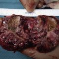
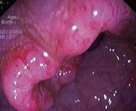
You explain to the patient that while the bleeding is most likely to be due to her haemorrhoids, a complete investigation of the large bowel is indicated. You arrange a colonoscopy. A lesion is found (Figure 19.1).

The tumour is indentified in the ascending colon. The colonoscopy was otherwise normal. The biopsy shows moderately differentiated adenocarcinoma.

Her haemoglobin is 100 g/L, her liver function tests and biochemistry are normal. A CT abdomen and chest is normal.

At operation a carcinoma of the ascending colon is found and a right hemicolectomy is performed. There is no evidence of adjacent or distant spread of the tumour. Apart from the tumour, there was nothing else abnormal in the resected specimen of bowel. There was no indication to perform a defunctioning ileostomy and she makes a good recovery. The histology is confirmed and the carcinoma involves the muscularis propria and extends into the pericolic fat. There is no perineural, vascular or lymph node involvement.

The patient understands that you will keep her under surveillance. She would like to know what this will involve.

The patient wants to know if any of her children are at risk for developing this cancer.

You explain to the patient that the risk to her children is slightly greater than it would be for the general population. Table 19.1 shows the risks.

Table 19.1 Risk factors in colorectal cancer

Family History Risk
Up to 2 fold
3 to 6 fold
1 in 2 lifetime

The patient’s son is with her. He understands that he is at slightly increased risk and would like to know what screening you would recommend for him. He is aged 23.

You advise him that if he is symptom free, he does not need any surveillance at this stage. If he does have symptoms, they should be investigated as appropriate. In any patient who has a positive occult blood test, it is recommended they have a colonoscopy. Similarly, you explain to him that any patient who presents with new rectal bleeding needs investigation. As a guiding rule, any patient who presents with an iron deficiency anaemia needs a full colonic investigation unless there are clear alternative causes such as menorrhagia.

The son has heard that ‘polyps in the bowel usually turn to cancer’.

The patient remains in good health and is kept under regular surveillance.

Answers

A.1 The patient has three symptoms that cause concern: increasing constipation, rectal blood loss and tiredness. You should ask further questions that may indicate why she is constipated. There are many possibilities in addition to colonic pathology, such as dietary changes, use of narcotic analgesia (codeine) or development of hypothyroidism. You should enquire about a personal history of polyps and family history of colon cancer. In the absence of an obvious cause for her constipation (confirmed by impacted faeces in the rectum), such as recent use of a codeine-containing compound, this patient will require examination of all of her large bowel. A sigmoidoscopy may be performed as part of the general physical examination and might clarify matters and identify a source for the bleeding in the anal canal or lower rectum. Even if the patient does have internal haemorrhoids, which may be the cause of the bleeding, she must be investigated further. Haemorrhoids are extremely common, but it is important to inspect the rest of the large intestine and so not overlook a tumour.

A.2 The most likely cause of bleeding in this type of patient is internal haemorrhoids, but it is essential to exclude a more sinister cause for the blood loss, especially as she has recently become constipated and tired for no obvious reason. Cancer of the colon, although more common in older patients, occurs in this age group and often presents with these symptoms. Despite the haemorrhoids, she must have a colonoscopy. In the absence of other symptoms and the visualization of a bleeding haemorrhoid it would be reasonable to treat the haemorrhoids and only investigate further if the symptoms persist. If a colonoscopy was not available (uncommon) then a barium enema would be an alternative but does not allow any diagnostic or therapeutic manoeuvres such as biopsy and snaring of polyps to be undertaken and if abnormal would require a colonoscopy for assesment and treatment.

A.3 This lesion has the typical appearance of an adenocarcinoma with raised, rolled edges and a central crater. The centre of the ulcer is likely to be ulcerated. The tumour occupies half the circumference of the bowel and is about 3 cm in length.

A.4 As part of the work-up, the following investigations are required:

Iron deficiency anaemia is common in cases of colorectal cancer and must be excluded. The patient may have secondary spread of her disease. Colorectal cancers tend to spread to the liver, but metastatic deposits can also be found in lung and bone. A CT abdomen/chest and liver enzymes may not affect the decision-making of how to manage the primary tumour, but will give an indication of possible spread of the disease. This is often useful knowledge to have before surgery. Staging of the disease will influence adjuvant chemotherapy.

In cases such as this the entire large bowel must be examined. This patient had a negative colonoscopy other than the lesion visualized. However, co-existent disease is often present and patients may have polyps or other primary tumours. Synchronous tumours occur in 5% of patients and polyps may be found in up to 20% of cases. The discovery of polyps or another tumour may influence the extent of surgical resection required.

A.5 You should explain the diagnosis and management to the patient, who needs to understand that she has a cancer of the colon, although at this stage there is no evidence that the disease has spread outside the bowel. Explain that she requires surgery to prevent total obstruction of the colon and attempt to cure her of the disease.

You must explain to the patient in simple language (and be prepared to repeat things several times) what the operation will involve and the risks and benefits of the proposed treatment. You must explain that:

Risks including infection, anastomotic leakage, thromboembolic risks and operative mortality must be discussed. You can emphasize that these should be kept in perspective, as the patient has little alternative as, untreated, she will develop complete intestinal obstruction.

While not applying to this patient, remember that fears of being left with a ‘bag’ (colostomy) are common and these concerns must be fully addressed. However, with modern stapling techniques and the realization that the incidence of local recurrence is no greater with a 2 cm margin of clearance than a 5 cm margin, fewer patients now undergo total excision of the rectum with the formation of a permanent colostomy. Most patients with carcinoma of the rectum do not need complete excision of the rectum and anus. Fifty years ago, only 15% of all rectal cancers were treated with a restorative procedure, whereas that figure currently exceeds 65%. Temporary or defunctioning colostomies are still fashioned, but it is unlikely that the case discussed in this problem would require one. Colostomies (or defunctioning ileostomies) may be required after a difficult dissection deep in the pelvis or after emergency surgery for perforated or obstructed colon. The aim of a colostomy or ileostomy in such circumstances is to reduce the risk of anastomotic leakage, or in the event of established peritoneal contamination, to prevent further soiling.

Surgery remains important in incurable metastatic disease when the primary tumour is producing troublesome symptoms such as bleeding, obstruction or tenesmus. In some circumstances colonic stents can be used to relieve obstruction and avoid surgery in patients with very advanced disease.

A.6 This patient must be given a prognosis. Her tumour has been staged as a Stage ACPS B or pT3N0M0 and she will have an approximate 85–90% 5-year survival rate. Expressed simply to the patient, she could be told that all the known cancer has been removed, she has a good chance of cure, but will be monitored to look for recurrence of her disease or development of polyps which might develop into a new cancer. The surveillance will require repeated colonoscopy.

Although chemotherapy has no role in the initial treatment of colorectal cancer, it is well accepted that adjuvant chemotherapy will improve survival for patients with more advanced stages.

A.7 The aim of any cancer follow-up programme is threefold and is to detect:

Surveillance Strategies

A.8 The cause of colorectal cancer is unknown, but diet is likely to play an important role. In certain instances there may be a genetic predisposition to cancer (e.g. familial adenomatous polyposis (FAP), hereditary non-polyposis colorectal cancer (HNPCC)), or the patient may have a condition such as ulcerative colitis that places them at high risk. It is also apparent that first-degree relatives have a two- to threefold increase in risk of developing colorectal cancer. A past history of colorectal cancer increases the risk of developing a further carcinoma. However, in most cases no obvious risk factor exists and the cancer is thought to arise from a pre-existing adenoma. Not all adenomas turn malignant but almost all cancers develop in an adenoma. This has led to screening strategies which are based around identification and removal of polyps.

In this instance no particular risk factors can be identified but the young age and right-sided tumour raise the issue of HNPCC. MSI (microsatellite instability) and immunohistochemistry testing and if positive subsequent assessment of the tumour for germline mutations in the DNA mismatch repair (MMR) genes may reveal a strong possibility of HNPCC. These assessments are generally only done when patients meet one of the Amsterdam criteria/Bethesda guidelines. Approximately 10–15% of sporadic colorectal cancers will be MSI positive. The patient should be counselled that her children would be at increased risk over the general population. Use of the chart indicated in the text (Table 19.1) can be helpful.

A.9 On the assumption that the son has no digestive tract symptoms (particularly rectal bleeding), then nothing needs to be done until he is 35 (i.e. 10 years earlier than his mother’s diagnosis). The following guidelines should be applied. These recommendations for screening of first-degree relatives of patients with colorectal cancer follow the NH and MRC guidelines (Table 19.2).

Table 19.2 Screening guidelines for colorectal cancer

Screening Guidelines Recommendation
Category 1

Category 2

Category 3

A.10 An adenoma is one of four types of polyps that can be found in the colon and rectum. Apart from neoplastic lesions, there are three benign groups which include hyperplastic and inflammatory polyps and hamartomas. Serrated adenomas are a variant of hyperplastic polyps and may have a faster progression from benign to malignant. All adenomas have malignant potential. Adenomas may be tubular (often pedunculated), villous (usually sessile) or tubulovillous and can be found in any part of the bowel. As with cancers, the most common site for adenomas is the distal colon and rectum. The larger the polyp, the greater the chance of it being malignant. Fifty per cent of all polyps greater than 2 cm contain a focus of malignancy.

Revision Points

Colorectal Cancer

Further Information

, National Health and Medical Research Council, Canberra (2005) Guidelines for the prevention, early detection and management of colorectal cancer (CRC).

, www.cancer.gov/cancertopics/types/colon-and-rectal. Information about colon and rectal cancer treatment, prevention, genetics, causes, screening, statistics

, www.gastrolab.net. Endoscopic images presented in an MCQ fashion

, www.nlm.nih.gov/medlineplus/colorectalcancer.html. Patient-oriented tutorial available via interactive link