Rare Tumors

Published on 25/03/2015 by admin

Filed under Pediatrics

Last modified 22/04/2025

Print this page

rate 1 star rate 2 star rate 3 star rate 4 star rate 5 star
Your rating: none, Average: 0 (0 votes)

This article have been viewed 1463 times

Chapter 500 Rare Tumors

500.1 Thyroid Tumors

See Chapter 562.

Malignant Thyroid Tumors

Pediatric thyroid malignancies are rare tumors that include medullary thyroid carcinoma (MTC) and the differentiated thyroid carcinomas (DTCs): papillary thyroid carcinoma (PTC) and follicular thyroid carcinoma (FTC). The vast majority of childhood thyroid cancers are PTC. The major established environmental risk factor for the development of DTC is exposure to ionizing radiation. MTC is a very uncommon disease in childhood that almost always occurs in the context of 1 of 2 hereditary endocrine tumor syndromes that arise secondary to activating mutations of the RET (REarranged during Transfection) proto-oncogene: multiple endocrine neoplasia type 2a (MEN2A) and type 2b (MEN2B). In addition to the almost complete penetrance of MTC, 50% of patients with MEN2A and MEN2B develop pheochromocytomas, and up to 30% of MEN2A patients have primary hyperparathyroidism. Patients with MEN2B have a distinct phenotype with a characteristic facial appearance, marfanoid body habitus, and a generalized ganglioneuromatosis, manifested most obviously by the presence of oral mucosa neuromas.

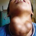
Children with thyroid cancer usually present with an asymptomatic thyroid mass and/or cervical lymphadenopathy. Lymph node metastases are present in the majority of PTC cases and lung metastases are identified in up to 20% of cases; metastases to other sites are rare. Despite a more advanced presentation, children with thyroid cancer usually have an excellent prognosis with anticipated survival over decades.

Total thyroidectomy and a compartment-oriented lymph node dissection, as indicated, are best accomplished by surgeons with extensive experience in the management of thyroid malignancies. In DTC, radioiodine (using 131I) is administered a few weeks after surgery to ablate any residual normal thyroid tissue and treat any residual thyroid cancer that is iodine-avid; children with MTC do not require radioiodine ablation. The TSH level is suppressed by giving supraphysiologic levothyroxine doses in DTC, but the TSH is kept normal in MTC. Long-term follow-up involves monitoring of tumor markers (thyroglobulin in DTC and calcitonin/carcinogenic embryonic antigen in MTC) as well as routine imaging based on the initial extent of disease presentation. Neck US is generally the most useful imaging modality. Traditional chemotherapy has generally not been shown to be effective in thyroid cancer, and newer anticancer agents, the oral tyrosine kinase inhibitors, are showing promise in the treatment of children with disease not amenable or responsive to standard therapeutic approaches.

In MEN2, there is a correlation between genotype and phenotype, and the biologic aggressiveness of MTC depends on the hereditary setting in which it develops. With the advent of genetic testing for RET mutations, MTC has become one of the few malignancies that can be prevented or cured via prophylactic thyroidectomy before it becomes clinically relevant. Recent guidelines have updated recommendations regarding the age of prophylactic thyroidectomy in children who are carriers of a RET mutation.

500.2 Melanoma

See Chapter 643.

The incidence of melanoma in persons <20 yr of age in the USA is 4.2 cases per million, with almost all of these cases occurring in adolescents (Chapter 643). Melanoma is more common among adolescent females than males. In the USA, incident rates of melanoma in younger age groups are increasing, although at a slower rate than in adults. Although sun exposure is a well-known risk factor for melanoma in adults, its role in pediatric melanoma is less clear. Pediatricians should counsel patients regarding avoidance of sun exposure to decrease the risk of later development of melanoma. Patients with fair skin and a family history of melanoma are at particularly high risk. Known risk factors for children are giant hairy nevus (>20 cm), dysplastic nevus syndrome, and xeroderma pigmentosum.

Findings of a rapidly enlarging nevus that is dark, has changed colors, has irregular borders, or bleeds easily should raise a concern of melanoma. However, many of the melanomas diagnosed in children have none of these features. Diagnosis is based on pathology. However, extra care must be taken in the diagnosis of melanoma in children because making the distinction from other lesions, particularly Spitz nevus, can be difficult.

Prognosis and treatment recommendations have previously been extrapolated from adult data; however, specific prognostic factors for pediatric melanoma are starting to accrue. The primary treatment is local excision with lymph node mapping and sentinel node biopsy for all but the most superficial melanomas. If the sentinel node is positive, a formal lymph node dissection is recommended. High-dose adjuvant interferon has shown some efficacy in the treatment of adult melanoma, whereas chemotherapy in combination with biologic agents and vaccine therapy has been used for treatment of distant metastases. The role of these treatments in pediatric melanoma is unclear.

500.3 Nasopharyngeal Carcinoma

Nasopharyngeal carcinoma is rare in the pediatric population, but is one of the most common nasopharyngeal tumors in pediatric patients. In adults, the incidence is highest in South China, but it is also high among the Inuit people and in North Africa and Northeast India. In China, it is rare in the pediatric population, but in other populations a substantial proportion of cases occur in the pediatric age group, primarily in adolescents. It occurs in males twice as often as in females and is more common in blacks. In the pediatric population the tumors are more commonly of undifferentiated histology and associated with Epstein-Barr virus (EBV). Nasopharyngeal carcinoma has been associated with specific HLA types, and other genetic factors may play a role, especially in low incidence populations.

Most pediatric patients present with advanced locoregional disease manifesting as cervical lymphadenopathy. Epistaxis, trismus, and cranial nerve deficits also may be present. The diagnosis is established from biopsy of the nasopharynx or cervical lymph nodes. In most cases the lactate dehydrogenase level is elevated, but this finding is nonspecific. CT or MRI evaluation of the head and neck is performed to determine the extent of locoregional disease. Chest radiography, CT, bone scan, and liver scan are used to evaluate for metastatic disease. Monitoring EBV IgA and ZEBRA (antibodies to the Epstein-Barr virus Bam H1Z transactivator protein) protein levels also may be useful.

Treatment is a combination of chemotherapy and irradiation. Cisplatin-based chemotherapy is given before or concurrent with irradiation. The outcome depends on the extent of disease; patients with distant metastases have a very poor prognosis. The use of intensity-modulated radiation therapy (IMRT) has improved local control and reduced the late adverse effects associated with radiation therapy, including hormonal dysfunction, dental caries, fibrosis, and second malignancies.

500.4 Adenocarcinoma of the Colon and Rectum

Colorectal carcinoma (CRC) is rare in the pediatric population. Even in patients with predisposing conditions, CRC usually does not present until adulthood. Hereditary nonpolyposis colon cancer (HNPCC) is an autosomal dominant disorder, with germline mutations in DNA mismatch repair genes (MMR) causing DNA repair errors and microsatellite instability. Familial adenomatous polyposis (FAP) and attenuated FAP (AFAP), are autosomal disorders, with germline mutations in the APC gene. In addition to CRC, patients with HNPCC, FAP, and AFAP are predisposed to a number of extracolonic cancers. MYH-associated polyposis (MAP), Peutz-Jeghers syndrome, and juvenile polyposis also predispose to CRC.

Genetic testing is available, and screening for cancer in HNPCC and FAP should begin during childhood or adolescence. Likewise, genetic evaluation for these conditions should be pursued in young patients presenting with colon cancer, even when there is no history of predisposing genetic conditions.

Presenting symptoms include bloody stools or melena, abdominal pain, weight loss, and changes in bowel patterns. Signs often are vague, often resulting in a delay in diagnosis and advanced disease. The histologic subtype differs from that seen in adults, with the majority of pediatric tumors being mucinous. Treatment consists of surgical resection when possible, with chemotherapy for unresectable tumors. Radiation therapy is useful in the treatment of rectal carcinomas.

500.5 Adrenal Tumors

See Chapter 575.

Adrenocortical tumors (ACT) arise from the outer adrenal cortex, whereas pheochromocytomas (PHEO) derive from the catecholamine-producing chromaffin cells of the adrenal medulla. The pathologic categorization of ACT in children as benign or malignant does not always correlate to the clinical behavior of these tumors, making it difficult to differentiate clinically significant adrenocortical carcinomas from those tumors that retain a good prognosis. ACT are very rare and tend to present at an age <5 yr. They have a female predominance and are functional tumors (producing androgens and/or glucocorticoids, typically) in >90% of cases. ACT may also present as an abdominal mass or pain. In children, ACT are associated with Li-Fraumeni syndrome (germ line inactivating mutations in the p53 tumor suppressor gene), Beckwith-Wiedemann syndrome (BWS), hemihypertrophy other than that seen as part of BWS, and rarely congenital adrenal hyperplasia. Other rare causes of nodular adrenocortical disease, which usually present with Cushing syndrome, include Carney complex and macronodular adrenal hyperplasia.

PHEO are more likely to be bilateral, extra-adrenal, malignant, and secondary to a heritable tumor syndrome in children. Most familial PHEO presenting in children are associated with von Hippel–Lindau disease, although MEN2, the familial paraganglioma syndromes due to mutations in the succinate dehydrogenase gene, and neurofibromatosis type 1 are also in the differential diagnosis. Compared with adults, hypertension is usually sustained in children and they may lack the typical triad of headache, palpitations, and diaphoresis. The initial test recommended for diagnosis is measurement of plasma and/or urine metanephrine levels. Children with PHEO who proceed to surgery are pretreated with alpha (and possibly beta) blockade.

Most children with ACT or PHEO should be treated with surgical resection. First-line medical therapy for malignant ACT includes mitotane and cisplatin-based chemotherapy, typically cisplatin, etoposide, and adriamycin. Endocrine therapy targeting hormonal overproduction may also be needed to palliate symptoms and improve quality of life. Metastatic PHEO is historically treated with cyclophosphamide, vincristine, and dacarbazine. Prognosis depends on tumor size, extent of tumor, resectability, and presence of distant metastases.

Bibliography

Armstrong R, Sridhar M, Greenhalgh KL, et al. Phaeochromocytoma in children. Arch Dis Child. 2008;93:899-904.

Baid SK, Lai EW, Wesley RA, et al. Brief communication: radiographic contrast infusion and catecholamine release in patients with pheochromocytoma. Ann Intern Med. 2009;150:27-32.

Barontini M, Levin G, Sanso G. Characteristics of pheochromocytoma in a 4- to 20-year-old population. Ann N Y Acad Sci. 2006;1073:30-37.

DeLellis RA, Lloyd RV, Heitz PU, et al. Pathology and genetics of tumours of endocrine organs World Health Organization Classification of Tumours (vol 8). France: Lyon; 2004.

Hanna AM, Pham TH, Askegard-Giesmann JR, et al. Outcome of adrenocortical tumors in children. J Pediatr Surg. 2008;43:843-849.

Ludwig AD, Feig DI, Brandt ML, et al. Recent advances in the diagnosis and treatment of pheochromocytoma in children. Am J Surg. 2007;194:792-796. discussion 796–797

Michalkiewicz E, Sandrini R, Figueiredo B, et al. Clinical and outcome characteristics of children with adrenocortical tumors: a report from the International Pediatric Adrenocortical Tumor Registry. J Clin Oncol. 2004;22:838-845.

Pham TH, Moir C, Thompson GB, et al. Pheochromocytoma and paraganglioma in children: a review of medical and surgical management at a tertiary care center. Pediatrics. 2006;118:1109-1117.

Rodriguez-Galindo C, Figueiredo BC, Zambetti GP, et al. Biology, clinical characteristics, and management of adrenocortical tumors in children. Pediatr Blood Cancer. 2005;45:265-273.

Waguespack SG, Rich T, Grubbs E, et al. A current review of the etiology, diagnosis, and treatment of pediatric pheochromocytoma and paraganglioma. J Clin Endocrinol Metab. 2010;95:2023-2037.

Young WFJr. Pheochromocytoma in children. UpToDate Online. 2008. 17.1