Rare Diffuse Interstitial Lung Diseases

Published on 29/05/2015 by admin

Filed under Pulmolory and Respiratory

Last modified 22/04/2025

Print this page

rate 1 star rate 2 star rate 3 star rate 4 star rate 5 star
Your rating: none, Average: 0 (0 votes)

This article have been viewed 2302 times

Chapter 54 Rare Diffuse Interstitial Lung Diseases

More than 7000 rare conditions exist, and although individually uncommon, together these diseases affect about 11% of all populations. Most rare diseases are chronic, often progressive, and can therefore form a significant portion of the workload for physicians. The term orphan disease is frequently applied to rare conditions and was originally used to suggest a lack of knowledge, research, or specific therapy. In many cases, however, significant progress in understanding the molecular basis of these diseases has resulted in biologically targeted treatments, making a definitive diagnosis especially important for these patients. This chapter describes select rare diffuse lung diseases, either primary or occurring in the context of systemic disease (secondary), likely to be encountered by most physicians, and particularly those for which a firm diagnosis can benefit patient management.

Rare Cystic Lung Diseases

Lymphangioleiomyomatosis

Lymphangioleiomyomatosis (LAM) is a cystic lung disease that affects almost exclusively women and has a prevalence of approximately 5 women per 1 million of most populations. LAM may occur sporadically or as part of the autosomal dominant genetic disorder, tuberous sclerosis complex (TSC). In both forms the lungs and lymphatics are infiltrated by LAM cells, an abnormal cell clone harboring biallelic inactivating mutations in the genes associated with TSC, either TSC-1 or more often TSC-2. LAM cells accumulate and cause progressive cystic destruction of the lungs, probably by the elaboration of proteolytic enzymes (Figure 54-1). LAM cells form small nodular clumps associated with lymphatic endothelial cells, which in turn form lymphatic channels that allow LAM cells to disseminate throughout the body. LAM cells also infiltrate the axial lymphatics and form the smooth muscle elements of angiomyolipoma, a rare tumor of the perivascular epithelioid cell family, present in up to 40% of women with sporadic LAM and almost all patients with TSC-LAM.

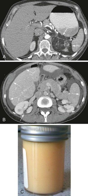
Other, extrapulmonary manifestations are related to lymphatic obstruction and include lymphadenopathy in up to 30% of patients and lymphangioleiomyomas, cystic lymphatic swellings usually in the abdomen, pelvis, or retroperitoneum occurring in up to 20% of patients (Figure 54-2). Thoracic and abdominal chylous collections are present in up to 10% of patients. LAM is also associated with a higher prevalence of meningioma than the general population. Cystic lung destruction results in recurrent pneumothorax and progressive airflow obstruction, with an average decline in forced expiratory volume in 1 second (FEV1) of 120 mL per year (normal, ~27 mL/yr). However, the clinical course of patients with LAM varies, with some remaining stable for many years. At present, no features definitively predict which patients will develop progressive disease; however, onset at a younger age, presentation with breathlessness, and poor lung function at presentation suggest more aggressive disease.

Patients with LAM most frequently present with breathlessness or pneumothorax, although chylous pleural effusions, complications of angiomyolipoma, or weight loss can also be the presenting feature. Recurrent pneumothorax or airflow obstruction in younger women, particularly nonsmokers, should raise the possibility of LAM. In most cases the computed tomography (CT) findings of multiple round thin-walled cysts with no zonal predominance suggest the diagnosis. Increasingly, patients are being identified with LAM after investigations for other problems where cystic lung disease is observed on high-resolution CT (HRCT) scanning. In addition, screening for LAM at 18 years of age by CT is recommended for women with TSC; many of these patients have evidence of LAM on CT, but only a minority have respiratory symptoms.

With diagnostic criteria recently formulated by the European Respiratory Society (ERS), a definite diagnosis of LAM can be made based on a characteristic HRCT scan with a renal angiomyolipoma, histologic evidence of LAM cells at other sites (e.g., lymphangioleiomyomas, chylous collections in abdomen or thorax), or the presence of TSC. If TSC or extrapulmonary manifestations are not present, lung biopsy is required for a definitive diagnosis. Transbronchial biopsy can occasionally be helpful, although in most patients, video-assisted thorascopic surgery (VATS) is preferred. LAM cells characteristically stain with the monoclonal antibody HMB-45 and also express α–smooth muscle actin, estrogen, and progesterone receptors. The use of these immunohistochemical markers improves identification of LAM cells in lung tissue, including smaller transbronchial biopsies. The recent observation that vascular endothelial growth factor type D (VEGF-D) serum level is increased in about two thirds of patients with LAM has led to its use as a diagnostic marker for LAM. Importantly, a serum VEGF-D level greater than 800 ng/mL can differentiate LAM from other cystic lung diseases, including Langerhans cell histiocytosis, Birt-Hogg-Dube disease, and emphysema.

Patients with sporadic LAM should undergo clinical examination and where necessary, formal screening to exclude tuberous sclerosis complex. Only one third of patients with TSC have the classical triad of epilepsy, learning difficulties, and facial angiofibromas. Many patients with TSC-LAM have normal intelligence and mild skin changes that may be overlooked. Almost all patients with TSC-LAM have renal angiomyolipomas, which tend to be larger and more frequently multiple and bilateral than in those with sporadic LAM. Making the diagnosis of TSC is important for the management of other aspects of TSC and for the patient’s offspring.

To confirm LAM in the presence of cystic lung disease or to evaluate patients for renal angiomyolipoma, a CT scan of the abdomen and pelvis should be performed. Angiomyolipomas greater than 4 cm are prone to growth, rupture, and hemorrhage and should be treated prophylactically when large or in the presence of symptoms.

General management of LAM includes avoidance of estrogen supplementation, including oral contraceptives and hormone replacement therapy. Prophylactic vaccination, pulmonary rehabilitation, and oxygen should be given when appropriate. Bronchodilators are helpful for those with airflow obstruction. Patients with LAM are prone to pneumothorax, which occurs in more than 70% of patients and is recurrent in the majority, and early surgical intervention is recommended. Patients considering pregnancy should be warned that the disease may be more active in pregnancy, particularly an increase in pneumothorax. Those with TSC should receive genetic counseling. For patients with severe disease, including those with TSC, lung transplantation can be performed.

Previously, hormone treatment with progesterone or other antiestrogen therapies have been used for these patients, but no evidence indicates there is a benefit for most patients. Studies show that rapamycin, an inhibitor of mTOR pathway, is effective in reducing the rate of decline of FEV1 in patients with impaired lung function. In those with renal angiomyolipoma, including those with TSC, a reduction in tumor volume is seen in those treated with rapamycin. On cessation of therapy, decline in lung function resumes and angiomyolipoma volume increases, and these patients may need long-term therapy. ERS recommends that mTOR inhibitors for LAM not be used routinely but likely have a role in patients with progressive disease.

Langerhans cell histiocytosis

Langerhans cell histiocytosis (LCH) is a bronchocentric disease categorized by a clonal proliferation of Langerhans cells in the lungs with focal granuloma and small airways destruction resulting in nodules and cystic change. LCH is part of a spectrum of diseases characterized by activated Langerhans cells, including Letterer-Siwe disease (acute disseminated LCH) and Hand-Schüller-Christian disease (chronic idiopathic xanthomatosis). The latter two entities are systemic cancerlike diseases of childhood and are not discussed further here. Adult LCH is generally restricted to the lung, although up to 30% of patients have extrapulmonary features; most often diabetes insipidus caused by pituitary involvement, skin lesions, or bone lesions causing pain and rarely, central nervous system lesions.

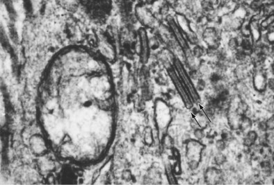
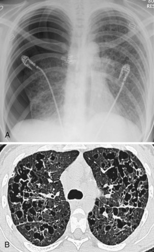
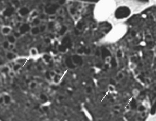
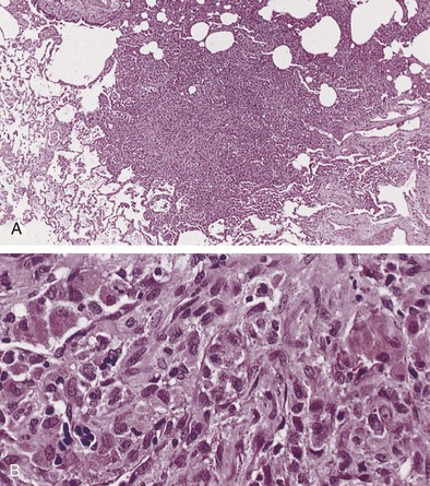
Langerhans cell histiocytosis typically affects individuals between 20 and 40 years of age, almost all of whom are current or recent smokers. The infiltrating Langerhans cell clone expresses the surface protein CD1a and has a characteristic appearance on electron microscopy with Birbeck inclusion granules (Figure 54-3). LCH may coexist with areas of desquamative interstitial pneumonitis, another smoking-related entity characterized by activation of macrophages. Similar to LAM, LCH is of variable severity, with approximately one quarter of LCH patients detected by imaging being asymptomatic. Pneumothorax is the presenting feature in about 20% of patients; the rest present with cough, dyspnea, or weight loss. The chest x-ray film is abnormal in most patients, showing reticular shadowing with midzone and upper-zone predominance (Figure 54-4). The CT scan shows a mixture of nodules, cavitating nodules, and cysts, often with thick, irregular walls forming bizarre shapes (Figure 54-5); characteristically, the bases of the lungs are relatively spared. As the disease progresses, the cysts tend to amalgamate, and nodules become less common. The diagnosis is usually suspected by the combination of these imaging appearances in younger smokers. Absolute confirmation often requires a surgical biopsy (Figure 54-6). However, bronchoalveolar lavage (BAL) fluid with greater than 5% Langerhans cells identified by CD1a positivity, often with increased macrophages and eosinophils, is supportive and can be diagnostic in the correct clinical context.

Lung function is normal in about 15% of LCH patients, although most have airflow obstruction with impaired gas transfer or a mixture of obstructive and restrictive changes. The course of LCH is variable, with spontaneous remission and even resolution in some patients; up to half of patients improve by CT criteria. However, some develop progressive respiratory failure, and a poor outcome is predicted by older age at onset, persistent constitutional symptoms, extrapulmonary involvement, and the presence of pulmonary hypertension. The overall survival for LCH patients is approximately 70% after 10 years, although some will require evaluation for pulmonary transplantation.

Management of LCH is generally supportive. Stopping smoking is the most important intervention and may significantly increase the chance of spontaneous resolution. As a particular feature of LCH, pulmonary hypertension should be screened for in those with active disease and may require specific treatment. In addition, many reports suggest an increased prevalence of lymphoma and lung cancer in patients with LCH. Persisting constitutional symptoms, hemoptysis, or other unexplained features should be thoroughly evaluated before being attributed to LCH alone. For those with active disease, steroids are frequently used, although no evidence supports this. More recently, in individual series, patients with active disease have been treated with 2-chlorodeoxyadenosine (Cladribine), a purine nucleoside analogue with activity against lymphocytes and monocytes, with evidence of response in many patients.

Birt-Hogg-DubE syndrome

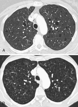
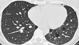
Birt-Hogg-Dube (BHD) syndrome was first described as the association of fibrofolliculomas, acrocordons, and trichodiscomas; benign skin-colored papules and skin tags of adult onset; presence of lung cysts; and a predisposition to renal cancer. It is now realized that BHD syndrome may be entirely restricted to the lungs, without cutaneous manifestations, and is caused by either germline or somatic mutations in the folliculin (FLCN) gene, a tumor suppressor of unknown function. Patients with pulmonary disease develop lung cysts predominantly in the central and lower zones of the lungs, characteristically around the heart (Figure 54-7). The cysts are 0.2 to 7 cm in size, have thin walls, are often ovoid in shape, and occasionally are multiseptated. The cyst walls have disrupted elastic fibers and are infiltrated by macrophages.

Diagnostic markers of BHD syndrome include a family history of pneumothorax, skin lesions, and early renal cancer. However, less than half of patients will have the triad of skin, renal, and lung features. The main respiratory manifestation is recurrent pneumothorax, generally occurring in adult life. Symptomatic disease is possibly more common in women and those harboring germline rather than somatic mutations. Diagnosis of BHD syndrome is usually made on clinical criteria and the presence of more than five fibrofolliculomas or trichodiscomas of adult onset or an FLCN mutation. Minor criteria are renal cancer before age 50, multiple lung cysts, or a first-degree relative with a germline FLCN mutation. In practical terms, particularly with isolated lung disease, diagnosis is best made by screening the patient’s blood for FLCN mutations in the correct clinical context.

Patients with BHD syndrome tend not to develop progressive respiratory failure. The important management is prevention of recurrent pneumothorax; screening for renal cancer, probably best done by yearly magnetic resonance imaging (MRI); and when necessary, cosmetic treatment of dermatologic manifestations.

Evaluation of Patients with Cystic Lung Diseases

In addition to the entities previously described, lung cysts are also seen in multiple cystic mesenchymal hamartoma, an entity categorized by a small number of larger, thick-walled cystic lesions prone to hemorrhage. Metastatic endometrial stromal sarcoma can present as lung cysts and can run a deceptively indolent course, in one case diagnosed only after lung transplantation for suspected LAM. Lung cysts are also seen in lymphocytic interstitial pneumonia (LIP) and patients with acquired immunodeficiency syndrome (AIDS). In these entities, cysts are generally less profuse and associated with interstitial changes.

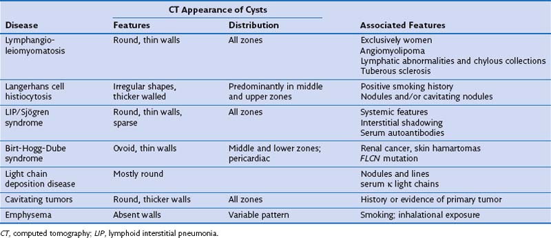
Diagnosis in patients with cystic lung disease can be difficult. Although imaging and associated features usually suggest the most likely diagnosis, imaging alone is not generally diagnostic (Table 54-1). Suspicion of LAM should be followed by abdominal CT, which will show extrapulmonary manifestations in two thirds of patients; LCH by BAL and CD1a staining, and BHD by FLCN mutation analysis and renal/skin evaluation. Nonspecific or mixed appearances can be evaluated by autoantibody screening, serum κ/λ light chain ratio and human immunodeficiency virus (HIV) serology. Surgical biopsy may be required but can be avoided in many cases. Because many of these diseases have genetic implications for the patient and family, and specific treatments and clinical trials are available, a definitive diagnosis is often necessary.

Other Rare Diffuse Lung Diseases

Pulmonary Alveolar Proteinosis

Also known as alveolar lipoproteinosis, alveolar phospholipidosis, and pulmonary alveolar phospholipoproteinosis, pulmonary alveolar proteinosis (PAP) is a disease caused by a failure of granulocyte-macrophage colony-stimulating factor (GM-CSF) signaling, which results in a failure of surfactant protein clearance by alveolar macrophages and the accumulation of lipoproteinaceous material in the alveolar spaces. Three categories of PAP have been described: idiopathic, secondary, and congenital.

Idiopathic PAP results from GM-CSF neutralizing autoantibodies and accounts for approximately 90% of cases, with overall prevalence or 4 to 40 per 1 million, dependent on the population studied. It is two to three times more common in men and smokers and has been putatively linked to exposure to flour, cement, wood, chlorine gas, gasoline, and plastics, but a causal link has not been established. Heavy silica dust exposure was known to cause silicoproteinosis, a similar condition, but this is now rarely seen as a result of reduced workplace exposure. Idiopathic PAP usually presents in adulthood, with a mean age at diagnosis of 39 years. Presentation is nonspecific, the most common symptoms being dyspnea, cough, chest pain, hemoptysis, and constitutional upset. Superimposed infection may also be the presenting feature. Examination can be normal, but lung crackles and finger clubbing may be present.

Secondary PAP is not related to GM-CSF autoantibodies but occurs in a small fraction of patients with hematologic disorders (e.g., chronic myelogenous leukemia, myelodysplastic syndromes), immunodeficiency (e.g., HIV, hypogammaglobulinemia, thymic aplasia, IgA deficiency) and lysinuric protein intolerance, a genetic disorder caused by an amino acid transporter mutation. Congenital PAP is a group of autosomal recessive disorders caused by a variety of GM-CSF receptor and surfactant protein mutations and is invariably fatal in childhood without transplantation.

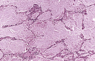
Chest radiography demonstrates consolidation with thickened intralobular septae (Figure 54-8). In a third of cases the changes are asymmetric or unilateral. The pattern is variable and in 50% of PAP patients may be perihilar, resembling the “bat wing” appearance seen in pulmonary edema. The CT appearance of air space shadowing in a geographic pattern with interlobular septal thickening is described as a “crazy paving” pattern (Figure 54-9). This appearance is not specific for PAP and may also be seen in cardiogenic pulmonary edema, bronchoalveolar cell carcinoma, Pneumocystis jirovecii pneumonia, alveolar hemorrhage, sarcoidosis, cryptogenic organizing pneumonia, nonspecific organizing pneumonia, exogenous lipoid pneumonia, and drug-induced lung diseases. Diagnosis is usually made on the basis of a compatible clinical history and typical radiology and a milky appearance on BAL fluid containing periodic acid–Schiff–positive granular material (Figure 54-10). Open-lung biopsy, although rarely essential, shows accumulation of lipoprotein in the alveolar spaces (Figure 54-11). Nonspecific increase in serum lactate dehydrogenase (LDH) may also be seen. Lung function is generally restrictive with decreased gas transfer. Anti-GM-CSF antibody titers also show promise as a diagnostic test in both serum and BAL fluid.
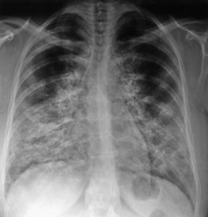
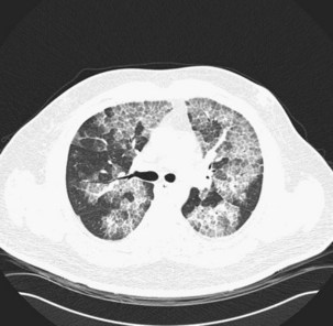
Opportunistic infections can complicate PAP because of impaired macrophage function and host defense. Common organisms include Nocardia species, fungi, and mycobacteria. Pulmonary fibrosis has been documented to occur in PAP, but this is rare, and it is unclear as to whether this process is progressive. The treatment of choice for idiopathic PAP is whole-lung lavage (WLL) with warmed 0.9% saline performed at specialist centers under general anesthesia using double-lumen endotracheal tube. Lavage is continued until fluid runs clear, typically taking 10 to 12 L of saline per lung. Chest percussion is also sometimes employed to aid clearance of lipoproteinaceous material. Both multisession WLL and single-session, sequential bilateral WLL are performed. In PAP patients with severe hypoxia, WLL has been performed with extracorporeal membrane oxygenation and in hyperbaric chambers. Complications of WLL include barotrauma and saline thorax. Although there are no randomized controlled trials (RCTs) of WLL, case series report 85% of patients treated have some improvement in symptoms, lung function, radiology, and survival. The median duration of clinical benefit is 15 months and 60% to 70% will require a repeat lavage. WLL may still be required for secondary PAP, but the mainstay of therapy is to treat the underlying condition. Congenital PAP does not appear to respond well to WLL.

Several small, nonrandomized studies of subcutaneous GM-CSF for idiopathic PAP show an improvement in up to 50% of patients. Inhaled GM-CSF has also undergone trials, but further studies are needed.

In the adult forms of PAP, estimated 5-year and 10-year survival are 75% and 68%, respectively. Approximately one third of cases resolve spontaneously over months to years, one third of patients have stable disease, and another third have progressive disease, with lung transplantation as an option. The overall risk of respiratory death is 10% to 15%. The prognosis is much worse in those under age 5 years with congenital PAP, with 5-year survival of about 14%.

Amyloidosis

The term amyloidosis encompasses a complex group of multisystem conditions that result in the extracellular deposition of insoluble amyloid, abnormally folded low-molecular-weight protein molecules, resulting in progressive organ damage. It may be acquired or inherited and present with multisystem or focal disease. Amyloidosis may present to respiratory physicians in a variety of ways. Chronic pulmonary conditions such as bronchiectasis may give rise to systemic AA amyloidosis; systemic amyloidosis itself may result in respiratory symptoms and referral; and localized respiratory tract amyloid deposits may either present with respiratory symptoms or may be found incidentally on chest radiography.

The two most common forms of amyloid to affect the respiratory system are AL light chain (AL) amyloidosis, which occurs as a result of deposition of excess immunoglobulin light chain fragments, usually secondary to a plasma cell dyscrasia, or reactive systemic AA amyloidosis caused by accumulation of the acute-phase reactant, serum amyloid A, secondary to a variety of other chronic inflammatory conditions. Almost all clinically significant respiratory tract disease is related to AL amyloid. The diagnosis of amyloidosis should be considered with unusual upper airways symptoms in patients with parenchymal changes, unexplained congestive cardiac failure, or nephrotic syndrome suggesting a multisystem disease.

Laryngeal amyloid represents 0.5% to 1% of benign laryngeal disease and may present as discrete nodules or diffuse infiltration; it is often localized and usually relatively benign. Laryngeal amyloid usually affects the supraglottic larynx, and patients present with hoarse voice or stridor, although choking or exertional dyspnea also occur. Tracheobronchial amyloid is uncommon, and presenting symptoms include dyspnea, persistent cough, and hemoptysis. Airway narrowing can also cause wheeze, distal atelectasis, recurrent pneumonia, or lobar collapse, and solitary nodules have been mistaken for neoplasia. Amyloidosis can also present with mediastinal and hilar lymphadenopathy although parenchymal amyloidosis presenting as either solitary or multiple nodules or a diffuse alveolar-septal pattern is the most frequently detected respiratory manifestation.

The “gold standard” for diagnosis remains biopsy demonstrating apple-green birefringence with polarized microscopy after staining with Congo red. In some centers, radiolabeled serum amyloid P (SAP) has been used to identify distribution and burden of disease, particularly for solid-organ involvement. With pulmonary involvement, lung function tests may show a restrictive pattern with reduced transfer factor, and in the case of tracheobronchial disease, large airways obstruction may be detected by flow-volume loop. Routine workup excludes an underlying blood cell dyscrasia (e.g., myeloma) and looks for other organ involvement.

Studies on management of respiratory tract amyloid are limited, and management decisions are often made empirically based on symptom management. No treatment may be needed, but local measures such as laser therapy, stenting, and surgical resection have been used for endobronchial and tracheobronchial disease. No evidence shows that steroids have any effect on laryngeal amyloid, and it is thought that repeated endoscopic procedures are safer than repeated open surgery. Many modalities used for myeloma, including chemotherapeutic agents (e.g., melphalan, thalidomide) and autologous bone marrow transplantation, are being tried in systemic AL amyloid, but there have been limited clinical trials.

Pulmonary Alveolar Microlithiasis

Pulmonary alveolar microlithiasis (PAM), first described in 1957, is characterized by the extensive accumulation of intraalveolar calcium phosphate deposits, or “microliths.” PAM is an autosomal recessive disease and, although reported worldwide, is most common in Japan, Turkey, and Italy. Presentation is most common in the mid-30s with dyspnea, cough, and chest pain. However, half of patients are asymptomatic, and PAM is detected as an incidental finding on chest radiography or by family screening.

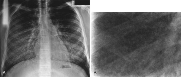
Diagnosis is usually made on the basis of a distinctive calcified micronodular “sandstorm” appearance seen on chest radiography with mid/lower-zone predominance, often obliterating the mediastinal and diaphragmatic contour (Figure 54-12). CT scanning, particularly in advanced disease, demonstrates calcified interlobular septa, pleura, and bronchovascular bundles; perilobular microliths; and often ground-glass attenuation, areas of consolidation, and subpleural cysts. Lung biopsy reveals characteristic lamellar intraalveolar microliths. Mutations in SLC34A2, a gene encoding a sodium-dependent phosphate transporter, highly expressed by alveolar type II cells have been identified in most patients, possibly leading to the aberrant accumulation of calcium phosphate in alveolar spaces.

Longer-term prognosis in patients with PAM is poor, with a mean age of death from respiratory failure of 46 years. Treatment is supportive, including oxygen when required and lung transplantation. Bisphosphonates have been used in children with PAM with some evidence of short-term radiologic and clinical improvement.

Pulmonary Manifestations of Rare Systemic Diseases

A number of rare systemic diseases associated with pulmonary manifestations may be encountered by respiratory physicians, including Hermansky-Pudlak syndrome, Erdheim-Chester disease, lysosomal storage disorders (e.g., Gaucher, Niemann-Pick, Fabry), and mucopolysaccharidoses.

Lysosomal storage disorders

Lysosomal storage disorders (LSDs) are a group of approximately 50 inherited conditions characterized by a defect in the functional expression of any of the lysosomal enzymes, resulting in the accumulation of their substrates both intracellularly and extracellularly and loss of function in a variety of organ systems, including the lungs. The clinical features depend greatly on the rate and magnitude of accumulation, with a wide spectrum of phenotypes observed, the vast majority presenting in childhood.

Gaucher disease (GD), the most prevalent LSD, is inherited in an autosomal dominant manner and results from a deficiency of glucocerebrosidase activity causing glucocerebroside to accumulate in phagocytic reticuloendothelial cells, called Gaucher cells. Pulmonary involvement is most common in the infantile form (type 2) although adult onset (type 1) is sometimes associated with an interstitial lung disease resulting from accumulation of Gaucher cells in the alveolar spaces and septa. Pulmonary hypertension and hepatopulmonary syndrome are also occasionally seen. Pulmonary involvement is more common in those with severe disease, particularly with neuropathic involvement. Enzyme replacement therapy is currently the mainstay of treatment, but respiratory involvement may not respond well to this.

Niemann-Pick disease is an autosomal recessive disorder characterized by the accumulation of lipid-laden macrophages (Niemann-Pick cells). Lung involvement is relatively frequent in infantile forms but less common in adult forms. The radiologic appearance is typically of reticular or reticulonodular infiltrates. Treatment by WLL may be of some benefit, and bone marrow transplantation has been attempted in some patients.

Fabry disease is an X-linked metabolic disease caused by the absence of α-galactosidase A, resulting in the accumulation of glycosphingolipids. Patients can present with a variety of pulmonary symptoms, including dyspnea, airway obstruction, wheeze, pneumothorax, and hemoptysis. The chest radiograph may be normal, but chest CT may show ground-glass change. Diagnosis is made by bronchial biopsy, bushings, or lavage. Enzyme replacement therapy has been shown to alleviate pulmonary dysfunction in some patients.

Suggested Readings

Allen TC, Chevez-Barrios P, Shetlar DJ, Cagle PT. Pulmonary and ophthalmic involvement with Erdheim-Chester disease: a case report and review of the literature. Arch Pathol Lab Med. 2004;128:1428–1431.

Colombat M, Stern M, Groussard O, et al. Pulmonary cystic disorder related to light chain deposition disease. Am J Respir Crit Care Med. 2006;173:777–780.

Duchateau F, Dechambre S, Coche E. Imaging of pulmonary manifestations in subtype B of Niemann-Pick disease. Br J Radiol. 2001;74:1059–1061.

Eng CM, Germain DP, Banikazemi M, et al. Fabry disease: guidelines for the evaluation and management of multi-organ system involvement. Gent Med. 2006;8:539–548.

Goitein O, Elstein D, Abrahamov A, et al. Lung involvement and enzyme replacement therapy in Gaucher’s disease. QJM. 2001;94:407–415.

Huqun IzumiS, Miyazawa H, et al. Mutations in the SLC34A2 gene are associated with pulmonary alveolar microlithiasis. Am J Respir Crit Care Med. 2007;175:263–268.

Johnson SR. Lymphangioleiomyomatosis. Eur Respir J. 2006;27:1056–1065.

Johnson SR, Cordier JF, Lazor R, et al. European Respiratory Society guidelines for the diagnosis and management of lymphangioleiomyomatosis. Eur Respir J. 2010;35:14–26.

Kamin W. Diagnosis and management of respiratory involvement in Hunter syndrome. Acta Paediatr Suppl. 2008;97:57–60.

Lachmann HJ, Hawkins PN. Amyloidosis and the lung. Chronic Respir Dis. 2006;3:203–214.

Pierson DM, Ionescu D, Qing G, et al. Pulmonary fibrosis in Hermansky-Pudlak syndrome: a case report and review. Respiration. 2006;73:382–395.

Toro JR, Pautler SE, Stewart L, et al. Lung cysts, spontaneous pneumothorax, and genetic associations in 89 families with Birt-Hogg-Dube syndrome. Am J Respir Crit Care Med. 2007;175:1044–1053.

Trapnell BC, Whitsett JA, Nakata K. Pulmonary alveolar proteinosis. N Engl J Med. 2003;349:2527–2539.

Vassallo R, Ryu JH, Schroeder DR, et al. Clinical outcomes of pulmonary Langerhans cell histiocytosis in adults. N Engl J Med. 2002;346:484–490.