Radiesse® / Radiesse® with lidocaine

Published on 16/03/2015 by admin

Filed under Dermatology

Last modified 22/04/2025

Print this page

rate 1 star rate 2 star rate 3 star rate 4 star rate 5 star
Your rating: none, Average: 0 (0 votes)

This article have been viewed 1519 times

6 Radiesse® / Radiesse® with lidocaine

Introduction

Soft tissue augmentation began in 1893 with free fat grafting, but became more popular with the introduction of bovine collagen injections in the late 1970s. Dermal filler injections are now the most common aesthetic treatments performed worldwide. The ideal soft tissue filler for facial augmentation should be non-allergenic, durable, cost-effective, and provide a natural look and feel. Also it should degrade naturally, require no reconstitution or refrigeration, have a long shelf-life, evoke minimal pain on injection, require minimal downtime, and exhibit few side effects.

Dermal fillers are used for injection into the mid- to deep dermis for the correction of moderate to severe wrinkles and folds, restore age-related facial soft tissue volume loss, augment existing facial structures, and improve nasal function. Some have limited indications for correction of facial lipoatrophy in HIV and acne scarring. There are many injectable types of filler on the international market, but only a small number have US Food and Drug Administration (FDA) approval. Fillers can be permanent or temporary and are classified into three categories: collagens, hyaluronic acids (HAs), and biosynthetic polymers.

The effects of biological products such as collagen and HA typically last for less than 1 year after treatment. Semipermanent fillers, including poly-l-lactic acid and calcium hydroxylapatite (CaHA), were observed to persist for 1–2 years in patients. Non-absorbable fillers such as polymethylmethacrylate, silicone oil, and polyacrylamide gel achieved the longest lasting aesthetic results, persisting for longer than 2 years.

CaHA, also known as Radiesse® (BioForm Medical Inc., San Mateo, CA), consists of a 30% concentration of 25–45 mm spherical particles suspended in sodium carboxymethylcellulose gel. Its unique profile makes it particularly well suited for facial augmentation and contouring. In particular, its biocompatibility, ease of use, and enhanced durability offer physicians flexibility in both injection and areas of application. CaHA was first used in 2002, and was initially approved for vocal cord augmentation, radiographic soft tissue marking, and maxillofacial defect correction. In these applications, it demonstrated a favorable safety profile and was rapidly adopted for off-label use in facial contouring. In December 2006 CaHA, Radiesse®, received FDA approval for soft tissue augmentation in the USA. The product may be stored for 2 years at room temperature. The correction ratio is 1 : 1 implant to tissue volume. The longevity is reported to be approximately 14–15 months in active tissues of the face and may approach 18–24 months in more static locations. In addition to the site of injection, factors influencing longevity include the patient’s age, ability to synthesize soft tissue, and rate of metabolism.

Specific characteristics of calcium hydroxylapatite

Tzikas reported that injection volumes required with CaHA are considerably less than those required with collagen or HA products. In addition, unlike collagen or HA filler materials, which typically require a certain degree of overcorrection, a conservative approach is best with CaHA injection, with no need for overcorrection, because it naturally provides for a 1 : 1 correction. However, Tzikas was unable to correlate the volume of filler injected with the duration of effect. Good longevity was noted, with more than 80% of patients reporting persistence of results at 12 months. The majority of patients return in 12–18 months for additional injection; Busso & Karlsberg have estimated that the average duration of effect is 14–15 months in patients treated with cheek augmentation. In a clinical trial for lipoatrophy by Silvers and colleagues, skin thickness measurements at 12 months remained statistically better than those at baseline. CaHA is highly biocompatible with minimal acute reactions to injection and no reported serious side effects; its particles become fixed in position over time, do not migrate, and take on the natural characteristics of the soft tissue as new collagen is deposited. In one study, CaHA was successfully used in patients up to 85 years old. However, because these patients have poorer skin elasticity with deeper lines and grooves, they may have a decreased fibroelastic response that may limit results. Therefore, they often need larger volumes of filler with repeat injections and should be counseled to help avoid unrealistic expectations.

Injection techniques

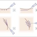
In general, for dermal fillers, the depth of the defect determines the depth of injection; the deeper the defect, the more viscous the filler. CaHA is typically placed in the mid- to deep dermis or subdermal planes. Four techniques for injection are reported: serial puncture, linear threading, fanning, and cross-hatching.

Cross-hatching / fanning

The fanning (Fig. 6.3) and cross-hatching techniques are variations of linear threading that allow filling of larger defects or facial contouring. In the fanning technique a single needle puncture allows ‘fan-like’ placement of successive linear threads by radially changing the needle direction. Cross-hatching delivers linear threads in a predetermined grid by multiple punctures. Both techniques are useful in the malar region and oral commissures to build a scaffold in deeper defects.

The traditional injection approach of CaHA is into the subdermal plane, using threading and cross-hatching of aliquots of dermal filler for treatment of nasolabial folds, oral commissure, and the marionette lines. A linear threading, cross-hatching, or fanning technique is used to deposit threads of the material in multiple layers as needed to bolster or support the fold or deficiency. The correct depth of the needle is at the deep dermal junction (i.e. subdermal plane). Following administration, the injection site is gently massaged and patients are observed for adverse reactions. Other injection techniques may be preferred for other areas of the face for volumizing and facial contouring.

In non-facial areas, CaHA mixed with lidocaine is used for treatment of aging hand and pedal defects, where the dorsal skin is first lifted to create a tent for injection away from tendons and vessels, and the hand is then immediately massaged.

Prior to introduction of lidocaine into CaHA, pain during injection was a limitation. Methods to minimize pain included application of nerve blocks, tissue infiltration with anesthetics, topical anesthetics, and physical aids such as vibration, icing, and skin cooling. In 2009, the FDA approved a novel technique using the addition of lidocaine into the CaHA syringe just before injection. The approach involves 0.2 mL of plain 2% lidocaine drawn into a 3 mL syringe, then attached to a 1.3 mL syringe of CaHA through a Luer-lock connector. The CaHA and lidocaine are pushed from one syringe to the other, back and forth 10 times, to create a homogeneous mixture. In 2009, a study was performed by Marmur and co-workers of a cohort of 50 patients who received CaHA in one nasolabial fold and CaHA / lidocaine blend in the other; all patients preferred pre-mixed CaHA with lidocaine compared with CaHA with no anesthetic. In 2008, a study by Busso & Voigts showed that the CaHA / lidocaine blend exhibited decreased viscosity, elasticity, and extrusion force, while it did not affect the longevity of the clinical benefits. The addition of lidocaine to CaHA has significantly decreased injection site pain and decreased the need for adjuvant anesthetic techniques.

Adverse events

In general, complications tend to be mild and technique related. Adverse events are classified into immediate (pain, hypersensitivity, anaphylaxis), early (swelling, ecchymosis, erythema, infection, overcorrection, necrosis), late (activation of herpes simplex virus, nodules, granuloma formation), and permanent (scarring). Patients’ expectations should be addressed prior to treatment and they should be informed of the risks, alternatives, and aftercare instructions.

Five uncontrolled clinical trials that examined the effect of CaHA in the cheek area were identified, all of which reported improved cosmetic outcomes and minimal adverse events. One of these studies (n = 100) was conducted in patients with HIV-associated lipoatrophy. The 18-month open-label clinical trial by Beer et al enrolled 94 men and 6 women who received CaHA injections into the submalar region. All patients were rated as improved or better on the Global Aesthetic Improvement Scale (GAIS) at every timepoint through 12 months; 91% were rated as improved or better at 18 months. In addition, skin thickness measurements were statistically improved at 12 months compared with baseline. Adverse events resulting from the treatment were mild (e.g. ecchymosis, edema, erythema, pain, pruritus) and of short duration. The other four clinical trials were performed in healthy patients seeking a fuller appearance to the cheeks. At 6 months, physician-assessed GAIS ratings indicated that 15 of the remaining 16 subjects were improved, much improved, or very much improved. Patient-assessed GAIS ratings were similar, with 14 patients reporting their appearance was improved, much improved, or very much improved. Adverse events were reported in two patients; one reported mild edema and hematoma, and the other reported mild ecchymosis and edema.

Erythema and swelling may sometimes require steroid treatment. Non-steroidal anti-inflammatory drugs and other anticoagulants should be discontinued prior to treatment so as to minimize postprocedural ecchymosis. Use of anterograde injections, icing both pre- and post-injection, and slow injection technique may also decrease post-procedural bruising, particularly in the pre-jowl sulcus and marionette lines.

Injectors should be aware of facial danger zones, specifically embolization of the supratrochlear (glabella), angular branch of facial (alar–peri-alar) and dorsal nasal arteries. These injections should only be performed by experienced injectors, with a thorough patient understanding and written consent regarding potential risks, as well as appropriate tools for managing intra-arterial injections such as nitric oxide paste and massage.

With CaHA, no osteoblast activity, i.e. calcification or ossification, has been observed in soft tissue. CaHA is unlikely to provoke allergic or other adverse reactions. However, patients with a history of anaphylaxis to any substance or multiple severe allergies are contraindicated for CaHA injections for cosmetic indications.

Marmur and co-workers examined punch biopsies using standard light and electron microscopy techniques in a small number of patients at 1 and 6 months following injection of CaHA into the dermis. Biopsies at 1 and 6 months showed CaHA microspheres scattered in the dermal/subcutaneous junction with minimal inflammation and evidence of new fibroelastic fibers surrounding the microspheres, but no apparent migration. No granuloma formation, ossification, or foreign-body reactions were evident.

Nodules can occur, they are most common in patients undergoing augmentation of the lip, and may occur in up to 12.4% of this population. In this location they are an aggregation of material due to the sphincteric action of the orbicularis oris muscle. Regardless of location, nodules are treatable via massage, needle disruption of the nodules, or excision. Jansen & Graivier reported a reduced incidence of lip nodules by using a smaller injection volume in combination with a more conservative threading technique and by injecting at the proper depth (subdermal plane). At present, the authors do not recommend using CaHA for lip augmentation or for injections within the orbital rim.

Comparative research studies

Thirty-two studies on the use of CaHA were identified, including three randomized controlled trials, 15 uncontrolled clinical trials, two cohort studies, one cross-sectional study, and 11 case series/reports. The most frequently studied site across these studies was the nasolabial folds, followed by the cheeks. The three randomized controlled trials that examined the use of CaHA were all conducted in patients with moderate to severe nasolabial folds. Two studies by Moers-Carpi and colleagues comparing HA and CaHA were multicenter, blinded, split-face, randomized controlled trials conducted in Europe by the same investigators, and determined that CaHA was superior to HA fillers in the treatment of nasolabial folds. The first study (n = 205) compared three hyaluronic acid fillers (Juvéderm 24®, Juvéderm 24HV®, and Perlane®) with CaHA, with outcomes assessed at 4, 8, and 12 months. Analysis was performed using the GAIS; investigators found that CaHA showed the largest number of nasolabial folds rated ‘improved’ or better. This was statistically significant compared with all HA fillers and at all timepoints except one (at 12 months compared with Juvéderm 24HV®), and led the authors to conclude that the CaHA was more effective and longer lasting than each HA filler in maintaining nasolabial fold augmentation. There was no serious adverse event requiring intervention at any timepoint for any of the injected materials. The second study (n = 60) compared Restylane® and CaHA with outcomes assessed at 6, 9, and 12 months. CaHA was found to be significantly more effective than HA, showing greater improvement and longer lasting effects, as evidenced by blinded-evaluator WSRS ratings that showed a mean improvement at 12 months of 0.41 for CaHA and 0.14 for HA (p = 0.007). Both products were well tolerated, and no serious adverse events were reported with either treatment. Only four adverse events (two hematomas, one nodule, and one extrusion) were reported in 118 folds injected two times each during the course of treatment and were resolved without complications.

The third randomized controlled trial by Smith and co-workers (n = 117) compared CaHA with human-based collagen for 6 months. Photographs by blinded experts indicated that CaHA achieved superior improvement to collagen. Adverse event rates for both treatments were comparable, with some increase in bruising and edema at CaHA-treated sites. Other studies by Hanke and co-workers assessing the use of CaHA for the correction of nasolabial folds indicated that it was effective and well tolerated.

Large-particle calcium hydroxylapatite injection

Because permanent fillers have not become popular, possibly because of reports of troublesome adverse events after their use, there are no temporary fillers that last longer than poly-l-lactic acid or small-particle CaHA. The large-particle version of CaHA (Coaptite®, Merz; distributed by Boston Scientific) offers the promise of a better and longer lasting correction than small-particle CaHA. Alam and co-workers reported three cases of facial augmentation using large-particle CaHA. Based on their limited experience of three cases, no persistent bruising, contour abnormality, vascular occlusion, neuropathic pain, or implant migration was detected in any of the patients. The only difference versus small-particle CaHA was that the needle entry sites were faintly visible as round red spots on the skin for 1–2 days after injection before resolving completely. To minimize marking, authors used only one entry site per side of the face, and used needles of different lengths, aimed at varying angles, to access all portions of each defect. Compared with prior injections with small-particle CaHA, the correction appeared to be more persistent. The authors stated that just three case reports cannot definitively establish the safety of large-particle CaHA for facial wrinkle correction, nor can they demonstrate with any certainty that this material has improved longevity; further studies are needed to make conclusions and recommendations regarding this product.

Further reading

Alam M, Havey J, Pace N, et al. Large-particle calcium hydroxylapatite injection for correction of facial wrinkles and depressions. Journal of the American Academy of Dermatology. 2011;65:92–96.

Beer K, Yohn M, Cohen JL. Evaluation of injectable CaHA for the treatment of mid-face volume loss. Journal of Drugs in Dermatology. 2008;7:359–366.

Bray D, Hopkins C, Roberts DN. A review of dermal fillers in facial plastic surgery. Current Opinion in Otolaryngology & Head and Neck Surgery. 2010;18:295–302.

Busso M. Calcium hydroxylapatite (Radiesse): safety, techniques and pain reduction. Journal of Drugs in Dermatology. 2009;8:S21–S23.

Busso M, Applebaum D. Hand augmentation with Radiesse (calcium hydroxylapatite). Dermatologic Therapy. 2007;20:385–387.

Busso M, Karlsberg P. Cheek augmentation and rejuvenation using injectable calcium hydroxylapatite (Radiesse). Cosmetic Dermatologic Surgery. 2006;19:583–588.

Busso M, Voigts R. An investigation of changes in physical properties of injectable calcium hydroxylapatite in a carrier gel when mixed with lidocaine and with lidocaine/epinephrine. Dermatologic Surgery. 2008;34(suppl 1):S16–S23. discussion S4

Carruthers A, Liebeskind M, Carruthers J, et al. Radiographic and computed tomographic studies of calcium hydroxylapatite for treatment of HIV-associated facial lipoatrophy and correction of nasolabial folds. Dermatologic Surgery. 2008;34(suppl 1):S78–S84.

Fakhre GP, Perdikis G, Shaddix KK, et al. An evaluation of calcium hydroxylapatite (Radiesse) for cosmetic nasolabial fold correction: a meta-analysis and patient centric outcomes study. Annals of Plastic Surgery. 2009;63:486–489.

Glaich AS, Cohen JL, Goldberg LH. Injection necrosis of the glabella: protocol for prevention and treatment after use of dermal fillers. Dermatologic Surgery. 2006;32:276–281.

Hanke CW, Rohrich RJ, Busso M, et al. Facial Soft-Tissue Fillers conference: Assessing the State of the Science. Journal of the American Academy of Dermatology. 2011;64:S66–S85. e1-136

Jansen DA, Graivier MH. Evaluation of a calcium hydroxylapatite-based implant (Radiesse) for facial soft-tissue augmentation. Plastic and Reconstructive Surgery. 2006;118:S22–S30. discussion S1-S3

Lemperle G, Morhenn V, Charrier U. Human histology and persistence of various injectable filler substances for soft tissue augmentation. Aesthetic Plastic Surgery. 2003;27:354–366. discussion 67

Marmur ES, Phelps R, Goldberg DJ. Clinical, histologic and electron microscopic findings after injection of a calcium hydroxylapatite filler. Journal of Cosmetic and Laser Therapy. 2004;6:223–226.

Marmur E, Green L, Busso M. A controlled, randomized study of pain levels in subjects treated with calcium hydroxylapatite (Radiesse) premixed with lidocaine for correction of nasolabial folds. Dermatologic Surgery. 2009;2010; 36(3):309–315.

Marmur ES, Taylor SC, Grimes PE, et al. Six-month safety results of calcium hydroxylapatite for treatment of nasolabial folds in Fitzpatrick skin types IV to VI. Dermatologic Surgery. 2009;35(suppl 2):1641–1645.

Moers-Carpi MM, Tufet JO. Calcium hydroxylapatite versus nonanimal stabilized hyaluronic acid for the correction of nasolabial folds: a 12-month, multicenter, prospective, randomized, controlled, split-face trial. Dermatologic Surgery. 2008;34:210–215.

Moers-Carpi M, Vogt S, Santos BM, et al. A multicenter, randomized trial comparing calcium hydroxylapatite to two hyaluronic acids for treatment of nasolabial folds. Dermatologic Surgery. 2007;33(suppl 2):S144–S151.

Radiesse 2009 [package insert]. Bioform Medical I.

Ridenour B, Kontis TC. Injectable calcium hydroxylapatite microspheres (Radiesse). Facial Plastic Surgery. 2009;25:100–105.

Schanz S, Schippert W, Ulmer A, et al. Arterial embolization caused by injection of hyaluronic acid (Restylane). British Journal of Dermatology. 2002;146:928–929.

Silvers SL, Eviatar JA, Echavez MI, et al. Prospective, open-label, 18-month trial of calcium hydroxylapatite (Radiesse) for facial soft-tissue augmentation in patients with human immunodeficiency virus-associated lipoatrophy: one-year durability. Plastic and Reconstructive Surgery. 2006;118:S34–S45.

Smith S, Busso M, McClaren M, et al. A randomized, bilateral, prospective comparison of calcium hydroxylapatite microspheres versus human-based collagen for the correction of nasolabial folds. Dermatologic Surgery. 2007;33(suppl 2):S12–S21. discussion S21

Tzikas TL. A 52-month summary of results using calcium hydroxylapatite for facial soft tissue augmentation. Dermatologic Surgery. 2008;34(suppl 1):S9–S15.