Q Fever (Coxiella burnetii)

Published on 22/03/2015 by admin

Filed under Pediatrics

Last modified 22/04/2025

Print this page

rate 1 star rate 2 star rate 3 star rate 4 star rate 5 star
Your rating: none, Average: 0 (0 votes)

This article have been viewed 1571 times

Chapter 224 Q Fever (Coxiella burnetii)

Q fever (for query fever, the name given following an outbreak of febrile illness in an abattoir in Queensland, Australia) is rarely reported in children but is probably underdiagnosed. Symptomatic patients can have acute or chronic disease.

Epidemiology

The disease is reported worldwide, except in New Zealand. Although seroepidemiologic studies suggest that infection occurs just as often in children as in adults, children less often present with clinical disease than do adults. Approximately 60% of infections are asymptomatic, and only 5% of symptomatic patients require hospitalization. Seroprevalence surveys show that 6-70% of children in endemic European and African communities have evidence of past infection, and in France the overall incidence of Q fever is estimated to be 50 cases per 100,000 persons. Cases in Africa are likely misdiagnosed as malaria. Reported cases of Q fever in the USA have increased by 6.5-fold from 26 cases in 2001 to 171 cases in 2007, which might reflect an increase in incidence, increased reporting after September 11, 2001, improved diagnostic tools, or a combination of factors. Reported cases in Asia and Australia have also increased. Most infections in children are identified during the lamb birthing season in Europe (January through June), following farm visits, or after exposure to placentas of dogs, cats, and rabbits. The largest community outbreak ever described occurred in the southeastern part of the Netherlands in 2008 and was associated with intensive goat farming. More than 20% of cases of clinically recognized acute or chronic Q fever occur in immunosuppressed hosts or in persons with prosthetic valves or damaged native valves or vessels. Although infections are recognized more often in men than in women, reported cases in boys and girls are equal.

Clinical Manifestations and Complications

Only about 40-50% of people infected with C. burnetii develop symptoms. Two forms of symptomatic disease occur. Acute Q fever is more common and usually manifests as self-limited undifferentiated fever or an influenza-like illness with interstitial pneumonitis. Chronic Q fever in adults usually involves native heart valves, prosthetic valves, or other endovascular prostheses. Q fever osteomyelitis is less common but proportionally more common in children.

Acute Q Fever

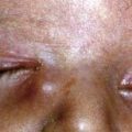
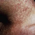
Acute Q fever develops about 3 wk (range, 14-39 days) after exposure to the causative agent. The severity of illness in children ranges from subclinical infection to a systemic illness of sudden onset characterized by high fever, severe frontal headache, nonproductive cough, chest pain, vomiting, diarrhea, abdominal pain, arthralgias, and myalgias. About 40% of children with acute Q fever present with fever, 25% with pneumonia or an influenza-like illness, >10% with meningoencephalitis, and >10% with myocarditis. Other manifestations include pericarditis, hepatitis, hemophagocytosis, rhabdomyolysis, and hemolytic uremic syndrome. Rash, ranging from maculopapular to purpuric lesions, is an unusual finding in adults with Q fever but is observed in ~50% of pediatric patients. Rigors and night sweats are common in adults with Q fever and occur less often in children. Prominent clinical findings that can create diagnostic confusion include fatigue, vomiting, abdominal pain, and meningismus. Hepatomegaly and splenomegaly may be detected in some patients.

Routine laboratory investigations in pediatric acute Q fever are usually normal but can reveal mild leukocytosis and thrombocytopenia. Up to 85% of children have modestly elevated serum hepatic transaminase levels that usually normalize within 10 days. Hyperbilirubinemia is uncommon in the absence of complications. C-reactive protein is uniformly elevated in pediatric Q fever. Chest x-ray films are abnormal in 27% of all patients; in children, the most common findings include single or multiple bilateral infiltrates with reticular markings in the lower lobes.

Acute Q fever in children is usually a self-limited illness, with fever persisting for only 7-10 days compared with 2-3 wk in adults. However, severe infections, such as myocarditis requiring cardiac transplantation, meningoencephalitis, pericarditis, and hemophagocytosis, have been reported.

Diagnosis and Differential Diagnosis

Although uncommonly diagnosed, Q fever should be considered in children who have fever of unknown origin, atypical pneumonia, myocarditis, meningoencephalitis, culture-negative endocarditis, or recurrent osteomyelitis and who live in rural areas or who are in close contact with domestic livestock, cats, or animal products.

The diagnosis of Q fever is most easily and commonly confirmed by testing acute and convalescent sera (2-4 wk apart), which show a 4-fold increase in indirect fluorescent antibody titers to phase I and phase II C. burnetii antigens. Predominant, elevated, or increasing titers of phase II antibody are characteristic of acute Q fever, and the appearance and persistence of titers of phase II antibody greater than phase I indicate chronic Q fever. Elevated titers of phase I immunoglobulin A (IgA) antibody are reported to be diagnostic for Q fever endocarditis; however, 1 evaluation showed that a phase I IgG titer of <800 is inconsistent with chronic Q fever. Cross reaction with antibodies to Legionella and Bartonella can occur.

Although culture has been considered the gold standard, sensitivity (compared with a composite standard including serology and PCR) is low. C. burnetii has been cultivated in tissue culture cells, which can become positive within 48 hr, but isolation and antimicrobial susceptibility testing of C. burnetii should be attempted only in specialized biohazard facilities. Testing by polymerase chain reaction (PCR) can be performed on blood, serum, and tissue samples and is available in some public health, reference, or research laboratories. Although PCR has been helpful in patients with equivocal titers; sensitivity has been improved by real-time methods and use of repeated sequences as targets. Immunohistochemical staining has also been used, but suffers from simila problems similar to PCR’s.

The differential diagnosis depends on the clinical presentation. In patients with respiratory disease, Mycoplasma pneumoniae, Chlamydophila pneumoniae, legionellosis, psittacosis, and Epstein-Barr virus infection should be considered. In patients with granulomatous hepatitis, tuberculous and nontuberculous mycobacterial infections, salmonellosis, visceral leishmaniasis, toxoplasmosis, Hodgkin disease, monocytic ehrlichiosis, granulocytic anaplasmosis, brucellosis, cat scratch disease (Bartonella henselae), or autoimmune disorders such as sarcoidosis should be considered. Culture-negative endocarditis suggests infection with Brucella, Bartonella, or HACEK organisms (Haemophilus, Aggregatibacter, Cardiobacterium hominis, Eikenella corrodens, Kingella), partially treated bacterial endocarditis, or nonbacterial endocarditis.

Treatment

Selection of an appropriate antimicrobial regimen for children is difficult owing to the lack of rigorous studies, the limited therapeutic window for drugs that are known to be efficacious, and the potential length of therapy required to preclude relapse.

Most pediatric patients with Q fever have a self-limited illness that is identified only on retrospective serologic evaluation. However, to prevent potential complications, patients with acute Q fever should be treated within 3 days of onset of symptoms, because therapy started >3 days after onset of illness has little effect on the course of acute Q fever. Because early confirmatory testing is not possible now, empirical therapy is warranted in clinically suspected cases. Doxycycline (4 mg/kg/day PO or IV divided every 12 hr, maximum 200 mg/day) is the drug of choice. Tetracycline and doxycycline may be associated with tooth discoloration in children <9 yr of age; however, most experts consider the benefits of treatment with doxycycline to be greater than the risks. Tooth discoloration is dose and duration dependent, and it is unlikely that children will require multiple courses. During pregnancy, Q fever is best treated with trimethoprim-sulfamethoxazole. The fluoroquinolones have also proved effective, and success with a combination of a fluoroquinolone and rifampin has also been achieved with prolonged therapy (16-21 days). Macrolides, including erythromycin and clarithromycin, are less effective alternatives.

For chronic Q fever, especially endocarditis, therapy for 18-36 mo is mandatory. The current recommended regimen for chronic Q fever endocarditis is a combination of doxycycline and hydroxychloroquine for ≥18 mo. For patients with heart failure, valve replacement may be necessary. Interferon-γ therapy has been used as adjunct therapy for intractable Q fever.

Bibliography

Centers for Disease Control and Prevention. Potential for Q fever infection among travelers returning from Iraq and the Netherlands. Official CDC health advisory (website). www2a.cdc.gov/han/archivesys/ViewMsgV.asp?AlertNum=00313. Accessed August 25, 2010

Delsing CE, Kullberg BJ. Q fever in the Netherlands: a concise overview and implications of the largest ongoing outbreak. Neth J Med. 2008;66:365-367.

Fournier PE, Etienne J, Harle JR, et al. Myocarditis, a rare but severe manifestation of Q fever: Report of 8 cases and review of the literature. Clin Infect Dis. 2001;32:1440-1447.

Kobbe R, Kramme S, Kreuels B, et al. Q fever in young children, Ghana. Emerg Infect Dis. 2008;14:344-346.

La Scola B, Maltezou HC. Legionella and Q fever community acquired pneumonia in children. Paediatr Resp Rev. 2004;5(Suppl):S171-S177.

Maltezou HC, Constantopoulou I, Kallergi C, et al. Q fever in children in Greece. Am J Trop Med Hyg. 2004;70:540-544.

Maltezou HC, Raoult D. Q fever in children. Lancet Infect Dis. 2002;2:686-691.

Maurin M, Raoult D. Q fever. Clin Microbiol Rev. 1999;12:518-553.

Nourse C, Allworth A, Jones A, et al. Three cases of Q fever osteomyelitis in children and a review of the literature. Clin Infect Dis. 2004;39:e61-e66.

Parker NR, Barralet JH, Bell AM. Q fever. Lancet. 2006;367:679-688.

Raoult D, Marrie TJ, Mege JL. Natural history and pathophysiology of Q fever. Lancet Infect Dis. 2005;5:219-226.

Raoult D, Tissot-Dupont H, Foucault C, et al. Q fever 1985–1998. Clinical and epidemiologic features of 1,383 infections. Medicine (Baltimore). 2000;79:109-123.

Ravid S, Shahar E, Genizi J. Acute Q fever in children presenting with encephalitis. Neurol. 2008;38:44-46.

Richardus JH, Dumas AM, Huisman J, et al. Q fever in infancy: a review of 18 cases. Pediatr Infect Dis. 1985;4:369-373.

Ruiz-Contreras J, Montero RG, Amador JT, et al. Q fever in children. Am J Dis Child. 1993;146:300-302.

Terheggen U, Leggat PA. Clinical manifestations of Q fever in adults and children. Travel Med Infect Dis. 2007;5:159-164.

Tissot-DuPont H, Raoult D, Brouqui P, et al. Epidemiologic features and clinical presentation of acute Q fever in hospitalized patients: 323 French cases. Am J Med. 1992;93:427-434.

Ughetto E, Gouriet F, Raoult D, et al. Three years experience of real-time PCR for the diagnosis of Q fever. Clin Microbiol Infect. 2009;15(Suppl 2):200-201.