145
Pyogenic granuloma
The fingers are a common location for pyogenic granulomas, leading to the theory that trauma is an important initiating factor.
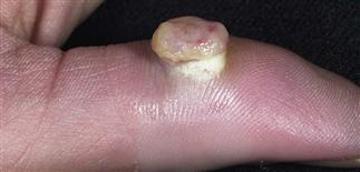
Pyogenic granuloma on the finger. Lesions on the fingers reoccur more frequently than in other locations.
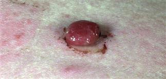
This dome-shaped papule has mucoid drainage; however, it is not infected.
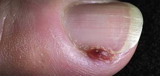
Pyogenic granuloma near the fingernail.
DESCRIPTION
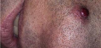
An exophytic, dome-shaped papule made up of proliferating capillaries separated by thick fibrous bands and surrounded by an epithelial collarette. Also called lobular capillary hemangioma.
HISTORY
• More common in children and young adults, less common in the elderly. • Cause unknown, but lesions occur at sites of trauma and during pregnancy, suggesting an important role for trauma and hormones. • Seen in increased frequency in acne patients treated with isotretinoin. • Pyogenic granulomas arise suddenly, attain a stable size, and persist without treatment, although some lesions spontaneously regress within 6 months. Larger, deep-seated lesions may recur with treatment. • Rarely, multiple satellite lesions can occur.
PHYSICAL FINDINGS
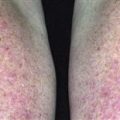
• Yellow to deep red, glistening, dome-shaped to polypoid papules of 3–10 mm. • Lesions grow rapidly, bleed profusely, and can be covered with yellow crust and surrounded by a collarette of scale. They can ‘fall off’ only to regrow. • Gingival lesions occurring during pregnancy are referred to as epulis gravidarum. • Lesions are more common on the head, neck, and fingers.
TREATMENT
• Treated by biopsy followed by electrodesiccation and curettage of the base and border of the lesion. • Most resolve with a single crateriform scar; recurrences occur in a few patients. • Rarely, multiple satellite lesions develop at and around the site of a previously treated pyogenic granuloma. This occurs most often on the shoulder and trunk in younger patients.