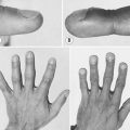
182 Pyoderma gangrenosum
Questions
What are the variants of this condition?
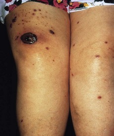
• Peristomal pyoderma gangrenosum at site of ileostomy (Fig. 182.2) or colostomy in ulcerative colitis or Crohn’s disease
• Penile or scrotal in adolescent adults
• Associated with preleukaemic state (the ulcer is shallow with bullous blue-grey border)
• Pyostomatitis vegetans is an oral variant with chronic pustular and eventually vegetative lesions in the mucous membrane of the mouth.
How would you evaluate these patients?
• Detailed history and physical examination
• Skin biopsy (with cultures for bacteria, fungus and viral)
• Serology for anti-neutrophil cytoplasmic antibodies (ANCA), anti-nuclear antibodies (ANA), anti-phospholipid antibody and serum protein electrophoresis
• Complete blood count, peripheral smear and bone marrow examination.
Advanced-level questions
What conditions can be mistaken for pyoderma gangrenosum?
Six broad disease categories may simulate pyoderma gangrenosum (N Engl J Med 2002;347:1412–8.):
• Vascular occlusive or venous disease: anti-phospholipid antibody syndrome, livedoid vasculopathy, venous stasis ulceration, small-vessel occlusive arterial disease, type I cryoglobulinaemia, Klippel–Trénaunay–Weber syndrome
• Vasculitis: Wegener’s granulomatosis, polyarteritis nodosa, cryoglobulinemic (mixed) vasculitis, Takayasu’s arteritis, leukocytoclastic vasculitis plus secondary
• Infection: herpes simplex virus, cutaneous TB, Entamoeba histolytica (amoeba cutis), deep fungal infections such as Penicillium marneffei, Sporotrichosis, Aspergillosis, Cryptococcosis, Zygomycosis spp.
• Malignancy: lymphoma, Langerhans cell histiocytosis, leukaemia cutis
How would you exclude these lesions?
• History. Markedly painful ulcer, rapid progression of ulceration, type of skin lesion preceding the ulcer (papule, pustule, or vesicle), minor trauma (pathergy) preceding development of the ulcer, symptoms of an associated disease (e.g. inflammatory bowel disease or arthritis)
• Medication history (e.g. bromides, iodide, hydroxyurea, or granulocyte–macrophage colony-stimulating factor)
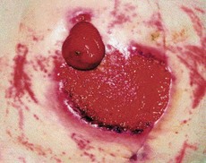
• Examination. Characteristic features of ulcer: tenderness, necrosis, irregular violaceous border, undermined, rolled edges
• Skin biopsy. Elliptical incisional biopsy preferable to punch biopsy (include inflamed border and ulcer edge at a depth that includes subcutaneous fat). The inflamed border is examined for routine histology (haematoxylin and eosin) and microorganisms (Gram, methenamine silver and Fite stains). The edge of ulcer is cultured in appropriate media to detect bacteria, fungi and atypical myobacteria
• Laboratory investigations. Complete blood count, ESR, blood chemistry (liver and kidney function tests), protein electrophoresis, coagulation panel (including anti-phospholipid antibody screening), ANCA, cryoglobulins