[level-membership-for-dermatology-category]
165
Pyoderma gangrenosum
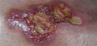
Large asymmetric ulceration with yellow fibrinous debris centrally and bright-red to violaceous inflamed and undermined border peripherally, characteristic of pyoderma gangrenosum.
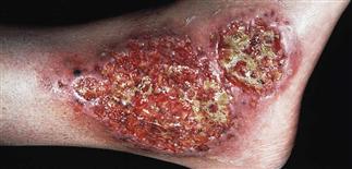
The pustule or papule will break down, evolving into erosions/ulcers with multiple crater-like holes. These ulcerated plaques consist of small fistulous tracks from which drainage occurs.
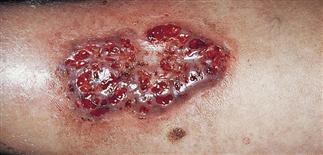
The legs are the most common site. New lesions form at sites of injury in a phenomenon called pathergy. Multiple lesions are typical.
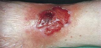
Differentiation from other diseases causing ulcers is sometimes very difficult. Malignancies may present as ulcerations with exactly the same appearance; biopsy uncertain.
DESCRIPTION
A necrotic, painful, non-infective, inflammatory skin disease characterized by rapidly enlarging, painful ulcerations on the legs.
HISTORY
• Ulcerative pyoderma gangrenosum lesions may begin spontaneously or at the site of trauma. • Most cases occur between 25 and 55. • The disease is commonly associated with inflammatory bowel disease (Crohn disease and ulcerative colitis); pyoderma gangrenosum patients should be evaluated for inflammatory bowel disease. • It is less commonly associated with rheumatoid arthritis, chronic active hepatitis, IgG monoclonal gammopathy, myelodysplasia, paraproteinemia, myeloid leukemias, and myeloma. • Pathergy (enlargement of the lesion) with trauma is typical in pyoderma gangrenosum. Postsurgical pyoderma gangrenosum may masquerade as wound infection, poor healing or dehiscence.
PHYSICAL FINDINGS
• Most common sites are lower legs, buttocks, abdomen. • Lesions begin as a very tender, red or dusky macule, papule, pustule, nodule, or bulla. The initial lesion is often described as a pustule or inflamed red nodule that then ulcerates, forming an extremely painful, sharply marginated, violaceous-bordered ulcer with a purulent base. • The edge of the ulcer is characteristically elevated or undermined. • Expansion occurs rapidly. • Multiple lesions are usually present. Eventually, lesions may coalesce into larger ulcers with crater-like holes with small fistulous tracks. • Lesions gradually heal with irregular, cribiform, or stellate scarring.
TREATMENT
• In general, many patients with pyoderma gangrenosum have a chronic relapsing course, even with adequate treatment. • Although eradication of the disease is the ultimate treatment goal, most regimens aim to reduce dependence on steroid therapy. • Hospitalization may be necessary for severe cases. • Analgesia is often required. • Most patients require combination therapy with multiple immunosuppressants. • High-dose systemic oral (1–3 mg/kg) or i.v. (1 g/day) steroids for 3–10 days should be administered initially to try to quickly suppress the immune response. • Ciclosporin (3–5 mg/kg) may be the best short term, non-steroidal immunosuppressant for pyoderma gangrenosum, and can be used in combination with systemic steroids in the appropriate patient. In recent years, tumor necrosis factor-alpha inhibitors, such as infliximab, adalimumab and etanercept have been used to treat pyoderma gangrenosum with dramatic success, and may allow reduction or complete cessation of systemic steroids. • Dapsone, mycophenolate mofetil, azathioprine, minocycline, clofazimine, and thalidomide have been used anecdotally with variable results. • Intravenous immunoglobulin is yet another drug used for pyoderma gangrenosum that is poorly studied but has been successful in some patients. • Intralesional steroids (Kenalog 10–40 mg/cc) can be used for small or single lesions, but care must be taken not to injure the skin. • Local compresses with Burow’s solution or silver nitrate 2 or 3 times daily. • Superpotent topical steroids and tacrolimus (Protopic) 0.1% ointment has been used with some success.
[/level-membership-for-dermatology-category][not-level-membership-for-dermatology-category]
165
Pyoderma gangrenosum
Large asymmetric ulceration with yellow fibrinous debris centrally and bright-red to violaceous inflamed and undermined border peripherally, characteristic of pyoderma gangrenosum.
The pustule or papule will break down, evolving into erosions/ulcers with multiple crater-like holes. These ulcerated plaques consist of small fistulous tracks from which drainage occurs.
The legs are the most common site. New lesions form at sites of injury in a phenomenon called pathergy. Multiple lesions are typical.
Differentiation from other diseases causing ulcers is sometimes very difficult. Malignancies may present as ulcerations with exactly the same appearance; biopsy uncertain.
[/not-level-membership-for-dermatology-category]
