Pulmonary Vasculitis and Hemorrhage

Published on 23/05/2015 by admin

Filed under Pulmolory and Respiratory

Last modified 22/04/2025

Print this page

rate 1 star rate 2 star rate 3 star rate 4 star rate 5 star
Your rating: none, Average: 0 (0 votes)

This article have been viewed 1235 times

Chapter 59 Pulmonary Vasculitis and Hemorrhage

Pulmonary Vasculitis

Pulmonary vasculitis is defined as inflammation of vessels in the lung of different sizes—pulmonary arteries, veins, and capillaries, as well as bronchial arteries. It usually is only one manifestation of a systemic disorder caused by any of a variety of immunologic mechanisms. Moreover, not all respiratory symptoms occurring in patients with vasculitis are caused by inflammation of pulmonary vessels.

Primary vasculitis is separated from secondary vasculitis. The primary systemic vasculitides are a heterogeneous group of syndromes, of unknown etiology, that share a clinical response to immunosuppressive therapy. Their wide spectrum of frequently overlapping clinical manifestations is defined by the size and location of the affected vessels as well as by the nature of the inflammatory infiltrate. Secondary vasculitis may occur in the context of a well-defined underlying disorder or may be attributable to a specific cause such as collagen vascular disease, infection, or therapeutic or illicit drug use.

Most classification schemes and definitions of the vasculitides are based on the size of the most prominently affected vessels. Definitions, nomenclature, and classification schemes have been and remain subject to change. The terms used in this chapter adhere to the nomenclature and definitions put forth by the Chapel Hill consensus conference in 1992.

Small Vessel Vasculitis

Granulomatosis with Polyangiitis and Microscopic Polyangiitis

Definitions and Nomenclature

GPA is characterized by necrotizing granulomatous inflammation involving the respiratory tract and necrotizing vasculitis affecting small to medium-sized vessels. The most commonly affected vessels are capillaries, venules, arterioles, and arteries, but the wall of the aorta also may rarely be affected by necrotizing granulomatous inflammation—hence the term polyangiitis. Histopathologic documentation of granulomatous involvement of the respiratory tract is not explicitly required. Radiologic evidence or clinical examination findings highly predictive of granulomatous pathology are sufficient. Consequently, the diagnosis of GPA depends on a correlation of clinical, histopathologic, and serologic features. MPA is defined as necrotizing vasculitis with few or no immune deposits, affecting small vessels including capillaries, venules, or arterioles (polyangiitis). Necrotizing arteritis involving small and medium-sized arteries may be present. Necrotizing glomerulonephritis is very common; pulmonary capillaritis resulting in alveolar hemorrhage occurs frequently. The vasculitis of MPA is indistinguishable from that of GPA, and substantial clinical overlap has been observed. For these reasons, the therapeutic approach to patients with GPA and to those with MPA is governed by the same principles, and most clinical studies and therapeutic trials have combined both diseases.

Histopathologic features that define GPA and separate it from MPA include discrete or confluent necrotizing granulomatous inflammation with vasculitis. Fibrinoid necrosis, microabscesses, focal vasculitis, thrombosis, and fibrous obliteration of the vascular lumen also may be seen. Giant cells are a hallmark of the necrotizing granulomatous inflammation of GPA. Atypical and rare histopathologic features of GPA include organizing pneumonia, bronchocentric inflammation, and a marked number of eosinophils in the inflammatory infiltrates.

For treatment stratification, clinical disease activity is categorized as “limited disease” or “severe disease.” Severe disease is either life-threatening or associated with involvement of an organ with potentially irreversible loss of function. Consequently, patients with any of the following disease manifestations should be labeled as having severe disease: alveolar hemorrhage, glomerulonephritis, eye involvement (except mere episcleritis), or nervous system involvement including sensorineural hearing loss. Limited disease includes essentially all patients who have nonsevere disease. The designation limited disease as used in the United States comprises what European investigators have referred to as “early-systemic disease” and “localized disease.” Even though this separation is not based on well-defined biologic distinctions, most disease manifestations leading to the categorization as severe disease are caused by capillaritis. By contrast, most symptoms leading to the classification as limited disease are the result of necrotizing granulomatous inflammation. Patients with limited GPA have a more protracted disease course, a greater likelihood of experiencing a disease relapse after a period of remission, and a higher prevalence of destructive upper respiratory tract disorders (e.g., saddle nose deformity). Tracheobronchial disease involvement also is a feature of GPA not shared with MPA; it may present unique treatment challenges (as discussed later on).

Diagnostic Evaluation

The diagnostic evaluation of patients suspected of having GPA or MPA should include imaging of the chest, pulmonary function testing if the patient has any respiratory symptoms or abnormalities on the chest radiograph, measurements of erythrocyte sedimentation rate, C-reactive protein, complete blood count, serum chemistry panel, urine analysis and microscopy, and testing for ANCA.

Respiratory symptoms usually are associated with unilateral or bilateral radiographic abnormalities including infiltrates, nodules, or mass lesions, which may or may not cavitate. The nodules range in size from a few millimeters to several centimeters across. Solitary nodules also may occur. Unusual manifestations include lymphadenopathy, lobar consolidation, and large pleural effusions.

Tracheobronchial lesions are common and may be asymptomatic or mistaken for evidence of asthma. Pulmonary function testing including inspiratory and expiratory flow-volume loops may provide important clues to the presence of airway narrowing and should be part of the initial evaluation. Bronchoscopic inspection of the tracheobronchial tree is recommended for patients with unexplained respiratory symptoms, abnormalities on pulmonary function tests, or radiographic abnormalities.

Patients with GPA or MPA exhibit variable degrees of elevation of erythrocyte sedimentation rate or C-reactive protein levels. If GPA or MPA is suspected, urine analysis and microscopy and serum creatinine determination are crucial, because early renal involvement may be clinically silent yet can progress rapidly.

In GPA and MPA, ANCA that cause a cytoplasmic immunofluorescence pattern (c-ANCA) on ethanol-fixed neutrophils are caused by the reaction of antibodies with proteinase 3 (PR3) (i.e., PR3-ANCA). By contrast, a variety of antibodies can cause a perinuclear immunofluorescence pattern (p-ANCA) on ethanol-fixed neutrophils. Only those that also react with myeloperoxidase (MPO) are of interest in the context of ANCA-associated vasculitis. Maximal diagnostic accuracy of ANCA testing requires corroboration of a positive target antigen–specific test result (PR3-ANCA or MPO-ANCA) by immunofluorescence assay, or vice versa. Only the PR3-ANCA with c-ANCA combination and the MPO-ANCA with p-ANCA combination are sensitive and specific for ANCA-associated vasculitis.

The clinical utility—as reflected in positive and negative predictive values—of ANCA testing for GPA and MPA is critically dependent on the pretest probability of the disease in the patient tested, as well as on the analytic accuracy of the test method. If applied in patients with clinical features indicating a high pretest probability of GPA or MPA, ANCA testing has a very high positive predictive value. However, occasional false-positive ANCA test results have been reported in a variety of infections. Particularly, subacute bacterial endocarditis may represent a diagnostic dilemma as it may mimic small-vessel vasculitis clinically and has been reported with c-ANCA/PR3-ANCA. With other infections reported with ANCA, either the right pairing of immunofluorescence test results with its corresponding antigen specificity is lacking or their clinical features are distinct from those of GPA or MPA. In patients undergoing evaluation for necrotizing glomerulonephritis with or without alveolar hemorrhage, ANCA may occur in conjunction with anti–glomerular basement membrane (anti-GBM) antibodies. These ANCA usually are of the MPO-ANCA variety. The presence of anti-GBM antibodies seems to determine the prognosis in such double-positive patients.

Most patients with severe GPA or MPA demonstrate a positive result on ANCA testing (for a sensitivity greater than 95%), but up to 30% of patients with limited GPA may not have detectable ANCA. Despite the recognized association between ANCA titers and disease activity, changes in ANCA levels do not reliably predict the disease activity in individual patients. Therefore, serial titers of ANCA should not be used to plan long-term therapy.

Therapy

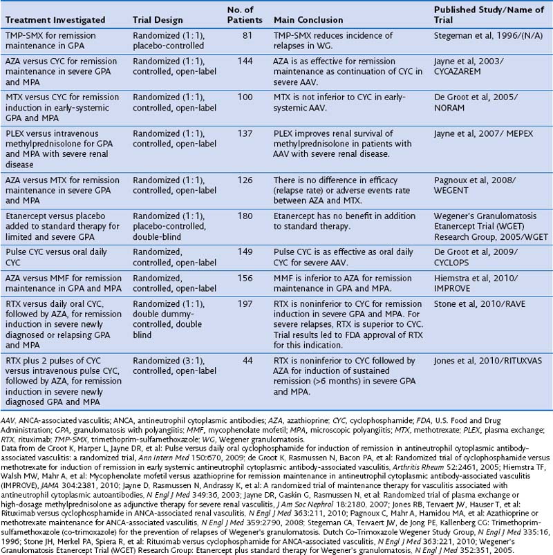
Standard therapy for GPA and MPA currently follows the same basic principles. Methotrexate (MTX) at a dose of up to 25 mg once a week in combination with oral prednisone is considered the standard of care for patients with limited GPA. However, only one prospective randomized trial has compared MTX and cyclophosphamide (CYC) for remission induction in such patients (Table 59-1). The trial, conducted by the European Vasculitis Study Group (EUVAS), showed that MTX is noninferior to CYC for remission induction, but the side effects were less frequent and less severe. The trial also documented that early discontinuation of immunosuppression in patients with ANCA-associated vasculitis is fraught with a high relapse rate. The largest reported group of patients with limited Wegener’s granulomatosis (WG) was treated with MTX for remission induction in the context of the Wegener’s Granulomatosis Trial (WGET). More than 90% of patients achieved remission with this regimen, and more than 70% achieved a sustained remission (lasting longer than 6 months). These rates are equivalent to those achieved with CYC in severe disease, as discussed next.

CYC at a dose of 2 mg/kg/day in combination with prednisone has been the standard of care for patients with severe GPA or MPA until recently. In contrast with the original regimen introduced by Fauci 40 years ago, the current consensus is to limit the duration of CYC therapy to the first 3 to 6 months of remission induction.

Once remission has been induced and the prednisone taper is well under way, CYC should be switched to either azathioprine (AZA), preferred in patients with renal involvement and any degree of renal insufficiency, or MTX. The first option is supported by the results of a randomized trial showing that AZA is as good as CYC for remission maintenance to 18 months. Another randomized controlled trial has shown that MTX and AZA are equivalent for remission maintenance. By contrast, a recent randomized controlled trial that compared mycophenolate mofetil (MMF) with AZA for remission maintenance has shown that MMF is inferior to AZA for this purpose. Thus, the use of MMF for remission maintenance can be supported only for patients who have failed MTX and AZA, or who have contraindications to both agents. The WGET study, in which MTX was used for remission maintenance, confirmed that long-term remission remains an elusive goal for many patients, because remission was maintained in less than half of the patients.

Whenever CYC is used for remission induction, consideration should be given to the patient’s fertility. Young men should be offered sperm banking before therapy is initiated. If time allows, ovarian protection should be offered to young women, in addition to minimizing the cumulative exposure as much as possible.

One randomized controlled trial has evaluated a regimen of intravenous pulse application of CYC compared with daily oral use. The pulse regimen was noninferior to the oral application of CYC for remission, and the frequency of leukopenia, but not infection, was lower. Even though the trial was not powered to detect a difference in relapses, however, the relapse rate was higher after remission induction with the intermittent pulse regimen than with the oral application of CYC.

A metaanalysis of earlier cohort studies had also indicated that intravenous pulse CYC therapy may be safer because of a lower cumulative dose, but a higher relapse rate also was observed after discontinuation. In my own experience, intravenous CYC is best avoided in the intensive care unit setting. However, its use is preferred over oral CYC in patients with questionable compliance, in young women with fertility issues, and in patients who have gastrointestinal problems with oral CYC application.

The four-decade-old standard use of CYC for remission induction in patients with severe GPA and MPA recently has been challenged by the results of two randomized controlled trials. The RAVE (rituximab for ANCA-associated vasculitis) trial was a randomized double-blind, double-placebo-controlled, multicenter trial that compared oral CYC (2 mg/kg/day) to rituximab (RTX) (375 mg/m2 body surface area/week for 4 weeks) for remission induction in severe GPA or MPA in 197 patients. Once remission was achieved between 3 and 6 months, patients randomized to receive CYC were switched to AZA for remission maintenance for 18 months, whereas patients in the original RTX group then received placebo. No difference was observed in rates of achieving remission at the end of 6 months and maintaining remission at 18 months between the two treatment arms. Among the 101 patients who entered the trial with severe relapsing disease, however, RTX proved superior to CYC. The results of the RAVE trial led to approval by the U.S. Food and Drug Administration (FDA) of RTX for remission induction in severe GPA and MPA.

Another randomized controlled open-label trial conducted in 44 patients with newly diagnosed severe ANCA-associated vasculitis with active renal disease, RITUXVAS (Rituximab versus Cyclophosphamide for ANCA-Associated Renal Vasculitis), showed results complementary to those of the RAVE trial. In the RITUXVAS trial, patients were randomized 3 : 1 to receive RTX (together with two pulses of CYC) compared with standard intravenous pulse CYC therapy followed by oral AZA. The primary outcome was sustained remission (of more than 6 months’ duration) at month 12. No difference between the treatment groups was found (RTX 76% versus CYC 82%).

Mycophenolate mofetil (MMF) may represent an alternative to CYC (and RTX) for patients with MPA who have MPO-ANCA and mild renal disease (as defined by creatinine levels less than 3.5 mg/day) and no other life- or organ-threatening disease manifestation. This claim is supported by data from a randomized controlled trial in 35 patients from China comparing oral MMF (1.5 to 2 g/day) with intravenous CYC (0.75 to 1.0 g/m2 once monthly). In addition, all patients received intravenous methylprednisolone bolus therapy (0.5 g/day for 3 days) followed by oral prednisone (0.6 to 0.8 mg/kg for 4 weeks tapered by 5 mg every week to reach 10 mg/day). These regimens demonstrated equivalent efficacy, but MMF was better tolerated than CYC. A prospective pilot trial in 17 patients conducted at the Mayo Clinic achieved similar results.

For some patients with Wegener granulomatosis (WG) and MPA, the combination of glucocorticoids and cyclophosphamide may not be sufficient to induce a remission quickly. Plasma exchange should be considered early in patients who present with rapidly progressive glomerulonephritis and renal failure, as well as in patients who present with diffuse alveolar hemorrhage (DAH). The use of plasma exchange currently is supported by two studies: The MEPEX (Methylprednisolone Versus Plasma Exchange) trial in 156 patients who presented with a serum creatinine level of 5.5 mg/dL or greater was conducted to compare three pulses of intravenous methylprednisolone with 2 weeks of plasma exchange (seven exchanges at rate of 60 mL/kg) in addition to standard therapy for severe disease (oral prednisone and CYC). Plasma exchange was superior to methylprednisolone with respect to renal recovery. A single-center cohort study of 20 patients presenting with alveolar hemorrhage described 100% survival when plasma exchange was added to standard immunosuppressive therapy. If alveolar hemorrhage is uncontrolled despite aggressive immunosuppressive therapy and plasma exchange, the use of recombinant activated factor VII may be considered.

The term refractory disease commonly is used to describe persistent disease activity in patients on the maximal tolerated dose of CYC or in those with contraindications to the use of CYC. A variety of agents have been proposed for use in addition to or instead of the failing regimen in such patients. Over the past decade, RTX has emerged as the agent of choice for treatment of refractory disease, based on over 20 case series and cohort studies comprising more than 200 patients.

Infliximab also has been used in small case series and uncontrolled trials. Of note, however, infliximab therapy was associated with a high frequency of infections with bad outcomes. The WGET showed that etanercept had no effect on remission induction or maintenance when used in addition to standard therapy. Moreover, significantly more malignancies were observed among patients who had received CYC in the etanercept treatment group. Accordingly, the use of etanercept in patients who have received or are receiving CYC is contraindicated. Consequently, no convincing rationale has emerged for the use of antitumor necrosis factor-α (anti-TNF) therapy in patients with GPA.

Several reports have described the use of other agents in patients with refractory disease. Antithymoglobulin has some efficacy but significant side effects. Dispergualine is an agent available in Japan and Europe but not in the United States.

The management of large airway involvement in GPA may present unique challenges. Subglottic stenosis may necessitate dilation procedures paired with local injection of long-acting glucocorticoids with or without mitomycin C, and stenosis of the large airways may warrant bronchoscopic interventions, including dilation by rigid bronchoscopy, yttrium-aluminum-garnet (YAG) laser treatment, and placement of silicone airway stents.

In patients with GPA and MPA, significant morbidity and mortality related to organ damage is not only caused by the disease itself but also attributable to treatment toxicities. Permanent complications have been reported to occur in 86% of patients from GPA itself, including end-stage renal disease, chronic pulmonary dysfunction, diminished hearing, destructive sinus disease, saddle nose deformities, proptosis, and blindness. Among the treated patients, 42% experienced permanent treatment-related problems including chemical (drug-induced) cystitis, osteoporotic fracture, bladder cancer, myelodysplasia, and avascular necrosis. In the 180 patients in the WGET, which reflects more recent standard practice, damage that occurred despite (or because of) therapy included visual impairment, hearing loss, nasal blockage, pulmonary fibrosis, hypertension, renal insufficiency, peripheral neuropathy, gonadal failure, and diabetes mellitus. Only 11% of the enrolled patients did not exhibit a single point on the vascular damage index (which was developed from 61 various items) after 1 year of study enrollment. The WGET investigators also concluded that patients with limited GPA are at a higher risk for GPA-related damage than are those with severe disease.

In the same cohort of 180 patients with GPA, the incidence rate of venous thromboembolism was found to be high in comparison with reported rates in the general population, patients with lupus, and patients with rheumatoid arthritis. This increased risk for thromboembolic disease also has been documented for the other ANCA-associated vasculitides. A high frequency of echocardiographic abnormalities attributable to GPA and associated with an increased mortality was observed in a study of 85 patients with confirmed GPA. Lesions directly related to GPA included regional wall motion abnormalities not matching coronary distributions, left ventricular systolic dysfunction with decreased ejection fraction, pericardial effusions, valvulitis, left ventricular aneurysm, and a large intracardiac mass. Evaluation of unexplained dyspnea in patients with GPA should therefore include echocardiography.

Eosinophilic Granulomatosis With Polyangiitis (Churg-Strauss Syndrome)

The Chapel Hill Consensus definition for EGPA is “eosinophil-rich and granulomatous inflammation involving the respiratory tract, and necrotizing vasculitis affecting small to medium-sized vessels, and associated with asthma and eosinophilia.” EGPA is included among the ANCA-associated vasculitides, but only 40% to 70% of patients with active EGPA have detectable ANCA. EGPA is distinguished from GPA and MPA primarily by a high prevalence of asthma and peripheral blood and tissue eosinophilia.

Three distinct phases of the disease have been described: The first is a prodromal allergic phase with asthma. This phase may last for a number of years. Second is an eosinophilic phase with prominent peripheral and tissue eosinophilia. This phase also may last a number of years, and the manifestations may remit and recur over this period. The differential diagnosis in this phase of the disease includes parasitic infection and chronic eosinophilic pneumonia. The third vasculitic phase consists of systemic vasculitis and may be life-threatening. The three phases are not seen in all patients, do not necessarily occur in this order, and may even be concurrent. However, asthma usually predates vasculitic symptoms by a mean of 7 years (range, 0 to 61 years). “Formes frustes” of EGPA also have been described with eosinophilic vasculitis and/or eosinophilic granulomas in isolated organs without evidence of systemic disease.

Pulmonary parenchymal involvement occurs in 38% of patients. Transient alveolar-type infiltrates are most common. These have a predominantly peripheral distribution and are indistinguishable from infiltrates seen in chronic eosinophilic pneumonia. Occasionally, nodular lesions may be seen in EGPA. In contrast with GPA and MPA, alveolar hemorrhage is exceedingly rare in EGPA. Renal involvement is less prominent than in GPA or MPA and does not generally lead to renal failure. By contrast, peripheral nerve involvement, typically in the form of mononeuritis multiplex, is more frequent. Skin, heart, central nervous system, and abdominal viscera also may be involved.

The classic histopathologic picture consists of necrotizing vasculitis, eosinophilic tissue infiltration, and extravascular granulomas. Not all features are found in every case, however, and they are not pathognomonic for the condition. In particular, the finding of a “Churg-Strauss granuloma” on skin biopsy should not lead to confusion with the diagnosis of EGPA. This type of necrotizing extravascular granuloma may be seen in EGPA as well as in other systemic autoimmune diseases including GPA and rheumatoid arthritis.

If ANCA are present, they usually are p-ANCA reacting with MPO. The ANCA status appears to correlate with disease activity. Recent evidence points to a more vasculitic disease phenotype in the presence of ANCA, but this association was not confirmed by all studies. Substantial overlap of organ manifestations between patients with EGPA who are ANCA-positive and those who are ANCA-negative is characteristic.

In recent years, significant attention has been devoted to EGPA detected in patients taking leukotriene receptor antagonists. Available case studies and limited population-based incidence estimates suggest that these agents may lead to unmasking of vasculitic symptoms in asthmatic persons by allowing dose reductions or discontinuation of oral glucocorticoid therapy. There is no evidence suggesting that these agents directly cause the disease.

The prognosis with EGPA is better than that with GPA or MPA, because the overall mortality is lower and not significantly different from that for the normal population. Most deaths are secondary to cardiac involvement.

Systemic glucocorticoids remain the mainstay of therapy. No clinical trials have been conducted to provide clear guidance for therapy for EGPA. It seems most appropriate to treat EGPA according to the principles applied to the management of ANCA-associated vasculitis. Accordingly, CYC should be added to glucocorticoids for remission induction in all patients with disease manifestations that threaten either life or function of a vital organ—particularly those with central or peripheral nerve involvement, glomerulonephritis, heart involvement, or alveolar hemorrhage. MTX, AZA, and MMF have been used as glucocorticoid-sparing agents in less severe disease and for remission maintenance. Refractory disease and that dominated by difficult-to-control eosinophilic inflammation have been reported to respond to interferon-α therapy. Of note, however, continued long-term interferon-α therapy may be necessary, and this treatment carries the risk of substantial toxicity. RTX also has been used successfully in EGPA, particularly ANCA-positive patients with renal disease, but the data are still limited, and use of RTX instead of CYC for patients with severe EGPA cannot be recommended. Anti-interleukin 5 (IL-5) also has recently been reported as a promising agent for treatment of EGPA. The glucocorticoid-sparing effect of anti-IL5 in EGPA has been documented in two separate prospective pilot studies.

Large Vessel Vasculitis

Giant Cell Arteritis

Giant cell arteritis (GCA), also known as temporal arteritis, cranial arteritis, and granulomatous arteritis, is a generalized inflammatory disorder involving large and medium-sized arteries. It is the most common form of vasculitis in the Northern Hemisphere and affects predominantly elderly patients. The typical features of the disease include new-onset headache, a palpably tender or nodular temporal artery with decreased pulsation, and elevated erythrocyte sedimentation rate (ESR). The clinical illness emerges gradually, with the development of nonspecific systemic signs and symptoms such as low-grade fever, malaise, and weight loss. Headache, variable but often severe, is the most common symptom in GCA. Amaurosis fugax is observed in 20% of patients, and visual loss in 10%. Granulomatous inflammation of the vessel wall is found in 60% of temporal artery biopsy specimens. The aorta also may be affected by the disease, and GCA should be considered as the cause of thoracic aortic aneurysm occurring in elderly patients.

Respiratory symptoms have been reported in up to 25% of patients but rarely require management by a respiratory physician. However, respiratory symptoms may be the initial presentation of giant cell arteritis. Therefore, GCA should be considered in any elderly patient with new-onset cough, hoarseness, or throat pain without other identifiable cause, and the ESR should be measured in such patients. Cough, hoarseness, and throat pain usually resolve promptly with glucocorticoid therapy. Isolated cases of GCA with pleural effusion or multinodular pulmonary lesions also have been reported. Such findings are difficult to interpret in this setting, and GPA should be considered in the differential diagnosis, because it also may involve the temporal artery. Therapy of GCA continues to be based on the use of glucocorticoids without proven alternative. The glucocorticoid-sparing role of MTX for GCA remains controversial.

Takayasu Arteritis

Takayasu arteritis is a large vessel vasculitis affecting predominantly the aorta and its major branches in young patients. Takayasu arteritis also has been called pulseless disease, aortic arch syndrome, or reverse coarctation. The disease affects mostly young adult women. It is not limited to patients of Asian descent. Early disease manifestations include constitutional symptoms, low-grade fever, and arthralgias. Variable pulses of the extremities and claudication of affected vascular territories are typical. Renovascular hypertension, pulmonary hypertension, and ischemia of affected organs may be the more disabling complications of this chronically relapsing disease.

Pulmonary complications are the result of a unique arteriopathy predominantly involving the large and medium-size pulmonary vessels. Progressive defects in the outer media of the arteries and ingrowth of granulation tissue–like capillaries associated with thickened intima and subendothelial smooth muscle proliferation lead to pulmonary artery stenoses and occlusion, as well as pulmonary hypertension, in up to half of all patients. The inflammatory infiltrate of the vessel wall is predominantly lymphoplasmocytic, with variable amounts of giant cells. The involvement of pulmonary arteries is common but often asymptomatic. It is detectable by conventional angiography, perfusion scan, or magnetic resonance angiography (MRA). Chest radiographs often are normal in appearance, but computed tomography (CT) may show areas of low attenuation as a result of regional hypoperfusion, subpleural reticulolinear changes, and pleural thickening. Fistula formation between pulmonary artery branches and bronchial arteries, as well as nonspecific inflammatory interstitial lung disease, also has been reported.

Therapy for Takayasu arteritis consists primarily of immunosuppression with glucocorticoids. Other immunosuppressive agents including MTX are used in conjunction with glucocorticoids for remission induction and as glucocorticoid-sparing agents for remission maintenance. Unfortunately, many patients relapse when the glucocorticoid dose is reduced below 15 mg daily. The use of antitumor necrosis factor-α agents may be beneficial for patients with disease refractory to standard therapy. Vascular bypass procedures may restore perfusion to areas affected by severe arterial stenoses, but the improvement is only temporary.

Other Vasculitis Syndromes

Behçet Disease

Behçet disease is a rare, chronically relapsing systemic inflammatory disorder characterized by aphthous oral ulcers and at least two or more of the following: aphthous genital ulcers, uveitis, cutaneous nodules or pustules, and meningoencephalitis. Reported prevalence of the disease is 1 : 16,000 in Japan and 1 : 20,000 in the United States. A strong association with the major histocompatibility complex antigen HLA-B51 has been recognized. The mean age of patients at the onset of Behçet disease is 35 years. Most studies have reported a predominance of men with the disease over women. Respiratory manifestations are common in Behçet disease and include cough, hemoptysis, chest pain, and dyspnea. Hemoptysis often is massive and fatal. The vasculitis of Behçet disease is immune complex–mediated and may involve vessels of all sizes. Secondary thrombosis with major venous occlusion can occur. Thrombosis may not be preventable in Behçet disease by anticoagulation, but aspirin 80 mg/day has been advocated. Destruction of the elastic lamina of pulmonary arteries causing aneurysm formation, secondary erosion of bronchi, and arterial-bronchial fistulas may result in massive hemoptysis. CT or MRA can be used to detect pulmonary artery aneurysms. Recurrent pneumonia as well as bronchial obstruction secondary to mucosal inflammation also has been described.

Therapy of the underlying disease consists of immunosuppression. Prednisone alone may not be sufficient to control the vasculitis. The addition of other drugs, such as colchicine, chlorambucil, MTX, cyclosporine, or AZA, is recommended. The use of biologic agents, in particular anti-TNF agents, also has been reported recently. The addition of AZA or CYC to glucocorticoids may result in resolution of pulmonary aneurysms. Once pulmonary arteritis has been identified, anticoagulation should be avoided. The prognosis of pulmonary involvement is poor. About one third of patients die within 2 years of developing pulmonary involvement, most from fatal pulmonary hemorrhage. Embolization therapy may be used for treatment and prevention of hemorrhage from pulmonary artery aneurysms.

Henoch-Schönlein Purpura

Henoch-Schönlein purpura (HSP), also known as anaphylactoid purpura, allergic purpura, or, as more recently proposed, IgA-associated disease, is a syndrome characterized by acute-onset purpura, arthritis, colicky abdominal pain, and nephritis. Histopathologic findings include acute arteriolitis and venulitis in the superficial dermis and the bowel. Proliferative and necrotizing glomerulonephritis usually is mild. A similar type of renal lesion is seen in patients with infective subacute bacterial endocarditis, GPA, MPA, systemic lupus erythematosus (SLE), and anti-GBM disease.

Immunofluorescence microscopy shows large deposits of IgA in the skin and kidney. Although HSP is more common in children (mean age of patients, 17 years), adults also may be affected. Palpable purpuric lesions, which usually are distributed over the buttocks and lower extremities, and fever are generally the first clinical manifestations. The purpura may precede, accompany, or follow onset of arthralgias and abdominal colic. The triad of purpura, arthritis, and abdominal pain is present in approximately 80% of patients. Joint involvement typically is monoarticular and transient, involves the large joints, and causes pain that is out of proportion to the objective evidence of synovitis. Peritonitis and melena are common.

Pulmonary manifestations of HSP are rare. Only 26 cases have been reported to date, and capillaritis has been documented histopathologically only in a minority of them. IgA deposits along the pulmonary capillary walls, analogous to those found in vessels of the skin and glomeruli of affected kidneys, are pathognomonic for HSP.

Alveolar Hemorrhage Syndromes

Diffuse hemorrhage into the alveolar spaces often is referred to as diffuse alveolar hemorrhage (DAH) syndrome. The clinical course of DAH is unpredictable: It may progress rapidly to respiratory failure, and it is always potentially life-threatening. Consequently, this entity should be considered in the differential diagnostic evaluation of any patient with alveolar infiltrates on chest radiograph. The symptoms of DAH are nonspecific. Patients usually seek care because of dyspnea and cough, possibly associated with fever. Diffuse alveolar infiltrates on the chest radiograph constitute the initial diagnostic hallmark. Depending on the severity of the disease process at the time of evaluation, anemia and hypoxemia may be prominent. Hemoptysis is a common presenting manifestation of DAH but may be absent in up to one third of patients with DAH.

DAH can result from a variety of underlying or associated conditions that cause a disruption of the alveolar-capillary basement membrane integrity. Mechanisms leading to DAH include immunologic inflammatory conditions or agents causing immune complex deposition or capillaritis (e.g., anti-GBM disease or Goodpasture syndrome, SLE, ANCA-associated vasculitis), direct chemical or toxic injury (e.g., from toxic or chemical inhalation, abciximab use, all-trans-retinoic acid, trimellitic anhydride, or smoked crack cocaine), physical trauma (e.g., pulmonary contusion), and increased vascular pressure within the capillaries (e.g., mitral stenosis or severe left ventricular failure).

DAH is best confirmed by bronchoalveolar lavage (BAL). Progressively more bloody return fluid indicates alveolar origin of the blood; the presence of greater than 20% hemosiderin-laden macrophages among the total number of alveolar macrophages recovered by BAL is reported to indicate alveolar hemorrhage, even in the absence of ongoing active bleeding. Pulmonary alveolar hemorrhage is significantly associated with the following: thrombocytopenia (less than 50,000 cells/µL), other abnormal coagulation variables, renal failure (creatinine concentration of 2.5 mg/dL or greater), and a history of heavy smoking. The clinical approach to the patient presenting with DAH is aimed at rapid identification of the underlying cause and prompt implementation of appropriate therapy.

Exposure to inhalational toxins such as trimellitic anhydride or pyromellitic dianhydrate, drug abuse including crack cocaine abuse, and tobacco smoke should be identified. The past medical history also will uncover preexisting comorbid conditions, factors, and agents that can cause DAH, including mitral stenosis, coagulation disorders, recent bone marrow or hematopoetic stem cell transplantation, preexisting autoimmune disorders, and various therapeutic drugs. Similarly, the initial physical examination should include a careful search for signs of comorbidity and possible systemic autoimmune disorders.

Initial blood and urine testing should screen for other organ involvement, particularly kidney involvement (complete blood count, blood chemistry profile, urine analysis and microscopy), and determine the current coagulation status (activated partial prothrombin time [aPTT], international normalized ratio [INR]). Baseline markers of inflammation (erythrocyte sedimentation rate and C-reactive protein) are helpful to monitor subsequent responses to therapy. At the same time, specific serologic testing for potential underlying systemic disease processes should be initiated. This includes testing for ANCA, anti-GBM antibodies, antinuclear antibodies, anti–double-stranded DNA antibodies, rheumatoid factor, and antiphospholipid antibodies, as well as determination of complement and creatine kinase levels.

The decision to obtain a biopsy specimen needs to be considered carefully, weighing the risks of the biopsy procedure, the likelihood of obtaining a diagnostic piece of tissue, the likelihood of the biopsy findings to alter the therapeutic approach, and the risks associated with the chosen therapy.

The DAH syndromes can be broadly separated into those in which DAH is caused by or associated with pulmonary capillaritis and those in which pulmonary capillaritis is lacking (bland histologic pattern). Pulmonary capillaritis refers to the specific histopathologic finding of alveolar wall infiltration with inflammatory cells centered on capillary walls and small veins. The inflammatory cells are predominantly neutrophils, but eosinophils or monocytes also may be encountered. Capillaritis usually causes fibrinoid necrosis of alveolar and vessel walls and may culminate in the destruction of the underlying lung architecture. The interstitial infiltration by neutrophils seen in the context of capillaritis needs to be distinguished from the predominant intraalveolar neutrophilic infiltration commonly associated with active infections. Another hallmark of capillaritis is the presence of pyknotic cells and nuclear fragments from neutrophils undergoing apoptosis. This feature, referred to as leukocytoclasis, allows the distinction of true capillaritis from neutrophil margination related to surgical trauma, which can simulate capillaritis.

Most of the syndromes associated with pulmonary capillaritis leading to DAH have been discussed earlier in the chapter. A few unique syndromes or conditions that also may be associated with DAH are described next.

Anti–glomerular Basement Membrane Antibody Disease (Goodpasture Syndrome)

Anti–glomerular basement membrane antibody (anti-GBM) disease, or Goodpasture syndrome, is a rare autoimmune disease characterized by the presence of autoantibodies directed against the NC1-domain of the alpha-3 chain of basement membrane collagen type IV. This epitope is accessible only to autoantibodies in the basement membranes of kidneys and lungs. DAH occurs in about half of the patients with anti-GBM disease. An additional inhalational injury, particularly smoking, is thought to be required for development of the pulmonary manifestation of this disease. Isolated alveolar hemorrhage in the absence of renal disease is rare in anti-GBM disease. Circulating autoantibodies to basement membrane are detectable in the serum, but the diagnosis of anti-GBM disease hinges on the histopathologic documentation of linear immunoglobulin G (IgG) deposits along the basement membranes in lung or kidney.

Anti-GBM disease is arguably not a vasculitis. Bland pulmonary hemorrhage is the most frequently described histopathologic pattern in DAH associated with anti-GBM disease. Capillaritis occurring as a secondary histopathologic feature, however, has been encountered in some patients. Early implementation of immunosuppressive therapy in conjunction with plasma exchange is the key to a favorable outcome in patients with anti-GBM disease. The pulmonary outcome with anti-GBM disease generally is favorable, but chronic renal failure is common.

Systemic Lupus Erythematosus and Other Collagen Vascular Disorders

The disease manifestations of SLE are highly variable. Pulmonary capillaritis leading to diffuse DAH is rare in SLE, but usually a severe complication of the disease. Direct immunofluorescence microscopy reveals prominent immune complex deposits in the affected tissue of patients with SLE including the lungs. Hence, the development of pulmonary capillaritis in SLE is thought to be immune complex–mediated.

The onset of DAH in patients with SLE usually is abrupt, and it is seldom the first sign of the disease. In the overwhelming majority of patients, the rapid development of pulmonary infiltrates is associated with fever. Hemoptysis may be absent in up to half of the patients. Consequently, the differentiation of DAH from infection may be difficult in SLE and may require diagnostic BAL. Mechanical ventilation, infection, and CYC therapy were identified by univariate analysis as negative prognostic factors in one cohort of patients. The reported mortality rate for DAH in patients with SLE ranges from zero to 90%.

Treatment consists of glucocorticoids and CYC. The use of plasma exchange has been suggested, but its benefit remains unproved.

In all other types of collagen vascular disease or connective tissue disorders, respiratory complications are very common. Pulmonary capillaritis manifesting as DAH is rare, however. Isolated cases have been reported with polymyositis, rheumatoid arthritis, and mixed connective tissue disease. Consequently, serologic testing performed as part of an evaluation of DAH should include studies aimed at the identification of these potential underlying disease entities.

Antiphospholipid Syndrome

Antiphospholipid syndrome (APS) is defined by arterial and venous thromboses, or recurrent miscarriages occurring in patients with antiphospholipid antibodies (anticardiolipin antibodies, lupus anticoagulant, or both). If APS occurs in the context of another autoimmune disease, malignancy, or drug exposure, it is labeled secondary APS. In the absence of other coexisting disorders, it is considered primary. Hypercoagulability can cause pulmonary embolism and infarction, pulmonary microthrombosis, and pulmonary arterial thrombosis with secondary pulmonary hypertension as a consequence. Of note, however, primary pulmonary hypertension and acute respiratory distress syndrome (ARDS) also have been reported as complications of APS.

Diffuse DAH is rare in APS. The clinical presentation is nonspecific and consists of cough, dyspnea, fever, and bilateral pulmonary infiltrates. DAH also can occur in the context of ARDS, and hemoptysis is absent in more than half of the reported patients with APS and DAH. Therefore, early BAL may help in the differential diagnosis. Tissue necrosis from microthrombosis as well as pulmonary capillaritis has been implicated as a cause of DAH in APS. As in SLE, the capillaritis of APS is immune complex–mediated. Most patients respond to glucocorticoids. Yet the coexistence of thrombosis and capillaritis with DAH represents a therapeutic dilemma, in that anticoagulation may need to be interrupted to control the hemorrhage. Early plasma exchange in addition to immunosuppressive therapy should be considered in patients with APS and DAH.

Alveolar Hemorrhage after Bone Marrow Transplantation

DAH occurs in both allogeneic and autologous hematopoietic stem cell transplantation recipients—specifically those undergoing bone marrow transplantation (BMT)—usually during the first 30 days, with most episodes occurring around day 12 after BMT. Recipients of autologous marrow transplants are at higher risk than recipients of allogeneic marrow transplants. DAH and diffuse alveolar damage are the two major complications after BMT. The overall incidence of diffuse alveolar damage is 5% to 7%. An autopsy series has found DAH in 24% of recipients of bone marrow transplants. Risk factors for the development of DAH include intensive conditioning chemotherapy and older age (older than 40 years).

Post-BMT alveolar hemorrhage is a form of noninfectious pneumonitis characterized by sudden onset of dyspnea, nonproductive cough, fever, and hypoxemia. Hemoptysis is rare, and its absence may lead to incorrect diagnosis. Many cases are identified only at autopsy. The clinical presentation is nonspecific. Chest imaging shows diffuse but patchy alveolar densities with central predominance of distribution. Lung biopsy is rarely indicated. BAL is important to detect DAH and to exclude infections. DAH is a potentially fatal respiratory complication, with a reported mortality rate in excess of 50%. Data that support use of high-dose parenteral glucocorticoids for BMT-associated DAH as beneficial therapy are not convincing, yet it remains standard practice in this setting.

Suggested Readings

Cartin-Ceba R, Fervenza FC, Specks U. Treatment of antineutrophil cytoplasmic antibody-associated vasculitis with rituximab. Curr Opin Rheumatol. 2012;24:15–23.

De Groot K, Harper L, Jayne DR, et al. Pulse versus daily oral cyclophosphamide for induction of remission in antineutrophil cytoplasmic antibody–associated vasculitis: a randomized trial. Ann Intern Med. 2009;150:670.

Jayne D, Rasmussen N, Andrassy K, et al. A randomized trial of maintenance therapy for vasculitis associated with antineutrophil cytoplasmic autoantibodies. N Engl J Med. 2003;349:36.

Jayne DR, Gaskin G, Rasmussen N, et al. Randomized trial of plasma exchange or high-dosage methylprednisolone as adjunctive therapy for severe renal vasculitis. J Am Soc Nephrol. 2007;18:2180.

Keogh KA, Specks U. Churg-Strauss syndrome. Clinical presentation, antineutrophil cytoplasmic antibodies, and leukotriene receptor antagonists. Am J Med. 2003;115:284.

Lee AS, Specks U. Pulmonary capillaritis. Semin Respir Crit Care Med. 2004;25:547.

Polychronopoulos VS, Prakash UB, Golbin JM, et al. Airway involvement in Wegener’s granulomatosis. Rheum Dis Clin North Am. 2007;33:755.

Sinico RA, Di Toma L, Maggiore U, et al. Prevalence and clinical significance of antineutrophil cytoplasmic antibodies in Churg-Strauss syndrome. Arthritis Rheum. 2005;52:2926.

Specks U. Methotrexate for Wegener’s granulomatosis: what is the evidence? Arthritis Rheum. 2005;52:2237.

Stone JH, Merkel PA, Spiera R, et al. Rituximab versus cyclophosphamide for ANCA-associated vasculitis. N Engl J Med. 2010;363:221.