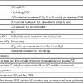
Assessment of the mechanical properties of the lung and chest wall and evaluation of the efficiency of gas exchange in the lungs are clinically important. When abnormalities are revealed early, impairment may still be reversible or at least treatable. Pulmonary function testing is also helpful in elucidating the basis for breathlessness, a common symptom of pulmonary disease, as well as important in characterizing the pathophysiology and providing a measure of the severity of pulmonary diseases. Pulmonary function testing is also an excellent measure of general health and the risk of mortality from all causes. The range of pulmonary function tests, their accepted symbols, techniques of performance, and interpretation are summarized in Plates 3-1 and 3-2.
RESPIRATORY MUSCLES
The chest expands and the lungs are filled with air by the contraction of the inspiratory muscles that create a negative pressure within the chest cavity and a negative pressure gradient down the airways (see Plate 2-1). The diaphragm is the principal muscle of inspiration and provides the pressure gradient for the movement of much of the air that enters the lungs during quiet breathing. Contraction of the diaphragm causes the left and right domes to descend downward and the chest to expand upward and outward. At the same time, because of the vertically oriented attachments of the diaphragm to the costal margins, diaphragmatic contraction also serves to elevate the lower ribs.
Contraction of the intercostal muscles, the external intercostal muscles, and the parasternal intercartilaginous muscles raises the ribs during inspiration. As the ribs are elevated, the anteroposterior and transverse dimensions of the chest enlarge because of the anatomic movement of the ribs around the axis of their necks. This is commonly referred to as the bucket handle effect. Upward displacement of the upper ribs is accompanied by an increase in the anteroposterior dimension similar to the motion of a “water pump handle,” and elevation of the lower ribs is associated with an increase in the transverse dimension of the chest.
In addition to the diaphragm and intercostal muscles, other accessory inspiratory muscles contribute to the movement of the chest in other situations. The scalene muscles make their major contribution during high levels of ventilation when the upper parts of the chest are maximally enlarged. These muscles arise from the transverse processes of the lower five cervical vertebrae and insert into the upper aspect of the first and second ribs. Contraction of these muscles elevates and fixes the uppermost part of the rib cage.
Another accessory muscle, the sternomastoid (sternocleidomastoid), normally also becomes active only at high levels of ventilation. Contraction of the sternomastoid muscle is frequently apparent during severe asthma and with other disorders that obstruct the movement of air into the lungs. The sternomastoid muscle elevates the sternum and slightly enlarges the anteroposterior and longitudinal dimensions of the chest.
In contrast to inspiration, expiration during quiet breathing occurs as a more passive process as a result of recoil of the lung. However, at higher levels of ventilation or when movement of air out of the lungs is impeded, expiration becomes active. Muscles involved in active expiration include the internal intercostal muscles, which depress the ribs; the external and internal oblique abdominal muscles; and the transversus and rectus abdominis muscles, which compress the abdominal contents, depress the lower ribs, and pull down the anterior part of the lower chest. These expiratory muscles also play important and complex roles in regulating breathing and lung volume during talking, singing, coughing, defecation, and parturition.
The strength of the respiratory muscles can be determined from maximal static respiratory pressures (i.e., maximal pressure generated during a forced inspiratory or expiratory maneuver against a manometer or pressure gauge). Pressure developed during an isometric contraction of the respiratory muscles is a function of the length of those muscles and is therefore related to the lung volume at which the maneuver is performed. Maximal inspiratory static pressure is measured when the inspiratory muscles are optimally lengthened after a complete expiration to residual volume (RV). Similarly, maximal static expiratory pressure is determined after a full inspiration to total lung capacity (TLC) when the expiratory muscles are in their most mechanically advantageous position.


