Pulmonary Complications of Hematopoietic Stem Cell Transplantation

Published on 23/05/2015 by admin

Filed under Pulmolory and Respiratory

Last modified 22/04/2025

Print this page

rate 1 star rate 2 star rate 3 star rate 4 star rate 5 star
Your rating: none, Average: 0 (0 votes)

This article have been viewed 1481 times

Chapter 77 Pulmonary Complications of Hematopoietic Stem Cell Transplantation

Hematopoietic stem cell transplantation (HSCT) refers to the transplantation of stem cells from bone marrow, growth factor–stimulated peripheral blood, and umbilical blood for the treatment of malignant and nonmalignant hematologic, autoimmune, and genetic diseases. Transplant recipients are at risk of serious complications as a result of pretransplant cytoreductive conditioning regimens, immunologic sequelae from engraftment of allogeneic lymphoid cells (which mediate graft-versus-host responses), the patient’s immunosuppressed status, and infections secondary to immunosuppression. Autopsy studies show that pulmonary complications are responsible for more than 70% of deaths in HSCT recipients.

Overview of Hematopoietic stem cell transplantation

Conditioning Regimens

Before undergoing HSCT, patients receive a conditioning regimen with the goals of reducing the tumor burden (with malignancy), ablating the bone marrow, and suppressing the recipient’s immune system, thereby allowing engraftment of stem cells. The three regimen types are myeloablative conditioning, reduced-intensity conditioning, and nonmyeloablative conditioning. This classification is based on the duration of cytopenia and the requirement for stem cell support. Myeloablative conditioning causes irreversible cytopenia, for which stem cell support is always necessary, whereas nonmyeloablative conditioning causes minimal cytopenia, for which stem cell support may not be needed. Reduced-intensity conditioning causes cytopenia of variable duration, and stem cell support should be given.

Conventional myeloablative conditioning regimens include cyclophosphamide and total-body irradiation (TBI) or busulfan. The more recent nonmyeloablative or reduced-intensity conditioning regimens have used fludarabine and reduced-dose alkylating agents or TBI. Although these less intensive regimens do not provide a strong cytoreductive effect, they allow engraftment of the donor stem cells with a subsequent potentially beneficial graft-versus-malignancy effect. The nonmyeloablative or reduced-intensity conditioning regimens are associated with reduced transplant-associated morbidity and lower incidence of pulmonary complications after transplantation.

Prophylaxis after allogeneic transplant to prevent GVHD usually involves methotrexate, cyclosporin, corticosteroids, or in vitro T cell depletion of the graft before infusion.

Posttransplant Pulmonary Complications

Pulmonary complications after HSCT are common, with an incidence of 40% to 60% and with up to one third of recipients requiring intensive care after transplantation. Respiratory failure is the most common cause of critical illness, and pneumonia is the leading infectious cause of death after HSCT (Figure 77-1). Pulmonary complications can occur early or late in the posttransplant course, can have infectious and noninfectious etiologies, and can present with assorted radiographic findings. The pulmonary complications of HSCT also vary depending on the indication for, type of, and preparative regimen preceding HSCT.

Cellular interactions between graft and host cells are essentially eliminated with autologous transplantation, obviating the need for immunosuppression to prevent or treat GVHD. As such, autologous transplantation is associated with lower incidence of infection, particularly viral pneumonias or cytomegalovirus (CMV) pneumonitis, invasive fungal disease, and other opportunistic infections (e.g., toxoplasmosis), as well as late airflow obstruction defects.

Risk Factors for Pulmonary Disease

Relapse status at transplant and donor-recipient HLA mismatching or nonidentity are risk factors for pulmonary complications and mortality after HSCT (Box 77-1). Active phase of malignancy, age over 21 years, and receipt of HLA-nonidentical donor marrow are risk factors for respiratory failure after HSCT.

Abnormalities in pretransplant pulmonary function tests (PFTs) may be predictive of subsequent risk of pulmonary complications and mortality. Reduced diffusing capacity and increased alveolar-arterial oxygen gradient are independent risk factors for interstitial pneumonitis and also independently associated with increased early mortality after HSCT.

Patients with elevated levels of transforming growth factor beta (TGF-β) in plasma, TGF-α in bronchoalveolar lavage (BAL) fluid, and granulocyte-macrophage colony-stimulating factor (GM-CSF) in BAL fluid seem to be at increased risk for pulmonary complications. One study showed that elevated pretransplant TGF-β levels in patients with breast cancer undergoing autologous HSCT were associated with increased posttransplant risk of pulmonary toxicity and hepatic venoocclusive disease.

Recipients of allogeneic transplantation have more infection complications than recipients of autografts, not only because of chronic immunosuppression, but also because GVHD itself causes an immunodeficient state by affecting the mucosal surfaces, the reticuloendothelial system, and bone marrow. These factors predispose allogeneic recipients to fatal viral pneumonias, multidrug-resistant bacteria, and invasive fungi. Similarly, bronchiolitis obliterans is almost exclusively seen after allogeneic HSCT.

Time Course

Specific pulmonary complications associated with HSCT tend to occur in a relatively well-defined timeline. The timing and intensity of cytoreductive therapies and the resulting pattern of immune reconstitution influence the duration of these intervals.

Infectious Complications

The overall risk of pulmonary infection in patients receiving HSCT depends on multiple risk factors, including chemotherapy and radiation-induced neutropenia, lung injury induced by the conditioning regimen, rejection in the form of GVHD, local disruption of host defenses, and intensity of pathogen exposure. In addition, HSCT recipients need to develop a functional immune system from donor-derived cells. Although the production of red blood cells, platelets, and granulocytes occurs soon after HSCT, production of lymphocytes, especially T cells, is considerably delayed. In the first 2 years after transplant, serious infections occur in 50% of otherwise uncomplicated transplants from histocompatible sibling donors and in 80% to 90% of matched unrelated donors or histocompatible recipients with GVHD.

Supportive care in the posttransplant period has changed the microbiology of pneumonia. Prophylactic administration of trimethoprim-sulfamethoxazole (TMP-SMX), antivirals, antifungals, and fluoroquinolones has decreased the incidence of Pneumocystis jirovecii, CMV, herpes simplex, and Candida albicans infections (Box 77-2). Resistant gram-negative and gram-positive bacteria, viruses, and other fungi remain important pathogens.

Fungal Infections

Fungal disease should be considered in patients with persistent focal radiographic abnormalities that do not respond to empirical antibiotic therapy and in those with nodular opacities on chest imaging, prolonged neutropenia, corticosteroid use, or a history of prior fungal infections.

Invasive aspergillosis is the leading cause of infectious death in HSCT patients, with a mortality of 70% to 90% in allogeneic recipients, despite treatment. The incidence of invasive aspergillosis in allogeneic recipients is 10% to 15%, with a bimodal distribution of cases. During the early preengraftment period that is characterized by profound neutropenia, both allogeneic and autologous recipients are at increased risk for invasive aspergillosis. Allogeneic patients experience a second period of vulnerability, however, during the postengraftment phase, coincident with the development of chronic GVHD, because of the need for augmented immunosuppression. The vulnerability to invasive aspergillosis is higher during the second period than during neutropenia.

Most cases of invasive aspergillosis are limited to the lungs, but sinus and central nervous system involvement can also be seen. Common presenting symptoms include cough and dyspnea, with fever absent in up to two thirds of patients. Concomitant pleuritic chest pain and hemoptysis are clues. Radiographic findings include single or multiple nodules, cavities, and consolidation. The infiltrates are usually focal. The air crescent sign describes a central nodule partially or fully surrounded by air, indicating a sequestrum of necrotic lung tissue that has separated from the surrounding parenchyma. The halo sign on computed tomography (CT) describes a rim of low attenuation representing edema or hemorrhage that surrounds a pulmonary nodule and is present in more than 90% of patients with neutropenia with invasive pulmonary aspergillosis. The diagnosis of invasive aspergillosis is confirmed by demonstration of fungal elements in diseased tissue and a positive culture result of a specimen from a normally sterile site. In clinical practice, however, biopsy is not usually feasible because of thrombocytopenia or coagulation deficits, and a presumptive diagnosis is often substantiated by high-resolution CT (HRCT) of chest, culture results, or indirect diagnostic tests, such as detection of galactomannan antigen in body fluids.

A randomized clinical trial comparing voriconazole with amphotericin B found that voriconazole led to better survival and improved responses of initial therapy. Therefore, voriconazole is recommended as the primary treatment for invasive aspergillosis. The voriconazole loading dosage is 6 mg/kg intravenously (IV) every 12 hours for 1 day, followed by 4 mg/kg IV every 12 hours. Although not the first choice, liposomal amphotericin B may be used as an alternative primary therapy. Posaconazole, 200 mg every 8 hours, is the first-choice agent for prophylaxis against invasive aspergillosis in high-risk patients, including HSCT recipients with GVHD.

Candida, Cryptococcus, and Zygomycetes (including Mucor and Rhizopus) are also important pathogens. The prevalence of invasive zygomycotic infection is 2% and bears a similar clinical course to that of Aspergillus. Zygomycetes are angioinvasive, leading to thrombosis, pulmonary infarction, and hemorrhage, with radiographs showing cavitation and halo sign. Mortality rates are high (60%-80%) despite treatment with amphotericin B and surgical resection.

The endemic fungi are encountered less frequently. Histoplasma and Coccidioides usually occur as reactivation of latent infection. Blastomyces usually represents primary disease. Other emerging fungal pathogens reported to cause pulmonary infections include Fusarium and Scedosporium spp.

Viral Infections

Viral infections, particularly CMV pneumonia, are an important cause of morbidity and mortality in the postengraftment period. Cytomegalovirus pneumonia has a nonspecific clinical presentation that includes fever, nonproductive cough, and hypoxia. Chest imaging demonstrates various abnormalities, most frequently bilateral interstitial opacities. A definitive diagnosis relies on demonstrating viral inclusion bodies in lung tissue (which can be difficult on transbronchial biopsy specimens) or detection of virus in BAL fluid by shell vial assay, polymerase chain reaction (PCR), or viral culture (in patients with a compatible clinical presentation). Mortality from CMV pneumonia in posttransplant patients exceeded 90% until the advent of combination therapy with ganciclovir and intravenous immune globulin (IVIG), which improved survival rates to as high as 70%. The use of prophylaxis has not only reduced the incidence but has also delayed the onset of disease, with more CMV infections now occurring 100 days after transplant.

Prophylactic regimens have taken two approaches: (1) universal prophylaxis to all high-risk patients for a defined period after engraftment and (2) preemptive treatment of patients only after detection of subclinical viremia by PCR assay. Both strategies reduce the risk of early CMV disease and are accepted practice guidelines. Seronegative patients who underwent allogeneic transplant from seropositive donors are at highest risk for CMV pneumonia.

The infection pattern of respiratory viruses in HSCT recipients is similar to that in the general population; however, HSCT recipients are more likely to develop lower respiratory tract infection, with mortality of 50% to 70%. Respiratory viruses classically recognized as causing disease in both immunocompetent and immunocompromised individuals include respiratory syncytial virus (RSV), parainfluenza, and influenza. New detection techniques have led to increasing identification of other respiratory viruses, such as human coronavirus, human metapneumovirus, human bocavirus, and human rhinovirus.

In a large prospective study, the 2-year incidence of a first episode of a viral respiratory infection was 29% in recipients of allogeneic HSCT and 15% in recipients of autologous HSCT. The most commonly isolated respiratory viruses were influenza A/B, human RSV, and human metapneumovirus.

Respiratory syncytial virus usually causes an upper respiratory infection, which may progress to fatal pneumonia in the immunocompromised patient. Late airflow obstruction has also been associated with RSV infection. Untreated, RSV pneumonia has a poor prognosis, with mortality approaching 80%. Treatment includes aerosolized ribavirin; other agents may be added, although a beneficial effect of concomitant therapy is not well established. Concomitant therapy includes IVIG, RSV-specific immunoglobulin, or palivizumab. Early initiation of treatment appears to be fundamental for the success of antiviral therapy. The role of preemptive therapy in the presence of isolated upper respiratory tract RSV infection is not well defined.

Adenovirus is an uncommon cause of pneumonia and can be isolated in 3% to 5% of patients after HSCT. The incidence is higher in children. It affects the upper and lower respiratory tracts, as well as the gastrointestinal and genitourinary systems. Infection usually develops within the first 3 months of transplantation, with a presentation that might include pharyngitis, tracheitis, bronchitis, pneumonitis, enteritis, hemorrhagic cystitis, or disseminated disease. Mortality with pulmonary involvement may exceed 50%. Cidofovir is used for preemptive (positive viremia despite absence of symptoms) and therapeutic treatment. Some centers have successfully used adenovirus-specific cytotoxic T cells for treatment.

Human metapneumovirus is an RNA paramyxovirus that causes infection in up to 5% of HSCT recipients. Patients usually present with upper respiratory tract symptoms but may also develop pneumonia. The role of antiviral therapy has not been defined in this infection.

Influenza types A and B can cause infection in both immunocompetent and immunocompromised patients, with type A being the most common. The 2009 influenza pandemic was caused by a novel H1N1 influenza A virus, a reassortment of classic swine H1N1, avian Eurasian swine H1N1, and North American human H2N2. Infection by novel H1N1 is characterized by severe disease in patients with specific risk factors, which include immunocompromised status. A cohort study from two medical centers found that HSCT recipients infected with novel H1N1 develop lower respiratory infection in 52% of the cases and have a 30-day mortality of 22%. Zanamivir and oseltamivir are neuraminidase inhibitors used to treat influenza types A and B. Amantadine and rimantadine are adamantanes, which are used only for influenza type A.

Other viral infections include herpes simplex virus (HSV), varicella-zoster virus (VZV), and human herpesvirus (HHV) types 6 and 7. HSV was a common cause of infection in HSCT recipients but incidence has been greatly reduced by acyclovir prophylaxis. HHV-6 has been associated with the idiopathic pneumonia syndrome. Epstein-Barr virus (EBV) infections usually manifest as posttransplant lymphoproliferative disorder.

Noninfectious Complications

Idiopathic Pneumonia Syndrome

Idiopathic pneumonia syndrome (IPS) refers to a form of severe lung injury in which infectious etiologies, cardiac dysfunction, acute kidney injury, and iatrogenic fluid overload have been excluded. The syndrome encompasses several entities with varying clinical presentation. The heterogeneous clinical manifestations reflect the many lung insults underlying the pathogenesis of IPS, including toxic effects of HSCT conditioning regimens, immunologic cell-mediated injury, and inflammatory cytokines.

The incidence of IPS varies between 2.2% and 15% after allogeneic HSCT; the lower incidence is observed with nonmyeloablative conditioning regimens. The median time of onset for IPS is 22 days after HSCT (range, 4-106 days). There are reports of IPS following autologous HSCT, but it is predominantly seen after allogeneic HSCT. Risk factors for IPS include conventional conditioning regimens, older age, diagnosis of acute leukemia or myelodysplastic syndrome, development of severe acute GVHD, and high-dose TBI.

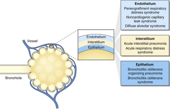
The insight obtained from animal models has stimulated the classification of the entities under the term idiopathic pneumonia syndrome according to the primary anatomic site of cellular damage: interstitial tissue, vascular endothelium, or airway epithelium (see Fig. 77-1). For example, in interstitial type of injury, there are increased numbers of cytotoxic T lymphocytes and cells expressing B7 family costimulatory ligands in the alveolar and interstitial spaces. In vascular endothelium type of injury, there are injured endothelial cells, and in airway epithelium type of injury, injured type II alveolar epithelial cells. This classification reflects complex interactive mechanisms with a pathogenic role attributed to several factors, including increased soluble inflammatory mediators (e.g., tumor necrosis factor alpha), increased host flora–derived lipopolysaccharide, enhanced oxidative stress, depletion of pulmonary surfactant, donor-derived T cell effectors, host antigen-presenting cells, donor accessory cells, and leukocyte recruitment to the lung.

Treatment of the HSCT patient with IPS includes supportive care and, more recently, the combination of corticosteroids. The use of subcutaneous etanercept has been associated with a high clinical response rate in a recent series.

Diffuse Alveolar Hemorrhage

Diffuse alveolar hemorrhage (DAH) occurs in approximately 5% of autologous and allogeneic recipients. Risk factors include high-dose chemotherapy received before transplant, irradiation, and advanced age. Rapid immune system reconstitution has also been theorized to contribute to DAH. Neutrophil influx into the lung may accentuate the injury and precipitate hemorrhage. The typical symptoms are dyspnea and hemoptysis. Cough and fever may also be present. Although highly suggestive of DAH in the appropriate clinical setting, hemoptysis is not always present. Patients usually are seen during the periengraftment period within the first month after transplantation, but up to 42% of patients with DAH are seen late, after day 30.

Diagnostic criteria proposed to diagnose DAH include alveolar injury, evident as diffuse lung infiltrates or increased alveolar-arterial oxygen gradient; absence of pulmonary infection; BAL fluid with increasingly bloodier return from three separate subsegments; or hemosiderin-laden macrophages at 20% or higher. The radiographic findings of DAH are often nonspecific and include an interstitial or alveolar pattern, usually of bilateral distribution and sparing the peripheral lung areas.

The mainstay of treatment of the HSCT patient with DAH is systemic steroid therapy, which should be instituted promptly. A reported regimen is methylprednisolone, 125 to 250 mg every 6 hours for 5 days, followed by a slow taper over 2 to 4 weeks.

Bronchiolitis Obliterans

Bronchiolitis obliterans (obliterative bronchiolitis) is a complication that follows allogeneic HSCT. Rarely, it may also occur after autologous HSCT. The incidence of bronchiolitis obliterans in different reports has ranged from 0% to 48%. Risk factors for obliterative bronchiolitis include chronic GVHD, older age, presence of obstructive disturbance before HSCT, viral infections of the respiratory tract, and busulfan-based conditioning regimens. The close association with GVHD has led some experts to claim that bronchiolitis obliterans may indeed represent the lung manifestation of GVHD.

The activation of donor-derived T lymphocytes plays an important role in the pathogenesis of bronchiolitis obliterans. T cells target the recipient epithelial cells in the bronchioles, leading to an inflammatory reaction. Toll-like receptor–4 signaling in donor-derived hematopoietic cells also appears to be an important factor in causing alloimmunity. Obliterative bronchiolitis tends to occur late, typically more than 100 days after HSCT. The most common symptoms are dry cough, dyspnea, and wheezing. The physical examination may reveal decreased breath sounds, wheezing, and inspiratory squeaks. Furthermore, physical examination often reveals signs of GVHD outside the lung such as skin manifestations. In a minority of patients, there are no respiratory manifestations, and the diagnosis is established by PFTs. The chest radiograph is usually normal, although patients with advanced disease may have signs of hyperinflation, bronchiectasis, and areas of scarring. The HRCT scan is more sensitive for all the previously mentioned abnormalities and valuable in showing air trapping and mosaic attenuation during expiration. PFTs show an obstructive pattern, with FEV1/FVC ratio less than 0.7 and a decrease in FEV1 greater than 20% from pretransplant value.

Treatment of the HSCT patient with bronchiolitis obliterans involves corticosteroids, such as prednisone at 1 to 1.5 mg/kg daily, and intensification of immunosuppressive therapy.

Pulmonary Venoocclusive Disease

Pulmonary venoocclusive disease (PVOD) is characterized by obliterative fibrotic vasculopathy, which affects mainly the small branches of the pulmonary venous circulation. PVOD may be idiopathic or secondary to another disease. Diseases and risk factors associated with development of PVOD include exposure to chemotherapeutic agents (e.g., carmustine, mitomycin C, bleomycin), autologous and allogeneic HSCT, solid-organ transplantation, radiotherapy, and autoimmune diseases. Patients are seen several months after transplantation with progressive dyspnea on exertion and fatigue. They may also have pleural effusions with right upper quadrant tenderness and ascites. Chest radiography reveals interstitial edema. Abnormalities on HRCT scan include ground-glass opacities with centrolobular distribution, septal thickening, and mediastinal lymphadenopathy.

The diagnosis of PVOD is definitively established by histologic analysis of lung biopsy specimens, but lung biopsy is not indicated because of the high mortality risk. Histologic analysis reveals fibrosis of the venules and small veins in the interlobular septae of the pulmonary circulation, with resultant intraluminal thrombosis. The pulmonary arteries may display medial hypertrophy and eccentric intimal fibrosis. Right-sided cardiac catheterization typically reveals a precapillary pattern of pulmonary hypertension, with an increase in the mean pulmonary artery pressure and normal-range pulmonary capillary wedge pressure. Pulmonary vasodilators can exacerbate the pulmonary edema. Anecdotal reports describe PVOD patient response to high-dose corticosteroids.

Diagnostic Evaluation

In the diagnostic evaluation of the HSCT patient, information should be obtained on the duration of the pulmonary complication, the radiographic abnormalities, and individual patient factors, such as exposure to toxic drugs, a history of receiving chest radiotherapy, current and previous immunosuppressive regimens and infection prophylaxis, CMV status of donor and recipient, and history of previous opportunistic invasive fungal disease. Useful information may be obtained from PFTs, sputum examination, chest imaging, and serologic studies.

Serologic Studies

The presence of galactomannan, a cell wall component released by Aspergillus spp., in serum indicates invasive aspergillosis, as detected by enzyme-linked immunosorbent assay. Using a cutoff optical density of 0.5, the test has a sensitivity of 97.4%, specificity of 90.5%, positive predictive value of 66.1%, and negative predictive value of 99.4%. A galactomannan assay for the diagnosis of invasive aspergillosis has also been evaluated in BAL fluid. Using a cutoff optical density of 0.5, the test showed sensitivity of 86% and specificity of 89%.

An elevated level of (1,3)-β-D-glucan in serum is typically associated with invasive aspergillosis and invasive candidiasis but is not specific for a particular fungal infection.

Newer diagnostic techniques have allowed not only a more accurate diagnosis of usual viral respiratory infections but also the detection of emerging viruses that were previously undetected. Nasopharyngeal swab assays employing multiplex PCR can detect up to 20 different respiratory virus types or subtypes in a single rapid test.

Determining CMV serologic status of both recipient and donor is essential in pretransplant screening. The serologic status of the recipient as opposed to the donor is the primary determinant for CMV conversion. A shell vial culture with monoclonal antibody to p72 requires only 48 hours, and tests for antigenemia are rapid, standardized, semiquantitative, and inexpensive. Nucleic acid amplification by PCR on DNA extracted from infected leukocytes also allows rapid diagnosis, detecting infection 2 weeks before viral cultures become positive and 1 week before positive antigenemia, and is highly sensitive.

Controversies and Pitfalls

Bronchoscopy with BAL has the potential to identify the cause of acute respiratory failure in HSCT patients; however, it also carries risks, especially in hypoxemic patients. The role of bronchoscopy was prospectively evaluated in a multicenter observational study in which cancer patients or HSCT recipients with acute respiratory failure received bronchoscopy with BAL, as well as noninvasive diagnostic tests. Noninvasive tests had a diagnostic yield of 66.7%, and BAL bronchoscopy, 50.5%. Although the only investigation that provided a diagnosis in one third of the patients, bronchoscopy with BAL was associated with respiratory deterioration in half the nonintubated patients. Thus, although the routine use of bronchoscopy in HSCT recipients with acute respiratory failure remains controversial, in select patients, bronchoscopy may provide the only means for accurately determining the etiology of lung disease.

Transthoracic needle aspiration under CT or fluoroscopic guidance has a high sensitivity (70%) for detecting pulmonary complications in HSCT patients, with the most common findings being infection and malignancy. However, pneumothorax is also a common complication and may require chest tube placement.

If no diagnosis is made with either bronchoscopy or transthoracic needle aspiration, surgical lung biopsy by either open thoracotomy or a video-assisted thoracoscopic approach may be considered next. Although surgical lung biopsy has a sensitivity of 60% to 80%, the procedure may be associated with worse outcomes and thus should be recommended primarily for hematopoietic stem cell transplant recipients whose test results likely will significantly alter their management.

Suggested Readings

Afessa B, Azoulay E. Critical care of the hematopoietic stem cell transplant recipient. Crit Care Clin. 2010;26:133–150.

Afessa B, Tefferi A, Litzow MR, et al. Diffuse alveolar hemorrhage in hematopoietic stem cell transplant recipients. Am J Respir Crit Care Med. 2002;166:641–645.

Azoulay E, Mokart D, Rabbat A, et al. Diagnostic bronchoscopy in hematology and oncology patients with acute respiratory failure: prospective multicenter data. Crit Care Med. 2008;36:100–107.

Bacigalupo A, Ballen K, Rizzo D, et al. Defining the intensity of conditioning regimens: working definitions. Biol Blood Marrow Transplant. 2009;15:1628–1633.

Boeckh M. The challenge of respiratory virus infections in hematopoietic cell transplant recipients. Br J Haematol. 2008;143:455–467.

Dickout WJ, Chan CK, Hyland RH, et al. Prevention of acute pulmonary edema after bone marrow transplantation. Chest. 1987;92:303–309.

Espinosa-Aguilar L, Green JS, Forrest GN, et al. Novel H1N1 influenza in hematopoietic stem cell transplantation recipients: two centers’ experiences. Biol Blood Marrow Transplant. 2010;17:566–573.

Gratwohl A, Baldomero H, Aljurf M, et al. Hematopoietic stem cell transplantation: a global perspective. JAMA. 2010;303:1617–1624.

Guo YL, Chen YQ, Wang K, et al. Accuracy of BAL galactomannan in diagnosing invasive aspergillosis: a bivariate metaanalysis and systematic review. Chest. 2010;138:817–824.

Herbrecht R, Denning DW, Patterson TF, et al. Voriconazole versus amphotericin B for primary therapy of invasive aspergillosis. N Engl J Med. 2002;347:408–415.

Kallianpur AR. Genomic screening and complications of hematopoietic stem cell transplantation: has the time come? Bone Marrow Transplant. 2005;35:1–16.

Maertens JA, Klont R, Masson C, et al. Optimization of the cutoff value for the Aspergillus double-sandwich enzyme immunoassay. Clin Infect Dis. 2007;44:1329–1336.

Mahony J, Chong S, Merante F, et al. Development of a respiratory virus panel test for detection of twenty human respiratory viruses by use of multiplex PCR and a fluid microbead-based assay. J Clin Microbiol. 2007;45:2965–2970.

Martino R, Porras RP, Rabella N, et al. Prospective study of the incidence, clinical features, and outcome of symptomatic upper and lower respiratory tract infections by respiratory viruses in adult recipients of hematopoietic stem cell transplants for hematologic malignancies. Biol Blood Marrow Transplant. 2005;11:781–796.

Montani D, O’Callaghan DS, Savale L. Pulmonary veno-occlusive disease: recent progress and current challenges. Respir Med. 2010;104:S23–S32.

Mullighan CG, Heatley S, Doherty K, et al. Mannose-binding lectin gene polymorphisms are associated with major infection following allogeneic hematopoietic stem cell transplantation. Blood. 2002;99:3524–3529.

Onizuka M, Kasai M, Oba T, et al. Increased frequency of the angiotensin-converting enzyme gene D-allele is associated with noninfectious pulmonary dysfunction following allogeneic stem cell transplant. Bone Marrow Transplant. 2005;36:617–620.

Pandya CM, Soubani AO. Bronchiolitis obliterans following hematopoietic stem cell transplantation: a clinical update. Clin Transplant. 2010;24:291–306.

Soubani AO, Pandya CM. The spectrum of noninfectious pulmonary complications following hematopoietic stem cell transplantation. Hematol Oncol Stem Cell Ther. 2010;3:143–157.

Sundin M, Le Blanc K, Ringdén O, et al. The role of HLA mismatch, splenectomy and recipient Epstein-Barr virus seronegativity as risk factors in post-transplant lymphoproliferative disorder following allogeneic hematopoietic stem cell transplantation. Haematologica. 2006;91:1059–1067.

Walsh TJ, Anaissie EJ, Denning DW, et al. Treatment of aspergillosis: clinical practice guidelines of the Infectious Diseases Society of America. Clin Infect Dis. 2008;46:327–360.

Wilczynski SW, Erasmus JJ, Petros WP, et al. Delayed pulmonary toxicity syndrome following high-dose chemotherapy and bone marrow transplantation for breast cancer. Am J Respir Crit Care Med. 1998;157:565–573.

Yanik GA, Ho VT, Levine JE, et al. The impact of soluble tumor necrosis factor receptor etanercept on the treatment of idiopathic pneumonia syndrome after allogeneic hematopoietic stem cell transplantation. Blood. 2008;112:3073–3081.