Pulmonary Circulation

Published on 23/05/2015 by admin

Filed under Pulmolory and Respiratory

Last modified 22/04/2025

Print this page

rate 1 star rate 2 star rate 3 star rate 4 star rate 5 star
Your rating: none, Average: 0 (0 votes)

This article have been viewed 2909 times

Chapter 4 Pulmonary Circulation

The lungs are served by two circulations—the pulmonary circulation, which accommodates the entire cardiac output from the right side of the heart through a low-pressure circulation, and the bronchial circulation, which arises from branches of the aorta with systemic pressure and usually carries less than 1% of the cardiac output.

Circulatory Structure

Pulmonary Circulation

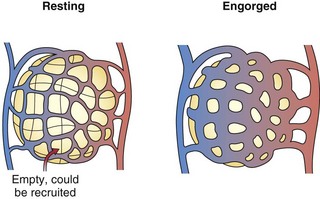
The pulmonary arteries lie near and branch in unison with the airways in the bronchovascular bundle. They are much thinner than systemic arteries and have proportionately more elastic tissue in their walls. The walls of the arterioles, with a diameter less than 100 µm, are so thin relative to those of their systemic counterparts that fluid and gas can move across them. Within the gas-exchanging zone, the arterioles give rise to a network of pulmonary capillaries in the alveolar walls that is continuous throughout the lungs. They are so numerous that, when distended, blood flows almost as an unbroken sheet between the air spaces (Figure 4-1). “Sheet flow” reduces vascular resistance and optimizes gas exchange by creating a very large surface area, estimated at over 100 m2. When the transmural pressure difference between the inside and outside of the vessels is low, many of the capillary segments are closed, but flow switches among segments frequently as some open and others close. Nonflowing segments are rapidly recruited into the pulmonary vascular bed as needed to accommodate increased flow and may be further distended by an increase in transmural pressure. Both recruitment and distention of the pulmonary capillary bed reduce resistance to blood flow and help to maintain a low pressure in the face of increased blood flow. This low pressure allows the capillary-alveolar membrane to be very thin (approximately 1 µm), facilitating diffusion of respiratory gases between blood and alveoli. A red cell that follows a capillary path from the pulmonary artery to a vein may cross several alveoli, with the average transit time through the vessels engaged in gas exchange calculated to be approximately 0.75 second. The capillaries unite to form larger alveolar microvessels, which become venules and then veins that run between the lobules toward the hila, where upper and lower pulmonary veins from each lung empty into the left atrium.

image

Figure 4-1 Alveolar capillaries. The normal cardiac output requires only a portion of the sheet of capillaries; any remaining vessels can be recruited when cardiac output rises during exercise.

(Modified with permission from Butler J: The circulation of the lung. In Culver BH, editor: The respiratory system, Seattle, 2006, University of Washington Publication Services, p 8.2.)

Circulatory Physiology

The pulmonary circulation conducts the entire cardiac output with a remarkably low driving pressure from the pulmonary artery (mean Ppa of 15 to 20 mm Hg) to the left atrium (Pla of 7 to 12 mm Hg). As in the airways, the branching pattern of vessels leads to an increase in total cross-sectional area as the alveolar vessels are approached, but unlike in the airways, this increase is not associated with a decrease in resistance. Total cross-sectional area increases at a branching point if the number of daughter branches (n) is greater than the ratio of the parent to daughter radii squared, (a/b)2, but resistance decreases only if n is greater than (a/b)4. The latter case occurs in the peripheral airways but not in the vessels, so although small peripheral airways contribute little to normal airflow resistance, pulmonary microvessels make up a substantial portion of vascular resistance. Efforts to partition the pressure drop longitudinally suggest that approximately 20% to 30% is in the arterial portion (including arterioles), 40% to 60% in the microvascular portion, and the remainder in the veins. With increases in flow, recruitment occurs mainly at the level of microvascular vessels, so their relative contribution to resistance becomes less.

The pulmonary circulation is a network of segmental resistors that share common upstream (i.e., Ppa) and downstream (i.e., Pla) pressures. Flow is distributed to the various segments in proportion to the reciprocal of the total serial resistance through any segment. The benefit of having the highest resistance at the microvascular level is that the control of blood flow distribution can occur at a finer level, allowing active mechanisms of flow regulation (see further on) to adjust blood flow to relatively small lung regions.

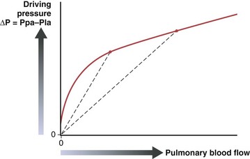
The pulmonary vascular resistance, PVR, is calculated as transvascular driving pressure, ΔP (mean upstream Ppa minus mean downstream Pla), divided by the flow: PVR = ΔP/Q. The calculated resistance must be interpreted in the context of flow, because the relationship of driving pressure to flow usually is not linear and its plotted curve does not pass through zero. As shown in Figure 4-2, pulmonary vascular resistance decreases as flow and pressure increase with the attendant recruitment and distention of vessels.

The resistance to flow through a vessel increases with its length, with the viscosity of the fluid, and, most important, with the inverse of the radius to the fourth power. In addition to muscle activity in the wall, the caliber of a distensible vessel depends passively on the transmural pressure difference between intravascular and extravascular pressures. This mechanism is particularly important in the lungs, where the vessels are embedded in expandable parenchyma. It is convenient to consider separately the effect of lung expansion on the extraalveolar arterial and venous vessels, which differs from the effect on the microvessels of the alveolar zone. With lung volume increase, extraalveolar vessels are distended as the pressure is lowered in the expanding perivascular space around them (Figure 4-3), and they are elongated as the lung expands.

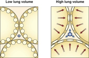
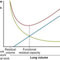
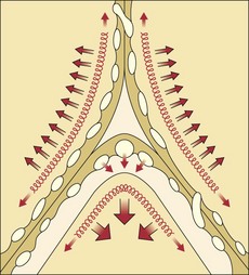
By contrast, the alveolar microvessels in the alveolar walls are elongated but partially collapsed by lung inflation, because the alveolar pressure that surrounds them tends to increase relative to the intravascular pressure. This effect is easy to recognize with positive-pressure ventilation, but it also occurs with spontaneous inspiration, because intravascular pressures fall relative to atmospheric and alveolar pressure. The sheets of capillaries in the alveolar walls are protected from the full compressive force of the alveolar pressure by the surface tension of the fluid that lines curved portions of the alveolar surface. Microvessels in the “corners” where alveolar walls meet are more fully protected from compression by the sharper curvature of the surface film and perhaps by local distending forces, analogous to the situation with extraalveolar vessels (Figure 4-4). The pulmonary vascular resistance is the sum of that through alveolar and extraalveolar vessels and thus has a complex relationship with lung volume. It is lowest at approximately the normal resting lung volume (functional residual capacity) but increases at higher and lower volumes.

image

Figure 4-4 Alveolar “corner” at the junction of three alveolar walls. Surface tension (depicted by “springs”) holds vessels open, particularly in corners, and promotes fluid transudation by lowering the pressure around vessels.

(Modified with permission from Butler J: The circulation of the lung. In Culver BH, editor: The respiratory system, Seattle, 2006, University of Washington Publication Services, p 8.5.)

Blood Flow Distribution

Both vascular geometry and gravity influence the distribution of blood flow within the lung. If the upright lung is viewed as a stacked series of slices, a vertical gradient occurs in which the average flow per slice rises progressively down the lung, consistent with the influence of gravity. Within each slice, however, a marked variability of blood flow is found among regions, with high-flow areas distributed dorsally. The tendency of blood flow to be higher in dorsal and basal regions is largely preserved even when the gravitational direction is reversed, which indicates that anatomic branching patterns are a major determinant of flow distribution.

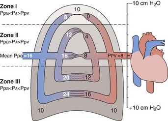
The gravitational effect has been conceptualized by dividing the lung into four zones, one above another, on the basis of the relationship of vascular and alveolar pressures (Figure 4-5). Intravascular pressures are higher at the bottom of the lung than at the top by an amount equal to a vertical hydrostatic column as high as the lung. Near the lung apex, zone I, the pressure in the alveoli (PA) exceeds that in both the pulmonary arteries (Ppa) and pulmonary vein (Ppv) and collapses the alveolar vessels, except those in the alveolar corners, which remain patent and allow some flow to continue. Below this, in zone II, Ppa exceeds PA, but PA is greater than Ppv, so flow depends on the pressure difference between Ppa and PA. The vessels remain open but are critically narrowed at the downstream end, where venous pressure is lower than alveolar pressure. This condition creates independence of flow from the downstream venous pressure, analogous to a waterfall in which a stream that flows over a precipice is unaffected by a rising level in the pool below until it rises above the level of the lip. In the middle to lower portion of the lung, zone III, both Ppa and Ppv exceed PA, the vessels are distended, and blood flow is the highest. Zone IV is restricted to a small area in the most dependent region, where flow diminishes. It has been postulated that this reduction is the result of increased vascular resistance secondary to low lung volume or perivascular edema in this area.

image

Figure 4-5 Perfusion in the lungs is influenced by the relationship of pulmonary arterial and venous pressures (Ppa and Ppv) to alveolar pressure (PA). In this example, the alveolar pressure is 10 cm H2O, as might be found in a patient who receives positive-pressure ventilation.

(Modified with permission from Culver BH: Hemodynamic monitoring: physiologic problems in interpretation. In Fallat RJ, Luce JM, editors: Cardiopulmonary critical care, Edinburgh, 1988, Churchill Livingstone.)

Although the gravitational effect expressed in the vertical zone concept contributes to the average increase in flow down the lung, it does not explain the observed large variability in flow within an isogravitational slice, which implies that other anatomic or vasoregulatory factors are important at this level. More recent studies have determined that the heterogeneous distribution of blood flow within horizontal (isogravitational) planes is due to asymmetric branching geometries (and hence resistances) of the vascular tree. Because the vascular tree is largely a dichotomous branching structure, differences in resistances between daughter branches cause flow to be distributed unevenly between the branches. With differences in resistances occurring at every bifurcation in the vascular tree, blood flow becomes progressively more heterogeneous, resulting in a broad distribution of flows at the terminal branches. Owing to the shared heritage up the vascular tree, neighboring lung regions have similar magnitudes of flow, with high-flow regions near other high-flow regions and low-flow regions neighboring other low-flow regions. Hence, the spatial distribution of pulmonary blood flow is not random but rather exhibits a clear pattern of high and low flows (Figure 4-6). Studies have demonstrated that the pattern of perfusion distribution is very stable over time and with growth, and that the pattern is genetically determined. These insights provide a new perspective on blood flow distribution in the lung. The traditional model of vertically stacked zones needs to be replaced by one in which the multiple zones can exist within horizontal planes. In addition, the large degree of heterogeneity within isogravitational planes suggests that mechanisms other than gravity must be responsible for the tight matching between regional ventilation and blood flow.

Regulation of Pulmonary Blood Flow

Besides their responses to passive mechanisms (anatomy, gravity, lung volume, alveolar pressure), the pulmonary vessels exhibit vasomotor activity regulated by both neural and non-neural factors. Motor efferents from three autonomic networks are in anatomic proximity to the vasculature: sympathetic, parasympathetic, and nonadrenergic noncholinergic fibers. The sympathetic efferents have a vasoconstrictor effect, whereas parasympathetic stimulation dilates constricted vessels. Although acetylcholine is a potent pulmonary vasodilator, there is little cholinergic innervation of the pulmonary resistance vessels. The nonadrenergic noncholinergic system is inhibitory, constantly releasing small vasodilatory peptides at the ganglia and postganglionic ends of its unique network. This vasodilator function is augmented with exercise.

Pulmonary arteries demonstrate an intrinsically low tone as they remain relaxed when isolated from the lung. This state reflects a balance between effects of endothelium-derived vasoconstrictor and vasodilator substances. Although their relative roles are yet to be clarified, many vasoactive peptides are found in the lung. Those exerting vasoconstrictor activity on the pulmonary circulation include angiotensin II, arginine vasopressin, endothelin 1, peptide tyrosine Y, and substance P. Vasodilatory peptides include adrenomedullin, atrial natriuretic peptide, calcitonin gene–related peptide, endothelin 3, somatostatin, and vasoactive intestinal peptide.

Nitric oxide is produced in endothelial cells in the pulmonary circulation and elsewhere and is now recognized as an important mediator of vasodilatation. The oxidation of a nitrogen from L-arginine is catalyzed by nitric oxide synthase, present in both a constitutive form and a form that is inducible by products of inflammation. Nitric oxide activates guanylate cyclase, which increases cyclic guanosine monophosphate (cGMP) within vascular smooth muscle cells. This in turn reduces intracellular Ca++ by several mechanisms, leading to vascular relaxation. Nitric oxide also is abundantly produced in the nasal sinuses, providing an intriguing mechanism whereby inhaled nitric oxide may enhance blood flow to the best-ventilated areas of lung.

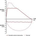
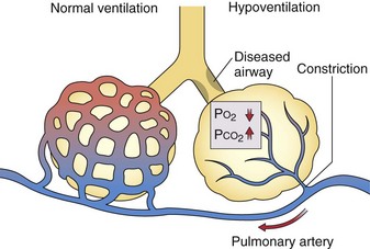
Although the role of nasal nitric oxide in ventilation-perfusion matching is still speculative, the role of alveolar hypoxia in vasoregulation has been recognized for more than 50 years, but the mechanisms involved are still uncertain. Pulmonary arterioles constrict when the PO2 in the alveoli they serve falls, and additional vasoconstriction results if alveolar PCO2 rises (Figure 4-7). Thus, when ventilation is decreased by an obstructed airway or other injury, local hypoxic pulmonary vasoregulation decreases blood flow to the affected region, which tends to restore the local ventilation-perfusion ratio (image) toward normal and thereby improve the PO2 of the blood flowing through that area. The diverted blood flow can be directed to better-ventilated regions, which further contributes to an improvement in overall matching. This hypoxic vasoconstriction seems to be a response to a low PO2 in the air spaces, rather than in the intraluminal blood, which normally is desaturated in these prealveolar vessels. The effector cell is thought to be pulmonary artery smooth muscle located at the entrance to the acinus, and the sensor may be the oxygen-consuming mitochondria within these cells. Several candidate signaling pathways to generate the increase in intracellular calcium necessary for muscle contraction are under investigation.

image

Figure 4-7 Hypoxic vasoconstriction reduces blood flow to poorly ventilated areas. This adaptation improves ventilation-perfusion matching and oxygenation but, if generalized, contributes to pulmonary hypertension.

(Modified with permission from Butler J: The circulation of the lung. In Culver BH, editor: The respiratory system, Seattle, 2006, University of Washington Publication Services, p 8.7.)

Considerable individual variability is found in the hypoxic vasoconstrictor response, and it may be diminished by vasodilating drugs. Diversion of blood flow is most effective in atelectatic areas of lung, in which hypoxic vasoconstriction is unopposed by the radial traction of surrounding expanded lung tissue. A reciprocal reflex in the airways also contributes to better matching: Small airways constrict when intraluminal PCO2 falls and dilate when it rises. Hypoxic vasoconstriction is a helpful, adaptive response to local or regional lung abnormalities, but when alveolar hypoxia is generalized (e.g., hypoventilation or altitude), the increased resistance can lead to pulmonary hypertension.

Nonrespiratory Functions of the Pulmonary Circulation

Filtering

Aggregates of blood elements and emboli of various types (e.g., fat, air, particulate matter) carried in the systemic venous return are continually filtered out, dissolved, or engulfed by the cells of the pulmonary capillary bed. This process provides vital protection for the cerebral, coronary, and other systemic vascular beds and is a benefit of the entire cardiac output from the right side of the heart passing through the lungs. Emboli that bypass the pulmonary circulation through anatomic shunts are called “paradoxical emboli” and are the most common cause of strokes in young persons. Larger emboli have the potential to cause ischemia in affected regions of the pulmonary vascular bed, but this may be countered in part by limited perfusion from the bronchial circulation through bronchopulmonary anastomoses and by exposure to oxygenated pulmonary venous blood that backflows into the occluded region during tidal lung volume changes. Thus, ischemic damage to the alveoli is prevented while a thrombus is lysed and the pulmonary flow restored.

Large numbers of white cells, mainly polymorphonuclear leukocytes, are sequestered in the small vessels of the pulmonary bed. The numbers of leukocytes sequestered in the lung increases markedly if the pulmonary endothelium expresses ligands that cause these cells to adhere and then traverse the vascular wall into the interstitial and alveolar spaces. Many reticuloendothelial cells are found in the lung, and some evidence suggests that the vascular endothelial cell itself can be phagocytic when stimulated.

Fluid Exchange in the Pulmonary Circulation

The fluid flux across the pulmonary vascular endothelium is influenced by the same pressure relationship as in the systemic capillaries, summarized in the modified Starling equation presented in Table 4-1. The hydrostatic pressure in the pulmonary microvessels (Pmv) exceeds the interstitial hydrostatic pressure outside the microvessels, i.e., the perimicrovascular pressure (Ppmv). This effect favors filtration. The interstitial tissue fluid protein osmotic pressure is approximately two thirds that in the vessel; thus, the net osmotic force is absorptive and inward. The components of this equation make it convenient to categorize abnormal fluid flux into the lung into two broad types: hydrostatic edema, when the primary abnormality is an increase in Pmv minus Ppmv, and permeability edema, when endothelial injury increases fluid conductivity across the membrane (incorporated into the permeability factor) and decreases the osmotic reflection coefficient and osmotic gradient. The terms cardiogenic and noncardiogenic also are commonly used for these two mechanisms of edema formation.

Table 4-1 Modified Starling Equation

F = Kf [(Pmv − Ppmv) − σ(pv − pt)]
Symbol Description
F Net fluid flux out of vessels
Kf Permeability factor (constant)
σ Reflection coefficient for oncotic agents
Pmv Pressure in microvessels
Ppmv Perimicrovascular pressure
pv Osmotic pressure in vessels
pt Osmotic pressure of tissues

Fluid flux is sensitive to small intravascular or perivascular pressure changes. Intravascular pressure rises may originate downstream (as with a failing left ventricle) or may follow overall vascular volume increments (as in overhydration) or displacement of blood from the systemic to the pulmonary vessels. The pulmonary capillary endothelium is much less permeable than that of systemic capillaries, and the interstitial space around alveolar microvessels is tightly restricted by the collagen network between the alveolar walls, forming an inexpansible space, so leakage at this site is limited. The extraalveolar arterioles and venules, which are not so confined and are more permeable, appear to constitute an additional important site of fluid leakage and reabsorption in the lungs.

Surface tension in the fluid film that lines the alveoli opposes alveolar pressure and tends to lower the interstitial pressure around pulmonary microvessels, particularly in corner areas (see Figure 4-4). An increase in surface tension may contribute to edema when surfactant is lost in an injured lung. Interstitial pressure around the extraalveolar vessels is close to intrathoracic (pleural) pressure and falls as the lungs are distended, which favors relatively more leakage from them at high rather than low lung volumes (see Figure 4-3).

Interstitial Edema

Several factors tend to keep the lung from becoming edematous. Normally, a net outflow of fluid from the upstream arterioles and capillaries is reabsorbed into the downstream capillaries and venules, where the intravascular pressure is lower. Fluid leakage causes local perivascular pressures to rise, particularly in the restricted space between the alveolar walls, which reduces the outward fluid flux. It also may compress the vessels, thereby reducing the total surface available for leakage. Because the fluid that leaks through intact endothelium is largely protein-free, it dilutes and washes out the interstitial protein. This alteration reduces the perivascular osmotic pressure of tissues and thus increases the inward osmotic pressure difference and reduces the local fluid leak. If excess leakage does occur, the fluid moves from the alveolar walls, where it could interfere with gas exchange, into the low-pressure interstitial zones around the bronchovascular bundles, where it forms relatively innocuous venous, arterial, and peribronchial cuffs. This fluid may be absorbed in part by the rich bronchial vascular network and by the many lymphatics in the adventitia of the airways and vessels. Edema fluid also may reach the pleural space, where it is absorbed by the pleural lymphatic and blood vessels. Finally, experimental data suggest that all the blood perfusing the capillaries in alveolar walls must first pass through capillaries located in alveolar corners and that the negative interstitial pressure surrounding these corner capillaries is critically dependent on alveolar surface tension. When surface tension is eliminated by alveolar flooding, interstitial pressure around these vessels increases, thus serving to compress the corner vessels and diminish flow through the capillaries in the alveolar wall of these flooded alveoli. This mechanism provides for much more precise control of perfusion, virtually on an alveolus-to-alveolus basis, compared with the effects of alveolar hypoxia, which are directed to much more proximal vessels.

When the capillary endothelium is injured, locally or through the effect of circulating mediators, the vascular permeability to fluids and solutes is increased; consequently, even a modest outward pressure gradient causes a large fluid leak. The ability to retain large molecules is lost, protein-rich plasma leaks out, and the osmotic pressure in tissues approaches that in vessels, so that the osmotic force opposing intravascular hydrostatic pressure is lost. This high-permeability or “leaky capillary” edema can be a fulminant process that leads to severe abnormalities in gas exchange.

Alveolar Edema

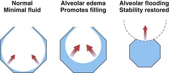
The epithelial cells that line the air spaces have tight junctions along their apical surface, so this membrane normally is even less permeable than the endothelial membrane, protecting alveolar spaces as interstitial edema increases. After total lung water has increased by approximately 50%, the edema fluid appears in the alveoli. A structural failure, at the epithelial cell junctions or elsewhere, is suspected, because there is no protein gradient between interstitial and alveolar edema fluid. Fluid initially is seen only in the corners of the alveoli, where surface tension causes the pressure below the curved fluid film to be lowest. As more fluid accumulates, the alveoli rapidly become completely filled, again because of surface tension effects. As alveoli fill, the radius of the curvature of the meniscus of the fluid becomes shorter, and the effect of surface tension becomes greater (Laplace’s law), which pulls fluid in more strongly (Figure 4-8). Thus, the sequence of edema development progresses from the perimicrovascular interstitium to peribronchovascular “sump” to patchy alveolar flooding.

Fluid and ions normally exchange across the bronchial and alveolar epithelial surfaces to regulate the character of the mucous blanket and maintain the subphase film beneath the surfactant that lines the alveoli. Alveolar edema can be cleared by an active process of sodium reabsorption with water following osmotic transport. The type II epithelial cells take in sodium through channels on their apical surface and move it by active Na+,K+-ATPase pumping on the basolateral surfaces into the interstitium. The type I cells seem to have similar, though less prominent, apparatus and, because they cover about 90% of the alveolar surface, might have a significant role. Fluid removal also may occur in distal airways, where epithelial and Clara cells actively transport sodium. These active mechanisms also are crucial in the initial clearance of fetal lung fluid at birth. In experimental models, fluid clearance from air spaces is enhanced by β2-agonists and blocked by antagonists such as propranolol.

High-Altitude Pulmonary Edema

Some persons traveling or climbing to high altitudes develop pulmonary edema that may be severe and life-threatening. The mechanisms are becoming better understood and seem to involve both hydrostatic and permeability factors. The underlying abnormality in people who are susceptible to high-altitude pulmonary edema (HAPE) and who are subject to repeated episodes with repeated exposures is an exaggerated elevation of pulmonary artery pressure in response to hypoxia. Susceptible persons may experience only slight elevations in Ppa at rest or during routine activities when breathing air at sea level but demonstrate a greater increase in response to exercise than that observed in control subjects. In response to a hypoxic challenge, HAPE-susceptible subjects experience a rise in Ppa that is three- to four-fold higher than that in control subjects. At altitude, typically greater than 3000 m above sea level, Ppa in susceptible persons would be expected to rise rapidly in response to alveolar hypoxia and to increase further with the exertion that is common to mountaineering activities. Symptomatic edema develops after 24 hours up to a few days later but rarely arises after 5 days at altitude. The few hemodynamic measurements made under these circumstances have shown marked elevation of pulmonary artery systolic pressure, as high as 80 to 100 mm Hg, but usually normal or only modestly elevated pulmonary artery occlusion pressure. Thus, although high hydrostatic forces are involved, this is not a typical cardiogenic mechanism, with elevation of left atrial pressure reflected into the pulmonary microvasculature. The site of hypoxic vasoconstriction is in small pulmonary arteries and arterioles, although some venoconstriction also occurs, which could contribute to a pressure increase at the capillary level. It has been hypothesized that a heterogeneous distribution of the increased pulmonary vascular resistance might divert relatively high blood flow to low-resistance arterioles, thereby increasing local microvascular pressure sufficiently to cause the patchy edema pattern typically seen on lung radiographs of patients with HAPE. Because some persons with a similarly exaggerated hypoxic vasoconstrictor response do not develop edema, it is suggested that a defect in alveolar epithelial fluid clearance also may contribute to HAPE.

Of interest, bronchoalveolar lavage fluid obtained from climbers on Mt. McKinley and elsewhere with symptomatic HAPE has shown high levels of protein, which is consistent with increased vascular permeability, and red cells, suggesting further loss of barrier function. Because granulocytes and inflammatory markers are seen in only modest quantities and tend to appear later in the course, the underlying mechanism is believed to be a noninflammatory permeability change. This may be explained by the stretching of pores under hydrostatic forces or, in more severe cases, by overt capillary stress failure with endothelial, epithelial, and basement membrane disruption, as described in experiments in rabbit lungs subjected to high intravascular pressure. Although the cellular mechanisms responsible for the exaggerated pulmonary vascular response are yet to be explained, a plausible sequence of events leading to pulmonary edema in HAPE-susceptible persons has now emerged, and pulmonary vasodilators have been shown to be protective.

Respiratory-Circulatory Interactions

Spontaneous Breathing

The phasic changes of intrathoracic pressure and lung volume of the respiratory cycle alter the preload and afterload of the right and left sides of the heart, which interact to vary cardiac output and blood pressure with the respiratory cycle. The changes are modest during normal tidal breathing, when intrathoracic pressure swings are small, but can be more notable in pathologic states. During inhalation, the decrease in intrathoracic pressure enhances systemic venous return to the chest. The right atrium and ventricle fill, and right heart output to the pulmonary vessels increases as the alveoli fill with air. Lung expansion dilates the extraalveolar pulmonary arterial vessels, which reduces their resistance and helps to accommodate the increased flow. Ppa stays almost constant relative to alveolar pressure. The increase in right ventricular volume tends to stiffen or compress the left ventricle within the common pericardium, but the surge of pulmonary flow reaches the left heart after two or three beats, so that systemic output and blood pressure begin to rise in late inspiration or early expiration. This preload effect normally is dominant, but the inspiratory drop in intrathoracic pressure also can add effective afterload to the left ventricle. When the pressure surrounding the heart is lower, the myocardium would have to generate a greater transmural pressure difference to maintain the same stroke volume and systemic arterial pressure. Accordingly, systemic blood pressure falls a few millimeters of mercury coincident with inspiration and rises a few millimeters of mercury during exhalation. Depending on the respiratory rate, this direct pressure effect may be enhanced or countered by the arrival at the left ventricle of the inspiratory surge of venous return.

When intrathoracic pressure swings are exaggerated, as occurs during an asthma attack or an exacerbation of chronic obstructive pulmonary disease, the inspiratory drop in blood pressure can be 20 to 30 mm Hg, creating the clinical observation of pulsus paradoxus. Of interest, such markedly negative inspiratory pressures do not generate a proportionate increase in systemic venous return because of a flow-limiting, or waterfall, mechanism in the central veins. When the intraluminal pressure falls in these intrathoracic veins, the vessels collapse at the point where they are first exposed to atmospheric pressure, in the neck, axilla, and abdomen, and their flow becomes independent of the increasingly negative downstream right atrial pressure.

When the pericardial space is limited (e.g., pericardial effusion, constrictive pericarditis, enlarged heart), the interaction between the two ventricles is more prominent. Inspiratory filling of the right side of the heart limits the diastolic expansion of the left side of the heart. This ventricular interaction contributes to an inspiratory decrease in systemic outflow and blood pressure and allows both to increase when the right side of the heart is less full during expiration.

Positive-Pressure Ventilation

When patients are mechanically ventilated with positive inspiratory pressure, the same mechanisms seen in spontaneous breathing are involved, but the pressure effects shift phase in the tidal cycle. For example, the pressure outside the left ventricle rises during inspiration, so the same contraction yields a higher blood pressure early in the inspiratory phase. This increase may be augmented by blood pushed out of the capillaries by the positive alveolar pressure (see Figure 4-3). During late inspiration or early expiration, the blood pressure decreases as the effect of an inspiratory decrease in venous return to the right heart reaches the left side. If the expiratory phase is long enough, the blood pressure will begin to rise, reflecting enhanced venous return to the right heart earlier in expiration.

In addition to the cyclic changes, overall effects on cardiac output are seen when spontaneous breathing is replaced by positive-pressure ventilation, particularly when positive end-expiratory pressure (PEEP) is added. The mean airway pressure and the mean intrathoracic pressure both are high, and the latter is reflected in the pressure outside the right heart. This in turn causes the right atrial pressure to be higher, which may decrease the pressure difference driving venous flow from the systemic capacitance vessels. In addition, the increase in lung volume may partially compress the inferior vena cava as it runs through the lung just above the diaphragm, thereby increasing resistance to venous return. A resultant decrease in cardiac output typically is seen, accompanied by a decrease in right atrial transmural pressure and a decrease in right ventricular end-diastolic volume, particularly if intravascular volume is low. This effect may be opposed by a rise in abdominal pressure as thoracic volume increases and by increased venous tone to help restore the driving pressure for venous return.

When an increase in end-expiratory lung volume is recruited by PEEP, the chest wall also must be passively expanded, and its pressure-volume relationship (see Chapter 3, Figure 3-7) would predict at least a modest increase in pleural pressure. As shown by direct measurement with suitable flat devices, however, when the lungs are distended with PEEP, the pressure in the cardiac fossa may rise more than that measured by an esophageal balloon, and the pressure in the pericardium may be still higher. Bedside measurements of a decreased cardiac output accompanied by a higher pulmonary arterial occlusion pressure may suggest a decrease in cardiac function or contractility, but when accurate measurements of juxtacardiac pressure or left ventricular end-diastolic volume are made, the ventricle is seen to be operating at a lower preload on the same function curve. The same phenomenon may be seen when patients with severe airflow obstruction develop dynamic hyperinflation with an associated increase in cardiac fossa pressure.

High levels of PEEP and of end-inspiratory alveolar pressure compress alveolar septal capillaries, outweighing any distention of extraalveolar vessels with the lung volume increase, and thus increase pulmonary vascular resistance and right ventricular afterload. If this effect becomes dominant, a decrease in cardiac output may be associated with an increase in right ventricular end-diastolic volume.

The increase in juxtacardiac pressure with PEEP decreases the stroke work the left ventricle must do to maintain any given systemic blood pressure, thus effectively decreasing left ventricular afterload. In most circumstances, the preload effect previously described dominates, but a failing ventricle is quite sensitive to afterload, and this effect becomes more important in patients with severe heart disease. In such patients, the benefit of continuous positive airway pressure (CPAP) may be more hemodynamic than ventilatory.

Controversies and Pitfalls

The “classic” three- (or four-) zone model of lung blood flow distribution with its emphasis on gravitational effects has evolved to a more complete understanding of this distribution, as discussed earlier in this chapter. No real controversy clouds the accuracy of either the earlier data or more recent work, but the role of gravity has been so extensively taught that some misconceptions persist. Studies measuring blood flow with radioactive tags and relatively large counters placed over the upper, middle, and lower thorax clearly demonstrated a vertical gradient of flow increasing toward the base. The effect of alveolar and intravascular pressures on blood flow, with the latter decreasing with vertical height due to gravitational hydrostatic force, provided a plausible explanation for this finding. More recent data measuring regional flow by various techniques confirm this vertical gradient in upright lungs, but studies of inverted lungs do not show the reversal expected of a gravitational effect, and studies in zero or increased gravity show less-than-expected redistribution of regional flow. With the development of techniques to measure regional flow on increasingly smaller anatomic scales, wide variation in local blood flow is observed within an isogravitational plane, whereas flow in a specific region remains consistent despite different gravitational states. These data indicate that the distribution of blood flow is dominated by the arrangement of serial vascular resistances, due to anatomic development and largely under genetic control, with a relatively small superimposed effect of gravity. The influences of alveolar and intravascular pressures described in the zone model remain important but should not be tied to a stacked vertical alignment, because the physiology of different zones may coexist at the same height in the lung. The measurement techniques used to measure flow on a small anatomic scale require subsequent lung excision and destructive sampling, so they have been applied only to animals, including primates; whatever controversy remains, therefore, is related to the less-substantiated proof in human lungs and continuing debate over the relative importance of vascular structure and hydrostatic gradients in determining regional blood flow.

Failure to appreciate this newer description of blood flow distribution in the lung can lead to misunderstanding of clinical events. For example, considerable interest has emerged in the improved oxygenation that commonly occurs with prone positioning in acute lung injury. The gravitational model would suggest that the improved ventilation-perfusion distribution could be attributed to a shift in perfusion, but available data show that it is predominantly the ventilation that shifts to better match the maintained higher perfusion of the dorsal-caudal regions of lung.

Suggested Readings