176 Pseudoxanthoma elasticum
Patient 1
Salient features
History
• Family history (either autosomal recessive, which is most common, or autosomal dominant; the gene for both forms has been mapped to chromosome 16, Human Mol Genet 1997;6:1823)
• Upper GI haemorrhage, myocardial infarction, stroke and intermittent claudication, visual loss
Examination
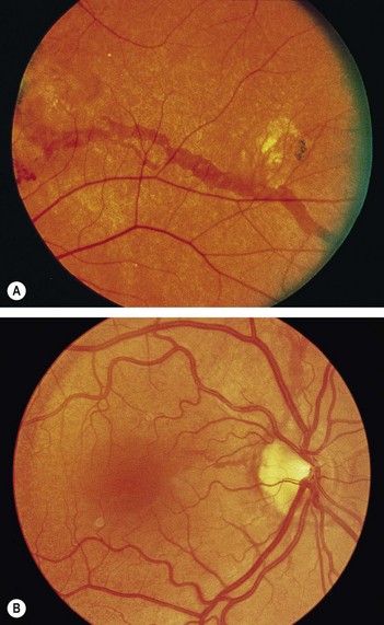
• Fundus shows angioid streaks: linear grey or dark red streaks with irregular edges lying beneath the retinal vessels (Fig. 176.1) (roughly 50% of patients with angioid streaks have pseudoxanthoma elasticum, whereas 85% of those with pseudoxanthoma have angioid streaks).
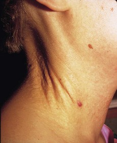
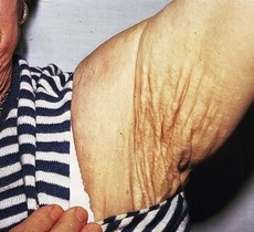
• Look at the neck (Fig. 176.2), antecubital fossae, axillae (Fig. 176.3), groin and periumbilical region for loose ‘chicken skin’ appearance of skin
• Examine peripheral pulses: absent pulses from peripheral arterial involvement but acral ischaemia is uncommon because of development of collaterals.
Patient 2
Advanced-level questions
What are angioid streaks caused by?
They are caused by abnormal elastic tissue in the Bruch’s membrane of the retina.
What are the cardiovascular manifestations of this condition?
• Premature coronary artery disease resembling accelerated atherosclerosis from calcification of the internal elastic laminae of arteries. Arterial grafts should not be used for coronary artery bypass surgery in these patients because of possible calcification of the internal elastic laminae of the internal mammary artery


