Prurigo nodularis
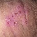
Erythematous, discrete, rounded plaques with central erosion and crust. There are sparse eczematous patches nearer the elbow, but most lesions are discrete.
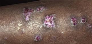
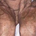
Lesions are thick and severely eroded by picking and scratching. The edges are hyperpigmented.
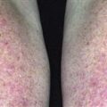
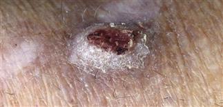
In neurotic excoriations, the linearity of the lesions, oriented in a direction of a natural scratch or ‘dig’, is common. These pink patches are scars, some with fresh but recurrent excoriation.
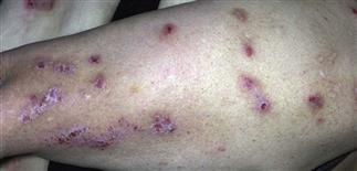
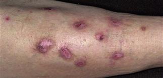
Prurigo nodules have been scattered here. There are old hypopigmented scars, hyperpigmentation, and some discrete papules with crust and pink nodularity. Intralesional steroid is often helpful.
DESCRIPTION
Prurigo may be considered an idiopathic, papular or nodular form of lichen simplex chronicus. In prurigo nodularis, there are very pruritic firm papules and nodules on easily accessed skin. Lesions are secondary to repeated, localized scratching and picking.
HISTORY
• Onset is usually gradual and occurs in setting of pruritus. • Prurigo nodularis occurs primarily in adults. • Stress is often anecdotally implicated. It may perpetuate the problem and seems to relate clinically to recurrence or flares. • Individuals with atopy and diabetes may be predisposed. • Affected patients may be compulsive ‘pickers’.
PHYSICAL FINDINGS
• Few to numerous dull, erythematous, or hyperpigmented nodules randomly distributed. Extensor arms and legs are typically affected; the lumbosacral area, nape of neck, and dorsal hands are reachable areas that are typically involved. • Lesions are created by repeated rubbing, picking, scratching. • The small papules and nodules are red or brown, hard, and often dome-shaped with a smooth, crusted, or warty surface. • Often, there are clues to a chronic process—hypopigmented scars or postinflammatory hyperpigmentation on skin accessible to repeated scratching and picking. • Skin biopsy is rarely necessary but may help confirm the diagnosis when in question. • In generalized prurigo of recent onset (less than 1 year), evaluate and exclude systemic causes of pruritus. • Causes of generalized pruritus include chronic renal disease, drug reactions, hypothyroidism, occult liver disease (including hepatitis C), HIV infection, occult malignancy (including solid-organ metastatic disease and leukemia or lymphoma). • Uncommon mimics of prurigo lesions have been reported and include dermatitis herpetiformis, nodular scabies, metastatic cancerous lesions, Langerhans cell histiocytosis, atypical lymphoproliferative diseases.
TREATMENT
• Medium- or high-potency topical corticosteroids (groups II–IV) with plastic wrap occlusion to enhance penetration and provide barrier to scratching. • Apply corticosteroid-impregnated tape (Cordran) to lesions every day. Covering lesions provides a barrier against trauma of repeated scratching while medicating underlying skin. • Apply topical superpotent steroids twice a day to individual lesions for several weeks. • Intralesional steroid injections (Kenalog 5–10 mg/mL) can be given and repeated every 4–6 weeks if needed. Hypopigmentation in dark skin may occur from such treatment. • Pramoxine with hydrocortisone (Pramosone ointment) or Sarna lotion may help relieve intense pruritus. • Light therapy with ultraviolet B, narrowband ultraviolet B, or psoralen plus ultraviolet A can be considered for severe, generalized cases. • Cryotherapy sometimes successful. • Excision of individual nodules is rarely performed but sometimes a valid option.