Retained Products of Conception

Published on 10/03/2015 by admin

Filed under Obstetrics & Gynecology

Last modified 22/04/2025

Print this page

rate 1 star rate 2 star rate 3 star rate 4 star rate 5 star
Your rating: none, Average: 0 (0 votes)

This article have been viewed 4240 times

Retained Products of Conception

Synonyms/Description

RPOC
Retained placenta
Incomplete abortion

Etiology

Retained products of conception (RPOC) may occur following an abortion, a vaginal delivery, or even after a Cesarean section. This obstetric complication most commonly results in prolonged or excess vaginal bleeding and has also been associated with endometritis. The retained tissue may include a portion of the placenta (such as a succenturiate lobe) or tissue from a pregnancy not completely evacuated.

Ultrasound Findings

Retained products of conception typically appear as an echogenic, vascular mass with ill-defined borders, centrally located within the endometrial cavity. The mass may contain areas of fluid but is largely solid. Color Doppler often shows an alarming amount of vascularity, which has been described as mimicking an arteriovenous malformation (AVM); however, this resolves completely after evacuation of the retained products. The sonographic appearance of the retained tissue can have characteristics of placenta, especially if the patient is postpartum. If the patient had an early pregnancy loss or termination, the tissue is more often echogenic with small cystic areas. Abundant vascularity is a characteristic feature of most types of retained products of conception.
Durfee and colleagues report that an endometrial mass is the most sensitive (79%) and specific (89%) sonographic finding in patients with retained products of conception. None of the patients in their study with this diagnosis had a completely normal scan.
Regarding vascularity, Atri and colleagues report that the presence of focal vascularity had sensitivity, specificity, negative predictive value, and positive predictive value of 94%, 67%, 95%, and 65%, respectively. Of the patients with pathologically confirmed retained products of conception, five had focal increased vascularity without a mass, but none had a mass without focal increased vascularity.

Differential Diagnosis

When observing a vascular mass in the endometrium, it is important to determine the clinical history. The sonographic appearance alone may suggest a large polyp or an abnormal endometrium such as hyperplasia or endometrial cancer. If the vascularity in the lesion is dramatic, an AVM may be suspected, although primary uterine AVMs are exceedingly rare. If the patient is postpartum or if there has been a recent pregnancy loss, the diagnosis is retained products of conception until proved otherwise.

Clinical Aspects and Recommendations

In patients who have had a first trimester pregnancy loss or termination, the presence of vascularity may be helpful in predicting the success of expectant management for RPOC. In the study by Casikar and colleagues, the absence of color flow was predictive of successful expectant management with a sensitivity, specificity, positive predictive value, negative predictive value, and positive likelihood ratio of 90.3%, 37.5%, 89%, 40.9%, and 1.445, respectively. There was no correlation between the volume of the retained products and the success of expectant management.
Active management includes both surgical and medical options. Medical management with misoprostol, a prostaglandin agonist, is a relatively new tool. Treatment with misoprostol for retained products from early pregnancy failure showed a greater than 90% success rate in women presenting with localized abdominal pain, Rh-negative blood type, or a combination of active bleeding and nulliparity. Thus, in a select group, the decision to pursue expectant, medical, or surgical management depends on multiple factors, including patient preference. Patients presenting with hemodynamically symptomatic bleeding or acute hemorrhage require surgical management with dilation and curettage (D&C). In these patients, swift action is needed to remove the cause of bleeding since such patients can rapidly develop disseminated intravascular coagulopathy (DIC).

Figures

image

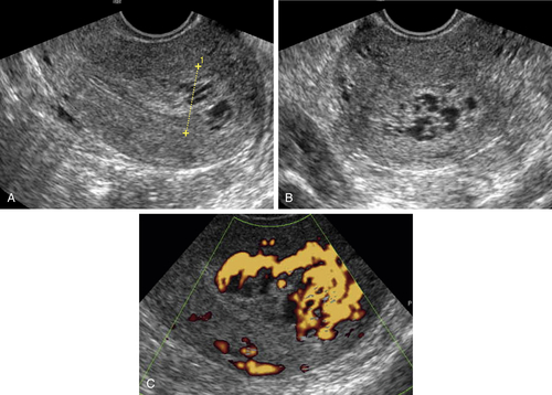
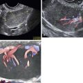
Figure R1-1 Typical case of RPOC after an incomplete first trimester pregnancy loss. A and B (gray-scale views) show the ill-defined margins of the endometrium at the fundus with irregular cystic spaces. The color Doppler in C shows that these spaces are blood vessels in this very vascular case of RPOC.


image

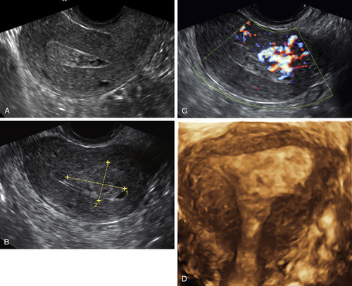
Figure R1-2 A to D, The uterine cavity contains an echogenic mass, filling the cavity (A) and extending to the edge of the endometrium, blurring the margins (B, calipers). C shows the abundant color flow at the site of the RPOC. D shows the 3-D appearance of the same case, demonstrating an asymmetric enlargement and deformity of the left fundus/cornu, representing the RPOC.


image

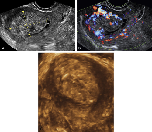
Figure R1-3 A to C, Sclerotic RPOC, 6 months after a term delivery (2-D, Doppler, and 3-D images). Note the complex-appearing vascular mass involving the endometrium. If this were seen in a postmenopausal patient with bleeding, the appearance would be consistent with endometrial cancer. The history of a symptomatic postpartum patient enabled the correct diagnosis of longstanding RPOC (sclerotic on pathologic examination).


image

Figure R1-4 Patient with an endometrial polyp. Note the similarity to the RPOCs. This patient had never been pregnant

image

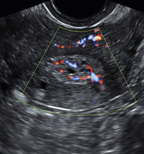
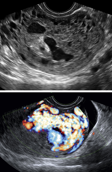
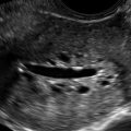
Figure R1-5 Rare case of spontaneous AVM unrelated to pregnancy. The entire uterus is replaced by cystic areas on gray-scale imaging. Color flow Doppler shows abnormal vessels reaching to the serosa and involving much of the myometrium. The pattern and clinical history were very different in this nulliparous patient.


Videos

Videos 1 and 2 on retained products of contraception are available online.

Suggested Reading

Atri M., Rao A., Boylan C., Rasty G., Gerber D. Best predictors of grayscale ultrasound combined with color Doppler in the diagnosis of retained products of conception. J Clin Ultrasound. 2011;39:122–127.

Casikar I., Lu C., Oates J., Bignardi T., Alhamdan D., Condous G. The use of power Doppler colour scoring to predict successful expectant management in women with an incomplete miscarriage. Hum Reprod. 2011;27:669–675.

Creinin M.D., Huang X., Gilles J., Barnhart K., Westhoff C., Zhang J. Medical management of early pregnancy failure. Obstet Gynecol. 2006;107:901–907.

Durfee S.M., Frates M.C., Luong A., Benson C.B. The sonographic and color Doppler features of retained products of conception. J Ultrasound Med. 2005;24:1181–1186.