Chapter 9 Production of X-rays


TARGET INTERACTIONS
BREMSSTRAHLUNG INTERACTIONS
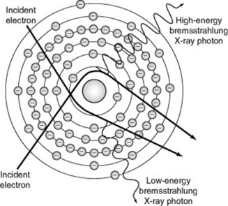
Bremsstrahlung in German means ‘to brake radiation’ or braking radiation. Bremsstrahlung target interaction occurs when projectile electrons pass by outer shell electrons of target atoms and interact with the force field of the nucleus of the atom. Because atomic nuclei are positively charged and electrons are negatively charged, there is a mutual attraction between them. The nuclear force field causes the entering electron to slow down (or brake) and change direction. The loss of kinetic energy that occurs when a projectile electron slows down is emitted as an X-ray photon. These X-ray photons are known as bremsstrahlung photons or brems radiation (Fig. 9.1). In the diagnostic range, approximately 85% of X-ray emissions are the result of bremsstrahlung interactions.
CHARACTERISTIC INTERACTIONS
Characteristic radiation is produced when projectile electrons with sufficient kinetic energy eject an inner orbital electron (Fig. 9.2). When this happens, the atom becomes unstable and temporarily ionised because of the missing electron. An electron from an outer shell instantly fills the void created by the missing electron and an X-ray photon is emitted. This process continues until the atom is stable. The energy of the emitted X-ray photon is equal to the difference between the binding energy of the two involved orbital electrons. Accordingly, each X-ray photon has a specific energy level. This explains why this type of emission is called characteristic radiation. The energy emitted is characteristic of the target element and the involved shells.
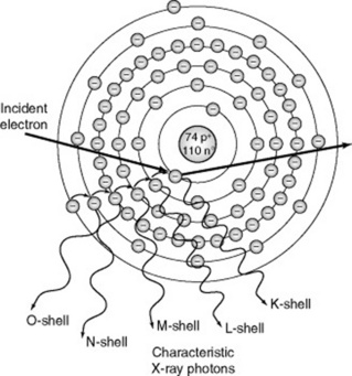
Higher energy X-ray photons result with target materials of a higher proton number and interactions that involve the ejection of inner shell electrons. Each target element emits characteristic radiation of a given energy. For example, the K shell binding energy for tungsten is 69.5 keV. Only projectile electrons with energies greater than the K shell binding energy are able to eject K shell electrons. Accordingly, K shell characteristic X-rays are only produced when the applied voltage exceeds 69.5 kVp. In comparison, the characteristic radiation of a molybdenum target (often used for mammography) is very different. The K shell binding energy for molybdenum is 20 keV,so K shell characteristic X-rays are produced when the applied voltage exceeds 20 kVp.
EMISSION SPECTRUM
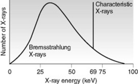
The emission spectrum is a graphic representation of the number of X-rays plotted against the energy of the radiation, which is measured in kiloelectron volts (keV) (Fig. 9.3). The emission spectrum for bremsstrahlung radiation is continuous because bremsstrahlung X-rays include a range of energies. The emission spectrum for characteristic radiation is discrete because characteristic X-rays consist of predictable energies that are specific to the target element.
CONTINUOUS X-RAY SPECTRUM
The size and shape of the emission spectrum reflects the quality and quantity of the X-ray beam. While the relative shape of the emission spectrum remains the same, its location along the horizontal axis can vary. Ranges located more towards the right represent X-ray beams of higher energy or quality. Graphically, the area under the curve represents the total number of X-rays emitted. A larger area represents X-ray beams with higher intensity or quantity. The greatest number of X-rays have approximately one-third to one-half of the maximum energy.1
DISCRETE X-RAY SPECTRUM
Characteristic radiation is graphically illustrated in the form of a line spectrum. Remember that the energy of a characteristic photon depends on the differences between the electron binding energies of a particular target material. As a result, the spectrum produced by characteristic X-rays is referred to as discrete or distinct. For example, there are only 15 specific energy levels of characteristic X-rays from tungsten: five from interactions at the K shell, four from interactions at the L shell, and the remainder from interactions at lower energy outer shells. In tungsten, only characteristic X-rays produced from the five K shell interactions are of sufficient energy to be of diagnostic value. The number of photons produced at each characteristic energy level is different because the likelihood for filling a K shell void varies from shell to shell. Often, the five energy levels are represented on the emission spectrum as a single line. As illustrated in Figure 9.3, the vertical line at 69 keV represents the characteristic K X-rays of tungsten.
X-RAY QUALITY AND QUANTITY
The quality of radiation in an X-ray beam is the penetrating ability of the beam. The quantity of radiation in an X-ray beam is the number of photons in the beam. The terms exposure and intensity may also be used to describe quantity.
While practitioners have little control over the selection of the target material and limited options for the use of added beam filtration, it is valuable to understand how the target material and beam filtration affect the quality and quantity of the X-ray beam. Practitioners are able to control distance and prime exposure factors. Consequently, it is essential to understand how these factors influence the quality and quantity of the X-ray beam (Table 9.1).
Table 9.1 Summary of factors affecting X-ray quality and quantity
| Factors affecting X-ray quality | Factors affecting X-ray quantity |
|---|---|
| Target material | Target material |
| Beam filtration | Beam filtration |
| kVp | Distance |
| kVp | |
| mA s |
kVp, kilovolt (peak); mA s, milliamp-second
DISTANCE
The distance of the anode from the image receptor (source–image distance, SID) affects the quantity of X-rays photons (see p. 91). The inverse square law governs the relationship between the quantity of X-ray photons and the distance from the target to the image receptor. The quantity of X-ray photons at the image receptor is inversely proportional to the square of the distance from the source (see p. 91). For example, if the SID is reduced by one-half, the number of X-ray photons quadruples.
PRIME EXPOSURE FACTORS
Kilovoltage (kVp)
The kilovoltage peak (kVp) set by the practitioner determines the voltage or potential difference applied across the cathode and anode during the exposure. This setting affects both the quality and quantity of the X-ray beam. As mentioned earlier, the kVp setting controls the speed of the electrons travelling from the cathode to the anode. An increase in kVp causes greater repulsion of electrons from the cathode and greater attraction of electrons towards the anode. This increased speed means projectile electrons possess greater potential energy.
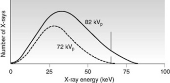
Changes in kVp affect the production of bremsstrahlung radiation, which influences both the quality and quantity of photons in the X-ray beam. An increase in kVp results in higher quality X-ray photons with a higher average energy and more penetrating ability. Keep in mind that the maximum energy of an X-ray beam remains equal to the kVp setting. With an increase in kVp there is also an increase in the quantity of X-ray photons at all energy levels. However, the increase is relatively greater for high energy X-rays than for low energy X-rays. The emission spectrum in Figure 9.4 illustrates how the area under the curve increases and shifts to the right as kVp is increased.
EXPOSURE MANIPULATION
Exposure manipulation includes those variables that practitioners most often employ to manage the quality and quantity of the X-ray beam. Distance, kVp, and mA s are the primary factors considered here.