nerve tissue
Nerve tissue and the nervous system
The nervous system has two structural components. Nerve tissue located in the brain and spinal cord is the major component of the central nervous system (CNS). Other primary tissues are present in the CNS though they are sparse, e.g. connective tissue provides some support. The peripheral nervous system (PNS) comprises nerve tissue which is not in the CNS, e.g. cranial and spinal nerves and their branches, and autonomic nerves (see below). Connective tissue also has a supporting role in the PNS.
The nervous system has two functional components, somatic and autonomic. The somatic nervous system involves the detection of sensations and the control of skeletal muscle contraction. Some sensations are not consciously perceived, such as a change in tension in a tendon, though the change detected can cause a response, such as contraction of skeletal muscle(s). The functions of the autonomic nervous system (ANS) involve the control of contraction of smooth and cardiac muscle cells and the secretory activity of some gland cells. The ANS, in general, is not under conscious control. However, an important exception is that the autonomic nerves involved in controlling the smooth muscle cells in the wall of the urinary bladder are normally under conscious control by the age of 2–3years.
Neurons
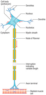
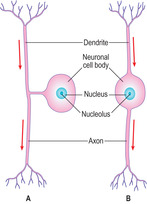
Neurons are derived from the ectoderm germ layer of the embryo (see Mitchell B, Sharma R. Embryology: An Illustrated Colour Text. Elsevier: 2004). Most neurons have a large mass of cytoplasm and large nucleus forming the body of the cell (the perikaryon) and a variable number of cytoplasmic processes (Fig. 6.1). Perikarya may measure up to 150μm in diameter, which is about 20 times larger than a red blood cell. The cytoplasmic processes are of two forms, dendrites and axons. Most neurons have several short dendrites that receive signals which they then pass to their own cell body. In general, each neuron has a long, single axon which takes signals away from the perikaryon either to other neurons or to other cell types, e.g. muscle cells. Axons may be up to about a metre in length in humans, i.e. the length from the lower regions of the spinal cord to the foot.
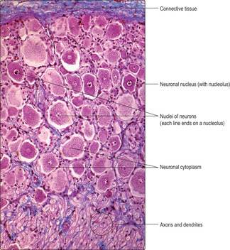
In routine H&E-stained sections, neuronal nuclei have large, palely stained areas (Fig. 6.2). Such areas represent uncoiled euchromatin, the hallmark of DNA transcription and a prerequisite for protein synthesis. The cytoplasm in neuronal cell bodies often appears granular, especially if special stains have been used (Fig. 6.3). The granules are referred to as Nissl granules and they are due to large accumulations of rough endoplasmic reticulum (RER), which are involved in the synthesis of various proteins. Some of these proteins provide the structural tubules and filaments in axons and dendrites. Others are enzymes involved in producing molecules which aid the transmission of signals.
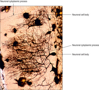
Neurons vary in shape and size, and according to function and location. The complexity of their cytoplasmic processes is vast and revealed by special stains (Fig. 6.4).
■ Variation in shape. Neurons are classified as multipolar (Fig. 6.1), bipolar or pseudounipolar (Fig. 6.5). The polarity is related to the number of cytoplasmic processes projecting from the perikaryon. Many neurons are multipolar and, typically, one axon and many dendrites extend from the perikaryon (Fig. 6.1). Some neurons in the brain and ventral regions (horns) of the spinal cord are multipolar. Bipolar neurons are present in organs of the special senses (e.g. the eye) and they have two cytoplasmic processes, one an axon and one a dendrite. Pseudounipolar neurons possess one process, though it bifurcates and one branch receives signals and the other branch transmits them away from the cell body. Examples of pseudounipolar neurons are those that transmit sensory information from the skin.
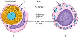
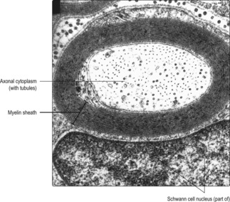
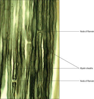
The rate at which nerve impulses are transmitted along axons varies. In general, the wider the diameter of an axon, the faster the nerve impulse travels. The speed of transmission of nerve impulses is also higher in axons that are sheathed in myelin (Fig. 6.1). Myelin is a lipid bilayer arranged as concentric, multiple layers around some axons (Figs 6.6A, 6.7 and 6.8). The layers are formed from the cell membranes of Schwann cells in the PNS and, in the CNS, from the cell membranes of certain glial cells (oligodendrocytes). In places, the myelin sheaths around axons are not layered and these regions are described as nodes of Ranvier (Figs 6.1 and 6.9). The nodes are the interface between two adjacent myelin-producing cells and they are important in ensuring a relatively rapid rate of transmission of the nerve impulse. Other axons are not ensheathed in myelin, but several axons are indented into the cytoplasm of one myelin-producing Schwann cell (Fig. 6.6B). Transmission of impulses along these non-myelinated axons is relatively slow.
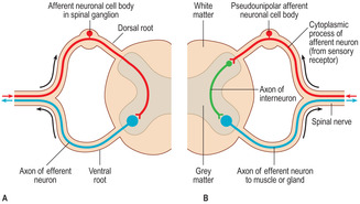
■ Variation in location. Afferent neurons carry sensory signals to the CNS and efferent neurons carry signals away from the CNS to various cells, e.g. muscle and gland cells. Afferent nerves enter the spinal cord by dorsal roots and efferent nerves leave by ventral roots (Fig. 6.10). Some afferent neurons communicate directly in the CNS with efferent neurons and form a two-neuron reflex arc (pathway) (Fig. 6.10A). Others transmit information to interneurons in the spinal cord which in turn communicate with efferent neurons in a three-neuron arc (Fig. 6.10B). In both types of reflex arc other neurons transmit impulses to the brain.
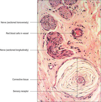
Afferent (sensory) neurons provide a variety of information. Some afferent neurons have specialised structures known as sensory receptors (Fig. 6.11) at the peripheral end of a cytoplasmic process; others end as ‘free nerve endings’. Sensory receptors in, for example, the skin detect sensations, and nerve impulses are transmitted to the CNS. Sensory receptors in skin can provide impulses that are perceived as pain, touch (differentiating type and pressure) and temperature. Signals from sensory receptors in some organs may be perceived as changes in pressure or amount of stretch. Other sensory receptors convey information which is not perceived consciously, e.g. changes in blood pressure, but the body responds to the changes detected. Highly specialised sensory neurons are present in the organs involved in sight, hearing, smell and taste; for information about these, a more detailed histology book should be consulted.
Efferent neurons transmit impulses away from the CNS and affect the function of other neurons and other cells, e.g. gland and muscle cells (efferent neurons which stimulate muscle contraction are known as motor neurons (Fig. 6.1)). There are two categories of efferent neuron:
■ Somatic efferent neurons. These stimulate skeletal muscle contraction and may involve a series of nerve impulses originating in the brain after conscious thought. Skeletal muscle contraction may also occur as a reflex response, e.g. to a painful stimulus. The sensory information passes in a reflex arc through the spinal cord (Fig. 6.10) and directly (or via an interneuron) to one or more efferent motor neurons which stimulate muscle contraction. This reflex pathway does not involve the brain but neurons relay the pain sensations rapidly to the brain and then the pain is perceived.
■ Autonomic efferent neurons. These control smooth muscle contraction, e.g. smooth muscle in the walls of blood vessels and the walls of the digestive, respiratory, reproductive and urinary tracts. Autonomic efferent nerves also control the contraction of cardiac muscle cells and gland cell secretion, e.g. by salivary and sweat glands.
Synapses
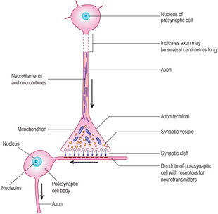
Synapses are specialised cell junctions between neurons or between neurons and other cell types, e.g. muscle cells. The commonest form of synapse is the axo-dendritic synapse formed between the axon of a neuron and the dendrite(s) of another neuron.
All synapses have similar characteristics (Fig. 6.12). In axo-dendritic synapses the membrane of the terminal part of the axon of the presynaptic cell lies in close apposition to the membrane of a dendrite of the postsynaptic cell. The space between the two cells is known as the synaptic cleft. The neuron transmitting the nerve impulse to a synapse has a presynaptic axonal swelling in which numerous cellular organelles and synaptic vesicles are present. The synaptic vesicles are small membrane-bound structures containing neurotransmitter molecules. The morphology of synaptic vesicles is related to their content. The main neurotransmitters include acetylcholine and catecholamines such as noradrenaline and dopamine.
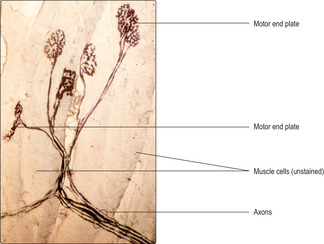
Synapses between axons and muscle cells are known as motor end plates (Fig. 6.13). Each axon usually branches at its peripheral end and forms synapses with several muscle cells. The presynaptic axonal membrane abuts the highly folded postsynaptic muscle cell membrane. The two membranes are separated by the synaptic cleft. The release of neurotransmitters triggers changes in the ionic permeability of the membrane of the muscle cells, which results in contraction. The motor neuron and the muscle cell (or cells) with which it synapses is described as a motor unit.
Ganglia and nuclei
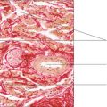
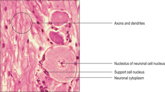
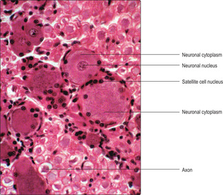
Ganglia are defined as collections of neuronal perikarya (cell bodies) not in the CNS. The equivalent aggregations of neuronal perikarya in the CNS are known as nuclei. In addition to neuronal cell bodies, ganglia also contain supporting (satellite) cells and parts of the cytoplasmic processes (axons and dendrites) of neurons (Figs 6.3 and 6.14). The satellite cells surround and support the perikarya in ganglia by providing electrical insulation and ensuring that nutrients reach the neuronal perikarya. Connective tissue surrounds each ganglion and a sparse network of connective tissue is present within each (Fig. 6.3).
There are two types of ganglion:
■ Spinal ganglia are attached to spinal nerves (Fig. 6.10) and located in intervertebral foramina. They are characterised by collections of closely packed sensory, pseudounipolar, neuronal perikarya. They receive and transmit signals about sensations through their single cytoplasmic process. Each cell body is surrounded by a layer of satellite cells (Fig. 6.14).
■ Autonomic ganglia are part of the ANS and occupy sites near to the spinal cord (in pre- and paravertebral ganglia) and in the walls of, or close to, some viscera. The neuronal cell bodies in autonomic ganglia are multipolar and receive signals, via synapses, from axons of preganglionic neurons. They transmit the signals away from ganglia via their axons.
Autonomic ganglia are classified into two functional types, sympathetic and parasympathetic.
■ Sympathetic ganglia. The neurons in these ganglia are involved in the body’s alarm responses (fright, fight and flight) and are in the sympathetic chain of paravertebral ganglia and in prevertebral ganglia.
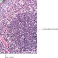
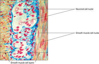
■ Parasympathetic ganglia. Parasympathetic neurons are involved in the ‘rest and digest’ activities of the body. They are similar in structure to sympathetic ganglia but are typically found in the walls of, or near to, the organs they affect (Fig. 6.15) and the number of perikarya present is often quite small.
Glial cells
Glial cells are present in the CNS and they are more numerous than the neurons. They function in maintenance, support and repair. There are three types: astrocytes, oligodendrocytes and microglial cells.
■ Astrocytes. Each astrocyte has a small cell body with numerous, radiating, small cytoplasmic processes like stars, hence their name. They act in two ways. Firstly, they have cytoplasmic processes that are wrapped around blood vessels; this is an important structural and functional relationship forming the blood–brain barrier. This barrier prevents the passage of molecules larger than about 500 daltons between blood and brain. Secondly, they repair damaged tissue, a process known as gliosis. The latter is the equivalent of repair by fibroblasts in the rest of the body.
■ Oligodendrocytes. These cells produce myelin which ensheaths some axons in the CNS.
■ Microglia. These are small cells and they are the only nerve tissue cells which originate from mesoderm. In response to inflammation, they undergo phagocytosis and aid repair; in this they resemble macrophages (Chapters 4 and 8).
Brain and spinal cord
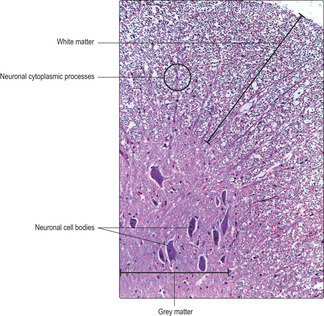
The arrangement of neurons in brain and spinal cord is complex (see Crossman AR, Neary D. Neuroanatomy: An Illustrated Colour Text, 3rd edn. Elsevier: 2005). Two principal areas are described in brain and spinal cord as white and grey matter. In the brain, grey matter forms the peripheral region, whereas in the spinal cord it is the central region (Fig. 6.10). Neuronal cell bodies are predominant in grey matter, and axons, particularly myelinated axons, are prominent in white matter. To the unaided eye, white matter appears white; it is the lipid content of myelin that gives this appearance. In histological sections the regions of grey and white matter can be clearly revealed (Fig. 6.16).
Peripheral nerves
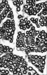
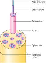
Peripheral nerves pass between the CNS and the rest of the body. The peripheral nerves attached to the brain are known as cranial nerves and those attached to the spinal cord as spinal nerves. Peripheral nerves may be up to 2cm in width and contain the cytoplasmic processes (axons and dendrites) of a very large number of neurons. Numerous ensheathing Schwann cells and connective tissue are also present. Peripheral nerves become smaller as they branch and pass around the body to the sites where the neuronal cell processes they contain initiate or receive signals. Their appearance in histological sections varies depending on the angle at which they are sectioned (Fig. 6.11). Individual cytoplasmic processes are difficult to distinguish in peripheral nerves but the nuclei of Schwann cells and supporting connective tissue cells can be seen, although not always distinguished (Fig. 6.11).
Peripheral nerves contain layers of connective tissue (Fig. 6.17). The connective tissue wraps around axons at three levels of organisation. Endoneurium surrounds individual axons. Groups of axons, with their coverings of endoneurium, are bundled together and surrounded by perineurium. The perineurium also packs the space between adjacent bundles of axons. Several bundles of axons within their investments of endoneurium and perineurium are surrounded by the connective tissue of the epineurium.
Most peripheral nerves contain neuronal cell processes which transmit impulses to the CNS and other cell processes which transmit impulses away from the CNS, respectively processes from afferent and efferent neurons. All peripheral nerves also carry cytoplasmic processes from sympathetic nerves, whereas some of those in the head and trunk also carry parasympathetic nerves.

Blood–brain barrier The blood–brain barrier presents a physical barrier to molecules over 500 daltons in size. If the barrier breaks down, the brain may swell. This condition, known as cerebral oedema, may also develop in response to a lesion in the brain, e.g. a tumour or an abscess, or after trauma, or after a stroke resulting in ischaemic brain damage.
Tumours of nervous tissue Such tumours are not usually derived from neurons. Neurons do not divide, thus we are born with the maximal number of neurons and many die as life progresses. Tumours of nerve tissue are mainly of the glial cell lineage. Of these, astrocytomas are the commonest. The most severe form is highly malignant and patients have a very poor prognosis.
Multiple sclerosis In this condition the immune system attacks the myelin sheaths of nerves in the CNS. The cause is unknown. The demyelination leads to lack of motor and sensory coordination. The attacks may be single or sequential, and the degree of disability varies between patients and at different times during the life of individual patients.
Nerve tissue
■ consists of neurons (nerve cells), glial cells and satellite cells.
The nervous system comprises:
■ the central nervous system (CNS) (brain and spinal cord)
■ the peripheral nervous system (PNS) (nerve tissue not in the CNS).
Neurons
■ Neurons mostly have large cell bodies (perikarya) with granular cytoplasm owing to RER involved in protein synthesis.
■ They have cytoplasmic processes (usually many dendrites which transmit nerve (electrical) impulses to their perikaryon and a single axon which transmits signals away from the perikaryon).
■ They transmit impulses fastest along axons ensheathed in myelin formed by Schwann cells in the PNS and oligodendrocytes in the CNS.
■ Sensory signals are transmitted by afferent neurons to the CNS and efferent neurons carry impulses away from the CNS which affect the function of, for example, muscles.
Synapses
■ These are specialised junctions between neurons or between neurons and other cell types.
■ A nerve impulse reaching a synapse stimulates the release of neurotransmitter molecules into the synaptic cleft between the pre- and postsynaptic cells. Neurotransmitters stimulate the postsynaptic cells to transmit a nerve impulse (if they are neurons), or to contract (if they are muscle cells), or to secrete or to stop secreting (if they are gland cells).
Ganglia and nuclei
■ Ganglia are groups of neuronal cells bodies in the PNS; nuclei are collections of neuronal cell bodies in the CNS.
■ Spinal ganglia are part of the somatic nervous system.
■ Autonomic ganglia are part of the autonomic nervous system. Sympathetic ganglia are involved in fright, fight and flight responses. Parasympathetic ganglia are involved in rest and digest activities.
Types of glial cell
■ Astrocytes are small cells involved in forming a structural and functional barrier between the blood and the brain.
■ Oligodendrocytes form myelin sheaths around some axons in the CNS.
■ Microglia are able to aid repair of damaged tissue.