Preoperative Pulmonary Evaluation

Published on 23/05/2015 by admin

Filed under Pulmolory and Respiratory

Last modified 22/04/2025

Print this page

rate 1 star rate 2 star rate 3 star rate 4 star rate 5 star
Your rating: none, Average: 0 (0 votes)

This article have been viewed 1818 times

Chapter 73 Preoperative Pulmonary Evaluation

Surgery can influence the patient’s pulmonary function. Pulmonary complications of surgery are common, costly, and increase morbidity and mortality in both the near term and the long term. The preoperative assessment of lung function and optimization of the management of preexisting lung conditions can improve outcomes. This chapter discusses surgery-related changes in pulmonary physiology, the impact of common postoperative pulmonary complications, risk factors for these complications, and recommendations for preoperative pulmonary assessment and management, including those related to the evaluation of the lung resection candidate.

Pulmonary Physiology

During and after surgery, ventilation, ventilation-perfusion matching, and airway clearance are altered by many mechanisms. The mechanisms responsible and the degree of impairment are influenced by the type of surgery and the patient’s underlying health. The following is a general description of potential physiologic changes related to surgery.

Postoperative Pulmonary Complications

The frequency of postoperative pulmonary complications (PPCs) varies with type of surgery, the patient’s health, and the definition of the complication. Pulmonary complications of surgery are at least as common as cardiac complications and may result in prolonged hospital stays, increased morbidity and mortality, and increased costs (Table 73-1).

Table 73-1 Postoperative Pulmonary Complications

Surgery in General Lung Resection Surgery

* Acute exacerbation of chronic obstructive pulmonary disease.

Risk Factors

The many risk factors for PPCs can be classified as patient or surgery related and assigned a simple rating based on the amount and quality of evidence available for support as a risk factor (Table 73-2).

Table 73-2 Selected Risk Factors for Developing Postoperative Pulmonary Complications

Risk Factor Level of Prediction
Systemic Disease  
Asthma (well controlled) Poor
Congestive heart failure Good
Chronic obstructive pulmonary disease Good
Diabetes mellitus Fair
Human immunodeficiency virus infection I
Obstructive sleep apnea Good
Pulmonary hypertension Fair
Signs and Symptoms  
Abnormal chest auscultation Good
Arrhythmia I
Functionally dependent Good
Impaired mental status Good
Poor exercise capacity Fair
Weight loss Good
Patient Features  
Alcohol user Good
Cigarette smoker Good
Corticosteroid use I
Objective Testing  
Albumin <35 g/L Good
Blood urea nitrogen >25 mg/dL Good
Chest radiography, abnormal finding Good
Spirometry I
Surgical Site  
Aortic aneurysm Good
Abdominal Good
Neurosurgery Good
Vascular Good
Hip Poor
Gynecologic or urologic Poor
Esophageal I
Surgical Factors  
Prolonged surgical duration Good
Emergent surgery Good
General anesthesia Fair
Perioperative transfusion Good

Good, ACP “A” or “B” rating or other data to support; Fair, ACP “C” rating or other data to support; Poor, ACP “D” rating, or is not a risk factor; I, indeterminate (insufficient data to support a rating.

Data from current American Academy of Chest Physicians (AACP) guidelines; Smetana GW et al: Cleve Clin J Med 73(Suppl 1):36–41, 2006; and other sources (see Suggested Readings).

Patient-Related Factors

In several studies, chronic obstructive pulmonary disease (COPD) has been found to double the risk of PPCs. The degree of risk is directly related to the severity of obstruction. One study of patients after upper abdominal surgery found that those with decreased breath sounds or other adventitious sounds (e.g., wheezing, rales) had a sixfold increased rate of PPCs.

In contrast, well-controlled asthma has not been found to be a risk factor for PPCs. A large retrospective analysis found that rates of bronchospasm, laryngospasm, and respiratory failure in patients with well-controlled asthma were comparable to healthy individuals. Those with poorly controlled asthma (e.g., more frequent albuterol use, recent emergency department visit for asthma) do have an increased risk for PPCs.

Pulmonary hypertension (PH), primary and secondary, may be associated with increased rates of PPCs and in-hospital mortality. In one study, 21% of patients with PH developed respiratory failure versus 3% in matched controls after noncardiac surgery.

A patient’s general health may be assessed by using the American Society of Anesthesiologists (ASA) classification system (Table 73-3). Studies have shown a direct correlation between a higher ASA class and increased pulmonary complications. Patients with an ASA Class III or higher have a twofold to threefold increase in PPCs compared with those with ASA Class II or lower. Difficulty or inability to perform activities of daily living has also been linked to increased complications.

Table 73-3 ASA Classification for Surgical Candidates

Class Patient Status
I Normal healthy patient
II Patient with mild systemic disease (e.g., mild asthma)
III Patient with severe systemic disease, but not incapacitated
IV Patient with life-threatening illness who is incapacitated
V Moribund patient
VI “Brain-dead” patient eligible for organ donation

ASA, American Society of Anesthesiologists.

Obesity alone does not seem to increase the risk of PPCs. A wide review of gastric and general surgeries shows that obese and morbidly obese patients have the same risk for PPCs as nonobese patients. However, the presence of a common comorbidity in obese patients, obstructive sleep apnea (OSA), does confer increased risk. In a study of patients after hip or knee replacement, those with OSA had longer hospital stays or more frequent transfers to the intensive care unit (ICU). A more recent study found that those with five or more desaturation episodes during the night had greatly increased rates of PPCs. Therefore, patients should be asked about symptoms of OSA preoperatively.

Age is the most significant patient-related predictor for PPCs. Even after adjustment for comorbid conditions, increasing age is an independent risk factor for increased PPCs. Congestive heart failure (CHF) is also an independent risk factor and should be of particular concern in the elderly population.

Cigarette smokers have a moderately increased risk for PPCs, independent of comorbid lung conditions. Smokers have higher rates of wound infections, oxygen desaturation, laryngospasm, and increased coughing with anesthesia, all of which can lead to longer hospital stays and contribute to pulmonary complications. Quitting smoking before surgery lowers the rate of PPCs. Although early data suggested that quitting smoking close to surgery may increase the rate of PPCs, a recent meta-analysis shows this is not true. In general, the sooner a patient can quit smoking, the better it is for the patient, but smoking cessation should be encouraged at any time before surgery.

Surgery-Related Factors

The planned surgery and surgical site have an impact on risk for developing PPCs that is often greater than patient-related factors. Surgical site was the most important predictor for developing postoperative pneumonia and respiratory failure in a cohort of 160,000 U.S. military veterans undergoing major noncardiac surgery. Those undergoing abdominal aortic aneurysm (AAA) repair or thoracic surgery had the highest risk, followed by upper abdominal, neurovascular, and neck surgeries.

Duration of surgery, extent of anesthesia, and invasiveness of the surgery all influence the risk of developing PPCs. Patients undergoing emergency surgeries or surgeries lasting longer than 3 hours are at greater risk for PPCs. Surgeries requiring general anesthesia (GA) carry a greater risk for PPCs than with peripheral nerve blocks. There is questionable evidence about the relative risk of PPCs from surgeries requiring GA versus neuraxial blockade (spinal or epidural anesthesia). A previous large review of various surgeries reported increased rates of pneumonia, respiratory failure, and mortality in patients who received GA compared to those receiving neuraxial blockade. A more recent small review of orthopedic surgeries found equal rates of pneumonias in these two groups.

A study of patients undergoing AAA repair found increased rates of PPCs (16%) in open repair compared to endovascular repair (3%). Video-assisted surgeries have reduced PPC rates compared with open approaches. Limited data suggest that patients undergoing cardiothoracic robotic surgery have lower rates of PPCs than those not receiving robotic surgery.

Surgery in General

The most important part of a preoperative pulmonary evaluation is taking a complete history and performing a complete physical examination. For those without pulmonary disease or symptoms, or those with well-controlled chronic lung conditions, routine pulmonary function tests (PFTs) and chest imaging are not indicated.

During the office visit, symptoms or signs of an underlying or undiagnosed pulmonary condition may be discovered. If unexplained dyspnea, coughing, wheezing, other abnormal lung sounds, or chest pain is revealed, testing before an elective surgery may be indicated to identify their cause. Historical clues should aid in distinguishing cardiovascular dyspnea from pulmonary dyspnea and guide which tests should be performed. Pulmonary function testing will help to identify and quantitate physiologic impairment of lung function. If a cardiac etiology is suspected, an electrocardiogram, echocardiogram, brain natriuretic peptide level, and stress test may be performed. Other cardiac risk assessment is beyond the scope of this chapter.

Further testing, such as a chest radiograph, complete blood count, serum chemistries, and thyroid-stimulating hormone (TSH) level can help to determine the cause of cardiopulmonary symptoms and the need for additional testing. If the cause of dyspnea remains unclear, a cardiopulmonary exercise test may help determine its nature and quantify its severity. Additional chest imaging may also be required if a pulmonary cause of a concerning symptom is likely. Screening for PH is not recommended in the preoperative setting, particularly if respiratory symptoms are not present or are otherwise well explained. Since most, if not all, of the conditions causing dyspnea result in increased rates of PPCs, proper diagnosis and treatment optimization of that condition will improve patient outcomes.

During the history and physical examination, patients should be screened for OSA by asking about snoring, witnessed apneas, and daytime somnolence. Neck circumference and obesity should be noted. Validated questionnaires are available for identifying those at risk for OSA (Box 73-1). Evidence is limited on the usefulness of diagnosing OSA preoperatively with polysomnography, with decisions based on degree of clinical concern and urgency of the surgery.

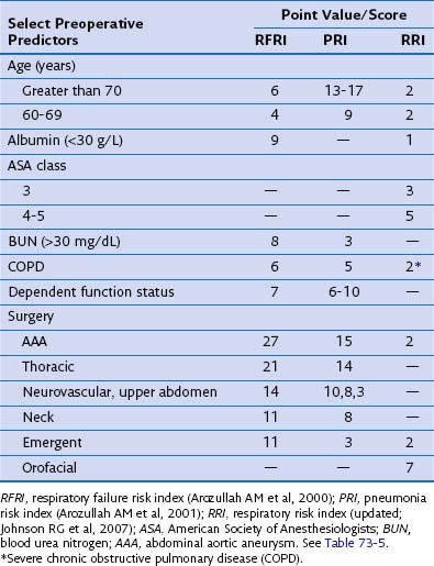
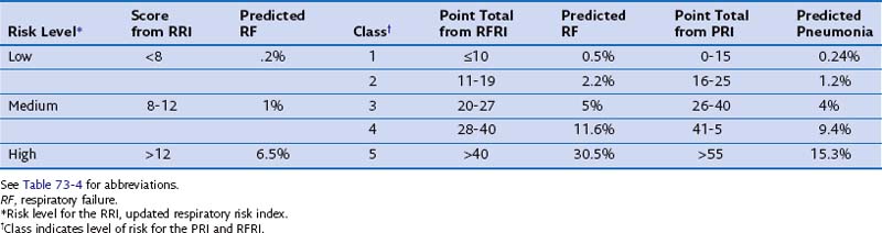
Validated indices are available for predicting postoperative pneumonia and respiratory failure after major noncardiac surgery. The investigators from three select reviews assigned point values to corresponding risk factors, as summarized in Tables 73-4 and 73-5. Clinicians may find these tools helpful in identifying those at risk for postoperative pneumonia and respiratory failure and for selecting perioperative testing and respiratory care for those at high risk.

Lung Resection Surgery

For patients with non–small cell lung cancer (NSCLC), surgical resection with a lobectomy or pneumonectomy offers the best chance at cure. Approximately 20% of lung cancer patients are deemed to be surgical candidates. However, these resections are not without serious risk. The mortality for lobectomy is 2% to 3%, and for pneumonectomy, 4% to 6%. Overall, PPCs range from 20% to 30%. Meticulous patient selection by a multidisciplinary team for these procedures is imperative. The clinician must identify patients who have a poor prognosis without surgery, a low risk of operative mortality, and a high likelihood of tolerating well the reduced lung function after resection. Lung resection surgery is unique in that the reduction in lung function related to surgery does not occur only in the immediate postoperative period but rather persists. Thus, preoperative evaluation must consider not only PPCs but also the long-term effect of the loss of lung function on the patient’s quality of life. The following general overview recognizes that decisions about lung resection need to be individualized, considering other treatment options, patient preferences, and comorbidities.

After a complete history and physical examination, the lung resection patient should undergo a thorough pulmonary assessment focusing on pulmonary reserve. All potential lung cancer resection patients should have spirometry performed. Patients with forced expiratory volume in 1 second (FEV1) measurements above 2 L and 80% of predicted normal values, who are free of cardiopulmonary symptoms, should tolerate resection without the need for additional testing. For all others, a diffusion capacity (DLCO) measurement should be obtained, and predicted postoperative (ppo) lung function should be calculated.

Estimating postoperative lung function can help determine the risks related to lung resection surgery and identify patients likely to tolerate resection who may not qualify by other measures. An anatomic segment method or radionuclide quantitative perfusion imaging may be used for patients undergoing surgery up to a lobectomy; the latter is preferred for those likely to require a pneumonectomy.

To calculate ppo lung function with the anatomic segment method, the fraction of total lung segments that will remain after the resection is multiplied by the preoperative lung function value. The two equations typically used are based on pulmonary segment total (19) or subsegment total (42). Using the former, the predictions correlate well with a lobectomy but less so with a pneumonectomy. Overall, these calculations tend to underestimate actual postoperative lung function, particularly in those with baseline impairment of lung function.

Radionuclide quantitative perfusion studies estimate the relative function of the lung to be resected by quantifying perfusion to the portion of lung to be resected as a fraction of total lung perfusion. Postoperative lung function can be calculated similar to the anatomic segment methods. Radionuclide studies also consistently underestimate FEV1 values in patients with lower baseline values. Although more accurate than segment methods for postpneumonectomy lung function prediction, the overall predictive value of quantitative perfusion studies is less than ideal.

Pulmonary Function Measures

Although clinicians cannot rely solely on pulmonary function measures to predict the risk of PPCs, perioperative mortality, or unacceptable levels of long-term dyspnea, the FEV1 value does correlate with these outcomes. As a general rule, a preoperative FEV1 value of 2 L or greater indicates that a patient without cardiopulmonary symptoms will tolerate a pneumonectomy, and with a value of 1.5 L or greater, a lobectomy. Because absolute values do not consider age, gender, or size, percent of predicted normal values are more useful. Values greater than 80% of predicted normal in those without symptoms support proceeding directly to resection without additional testing. As stated earlier, ppo values as a percentage of normal are also helpful in identifying patients capable of undergoing resection who might have otherwise been rejected based on absolute FEV1 values.

Guidelines recommend that patients with FEV1 values below the general parameters just listed and any symptomatic patient should have ppoFEV1 values calculated. It is difficult and probably inappropriate to apply cutoff values for ppoFEV1 to an individual patient. As expected, the lower the ppoFEV1 value, the higher is the rate of complications or unacceptable outcomes. One study noted that no postoperative deaths occurred in patients with ppoFEV1 greater than 40% after resection, whereas those with ppoFEV1 less than 40% had a 50% chance of postoperative mortality. Another study found that all patients with a ppoFEV1 of 30% or lower developed respiratory failure or died after resection.

The DLCO and ppoDLCO are also independent predictors for PPCs and mortality. The majority of patients undergoing lung resection should have DLCO measured. DLCO values may be low despite normal spirometry. As with FEV1, one cutoff value for resection should not be applied equally to all patients. Lower values correlate with worse outcomes. Those with an absolute DLCO of 60% predicted normal or lower had greater rates of postoperative respiratory failure and death in one study; ppoDLCO values of 40% predicted normal or greater have been used as cutoffs in published guidelines. One study found that patients with ppoFEV1 × ppoDLCO (predicted postoperative product) of less than 1650 have a high mortality rate.

Exercise Testing

When questions remain after standard measures of lung function have been performed and ppo values have been calculated, measures of exercise capacity can be helpful. Cardiopulmonary exercise testing (CPET) with cycle ergometry has been used to assess a patient’s fitness for resection. An individual’s exercise capacity can be measured as the peak oxygen consumption (VO2 peak). Values greater than 20 ml/kg/min have been used to suggest the ability to tolerate pneumonectomy, 15 ml/kg/min for lobectomy, and less than 10 ml/kg/min for a high-risk group. Many studies have found this value to be an independent predictor for PPCs and mortality. Formal oxygen desaturation studies may be performed as well. A 4% oxygen desaturation is associated with increased PPCs, including respiratory failure, increased ICU stays, and home oxygen requirement. Stair climbing, a 6-minute walk test, and shuttle walking also provide measures of cardiopulmonary capacity that can assist with decisions about resectability.

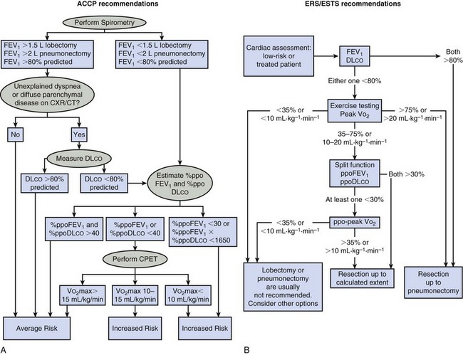
Box 73-2 summarizes the American College of Chest Physicians (ACCP) recommendations for the preoperative pulmonary evaluation of the lung resection candidate. Figure 73-1 outlines the ACCP and European Respiratory Society and European Society of Thoracic Surgeons (ERS/ESTS) recommended steps to follow when assessing a patient’s pulmonary fitness for lung resection. The differences in these guidelines highlight the lack of consensus in this area and the need to individualize patient decisions.

Box 73-2

Summary of ACCP Guidelines for Lung Cancer Resection

ACCP, American College of Chest Physicians; FEV1, forced expiratory volume in 1 second; DLCO, diffusion capacity; ILD, interstitial lung disease; PPCs, postoperative pulmonary complications; VO2 peak, peak oxygen consumption; ppo, predicted postoperative; LVRS, lung volume reduction surgery.

Modified from American College of Chest Physicians recommendations and Colice GL et al: Chest 132(3 suppl):161–177, 2007.

Management

Most patient-related and surgery-related risk factors are nonmodifiable. The following recommendations should be used in all patients regardless of their risk for PPCs.

Preoperative Measures

For patients with airway obstruction (asthma, COPD) the use of inhaled bronchodilators and inhaled and systemic corticosteroids should be based on standard management and optimization of these conditions. It is not necessary to add these treatments in the perioperative setting if the patient’s underlying condition is well controlled. There appears to be no risk of increased postoperative infections from a course of corticosteroids lasting less than 1 week. Antibiotics and cough expectorants should only be used in patients who are actively infected or are experiencing an acute exacerbation of COPD. All smokers should be strongly advised to quit smoking as soon as possible before surgery and should be provided with means available to help them quit. Breathing muscle exercises such as incentive spirometry, use of an inspiratory muscle trainer, deep-breathing exercises, or enrollment in a pulmonary rehabilitation program may be advised for those with underlying lung compromise undergoing high-risk surgery. Weight reduction for the obese patient is advised, particularly those with or at risk for OSA.

Patients at high risk for OSA should undergo a sleep study before their scheduled surgery if time permits. If OSA is proven, treatment with continuous positive airway pressure (CPAP) should begin as soon as possible. A recent study revealed that patients with newly diagnosed OSA had lower rates of respiratory failure if they had started CPAP therapy before surgery. Currently, data are insufficient to support postponing an elective surgery to optimize sleep apnea therapy.

Suggested Readings

Arozullah AM, Khuri SF, Henderson WG, Daley J. Participants in the National Veterans Affairs Surgical Quality Improvement Program: development and validation of a multifactorial risk index for predicting postoperative pneumonia after major noncardiac surgery. Ann Intern Med. 2001;135:847–857.

Chung F, Yegneswaran B, Liao P, et al. STOP questionnaire: a tool to screen patients for obstructive sleep apnea. Anesthesiology. 2008;108:812–821.

Colice GL, Shafazand S, Griffin JP, et al. Physiologic evaluation of the patient with lung cancer being considered for resectional surgery: American College of Chest Physicians evidenced-based clinical practice guidelines (2nd edition). Chest. 2007;132(3 suppl):161–177.

Johnson RG, Arozullah AM, Neumayer L, et al. Multivariable predictors of postoperative respiratory failure after general and vascular surgery: results from the patient safety in surgery study. J Am Coll Surg. 2007;204:1188–1198.

Lim E, Baldwin D, Beckles M, et al. Guidelines on the radical management of patients with lung cancer. British Thoracic Society; Society for Cardiothoracic Surgery in Great Britain and Ireland. Thorax. 2010;65(suppl 3):1–27.

Mazzone PJ, Arroliga AC. Lung cancer: preoperative pulmonary evaluation of the lung resection candidate. Am J Med. 2005;118:578–583.

Myers K, Hajek P, Hinds C, McRobbie H. Stopping smoking shortly before surgery and postoperative complications: a systematic review and meta-analysis. Arch Intern Med. 2011;171:983–989.

Poonyagariyagorn H, Mazzone PJ. Lung cancer: preoperative pulmonary evaluation of the lung resection candidate. Semin Respir Crit Care Med. 2008;29:271–284.

Smetana GW. Preoperative pulmonary evaluation: identifying and reducing risks for pulmonary complications. Cleve Clin J Med. 2006;73(suppl 1):36–41.

Smetana GW, Lawrence VA, Cornell JE. Preoperative pulmonary risk stratification for noncardiothoracic surgery: systematic review for the American College of Physicians. Ann Intern Med. 2006;144:581–595.

Sweitzer BJ, Smetana GW. Identification and evaluation of the patient with lung disease. Med Clin North Am. 2009;93:1017–1030.