Pregnancy

Published on 03/04/2015 by admin

Filed under Hematology, Oncology and Palliative Medicine

Last modified 22/04/2025

Print this page

rate 1 star rate 2 star rate 3 star rate 4 star rate 5 star
Your rating: none, Average: 0 (0 votes)

This article have been viewed 1573 times

44

Pregnancy

Haematological changes

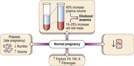
Several haematological changes occur in normal pregnancy (Fig 44.1). Beginning in the sixth week there is an increase in plasma volume accompanied by an increase in red cell mass. The plasma volume expansion peaks at around 24 weeks when it is approximately 40% greater than in a non-pregnant woman. As the increase in red cell mass is more modest (15–25%) a dilutional anaemia is inevitable. In practice the haematocrit and haemoglobin level start to fall at 6–8 weeks and reach a trough at around 20 weeks. It is unusual for the haemoglobin level to fall below 100 g/L and if this happens another cause for anaemia should be sought. Negative iron balance can be regarded as routine in pregnancy and as discussed below frank iron deficiency commonly occurs.

The other major changes which may be regarded as a physiological consequence of pregnancy affect the coagulation system. There are increases in the levels of the coagulation factors VII, VIII and X and a marked increase in plasma fibrinogen. The resulting hypercoagulability is helpful in limiting the likelihood of life-threatening bleeding at delivery but it does lead to an increased risk of thromboembolism. The platelet count falls about 10% during an uncomplicated pregnancy. Later in pregnancy there may also be an increase in mean platelet volume (MPV).

Anaemia in pregnancy

There are several causes of anaemia in pregnancy. The most common scenario is an exacerbation of the usual dilutional anaemia by deficiency of iron and/or folate. Erythropoietin levels increase less than in anaemic non-pregnant women, possibly suppressed by hormonal changes.

The identification of iron deficiency relies upon normal laboratory tests (p. 25). However, even in women with no overt clinical deficiency there is a progressive fall in serum iron through pregnancy. Routine dietary supplementation with modest amounts of iron (e.g. ferrous sulphate 200 mg daily) leads to a significant increase in haemoglobin level at term compared with women receiving no supplements. Parenteral iron is contraindicated in the first trimester.

The other major type of anaemia in pregnancy is megaloblastic anaemia. This usually results from deficiency of folate. As for iron, folate requirements are increased during pregnancy and the diet is frequently inadequate to meet this demand. Megaloblastic anaemia most often presents as a macrocytic anaemia in the third trimester or postpartum. It is normal practice to give folate supplements in pregnancy. The amount of folate routinely administered orally should be large enough to avoid megaloblastic anaemia but not so large as to risk masking pernicious anaemia with vitamin B12 deficiency which does occasionally occur in pregnancy. Folate deficiency in pregnancy has been linked with an increased incidence of neural tube defects in the fetus and recommendations for planned pregnancies are the use of folate supplements (400 µg daily) prior to conception and then particularly in the first 12 weeks. Larger doses of folate are recommended where women are at high risk of conceiving a child with a neural tube defect (e.g. previously affected pregnancy). There is no justification for the prescription of multi-ingredient vitamin preparations in pregnancy but a combined iron and folate tablet of adequate dosage may be prescribed.

It should be remembered that not all anaemia in pregnancy is caused by deficiency states. Other blood disorders may present in pregnancy and chronic blood diseases such as sickle cell anaemia can be especially difficult to manage at this time.

Thrombocytopenia in pregnancy

After anaemia, thrombocytopenia is the most common haematological disorder in pregnancy, occurring in 6–10% of all pregnant women. A few women have an obvious systemic disorder such as pre-eclampsia; disseminated intravascular coagulation (DIC) in pregnancy is further discussed below. However, the majority of women are systemically well with an apparently normal pregnancy. In these cases thrombocytopenia can be divided into two categories, with differing clinical implications for the mother and fetus.

ITP in pregnancy

The management of pregnancy in a woman with known chronic ITP can be problematic as severe thrombocytopenia may be a threat to the mother and there is also a risk of the child becoming thrombocytopenic. The latter complication arises as the causative IgG antiplatelet autoantibody in the mother freely crosses the placenta and can target fetal platelets. Fortunately, the majority of babies escape – severe thrombocytopenia (less than 50 × 109/L) occurs in around 10% of neonates and mortality from intracranial bleeding in those affected is less than 1%. All management decisions must thus acknowledge that fetal thrombocytopenia is uncommon and fetal mortality very rare. In this context, aggressive treatment of all mothers with ITP with corticosteroids and/or intravenous immunoglobulin and routine delivery by caesarean section are not justified. The fetal platelet count is not routinely measured. A conservative approach with normal delivery and an immediate neonatal cord blood platelet count is gaining support. If the baby’s count is low or falling, intravenous immunoglobulin can be given. In more severe thrombocytopenia, transcranial ultrasound can be performed to exclude intracranial haemorrhage. Because of its low incidence of side-effects, intravenous immunoglobulin is probably the treatment of choice for severe maternal thrombocytopenia.

Coagulation abnormalities in pregnancy

Thromboembolism and anticoagulant therapy

Pulmonary embolism (PE) remains a major cause of maternal death. Approximately half of fatal PEs occur antepartum and half postpartum, the majority of the latter in the first 2 weeks of the puerperium. About 70% of women who develop venous thromboembolism in pregnancy and the puerperium have major risk factors. These include increasing age, caesarean section, obesity, previous thrombotic problems and familial thrombophilia. Hereditary thrombophilia (see p. 78) has also been linked with recurrent fetal loss, intrauterine growth restriction, pre-eclampsia and placental abruption.

Both the anticoagulants commonly used in clinical practice, heparin and warfarin, require special consideration in pregnancy.

image Heparin. Neither unfractionated standard heparin nor low molecular weight heparin (LMWH) cross the placenta. LMWH is widely used and is both safe and effective in the prevention and treatment of venous thromboembolism in pregnancy, with significant bleeding, usually from primary obstetric causes, in less than 2% of cases. Anti-factor Xa levels can be monitored but the optimal therapeutic range is unclear.
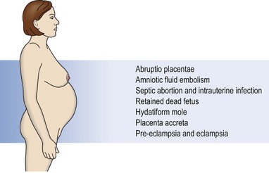
image Warfarin. Warfarin is not significantly secreted in breast milk and treatment is safe during lactation. However, it readily crosses the placenta and is a known teratogen, producing a specific warfarin embryopathy at around 6–12 weeks (approximately 5% incidence). Thus, heparin should be substituted for warfarin in the first trimester. There may be a risk of fetal haemorrhage secondary to warfarin throughout pregnancy, particularly if anticoagulant control is poor, and the risk to mother and fetus becomes unacceptable in the antepartum period. It should therefore be discontinued at 36 weeks and heparin substituted until after delivery. Current practice is to avoid use of oral anticoagulants in pregnancy wherever possible. There is currently no place for the newer oral anticoagulants (i.e. direct thrombin and anti-Xa inhibitors) due to concerns regarding toxicity.

DIC in pregnancy

DIC is associated with a wide variety of situations in pregnancy (Fig 44.2). The chief characteristics and pathogenesis of DIC are discussed on page 76. In pregnancy, DIC may manifest as a chronic compensated state or as life-threatening haemorrhage. The latter is a frightening medical emergency and there should be a planned regimen of management with input from an obstetrician, haematologist, physician, anaesthetist and nurse (Table 44.1). It is imperative that the source of bleeding is identified and addressed as soon as possible. It is often shock which triggers DIC with a resultant increase in bleeding.

HELLP syndrome

HELLP is an acronym for microangiopathic haemolysis (H), elevated liver enzymes (EL) and low platelets (LP). The syndrome complicates less than 1% of all pregnancies but develops in 10% of women with pre-eclampsia. Both disorders generally remit within several days after delivery and delivery of the fetus is therefore central to management.