Chapter 31 PRECOCIOUS PUBERTY
Normal puberty begins in girls between 8 and 14 years of age with breast buds and skeletal growth, followed by the arrival of pubic hair, axillary hair, and menarche. The age at which pubertal milestones are attained varies. In addition, it is influenced by activity level and nutritional status. Girls with low body fat may have a significant delay in menarche (up to a year or more), whereas obese girls may have earlier onset of puberty.
• Simple premature thelarche involves only breast development (unilateral or bilateral) without pubic hair growth, without accelerated bone maturation, and with a normal height outcome. No treatment is required. The disorder is usually self-limited and can resolve spontaneously or persist to normal puberty.
• Simple premature adrenarche involves only pubic hair development (pubic and axillary) without the other manifestations of puberty. No treatment is required, although the possibility of late-onset congenital adrenal hyperplasia should be investigated with a measurement of the serum 17α-hydroxyprogesterone level. There is an increased incidence of simple premature adrenarche in patients with CNS abnormalities.
Key Physical Findings
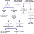
Suggested Work-Up
| Radiograph of the left wrist | To estimate physiologic age for comparison with the child’s chronologic age |
| Measurement of serum follicle-stimulating hormone (FSH), luteinizing hormone (LH), estradiol, testosterone, thyroid-stimulating hormone (TSH), thyroxine (T4), and human chorionic gonadotropin (hCG) | To confirm the impression of idiopathic precocious puberty, to localize the abnormality of the pathologic cause of precocious puberty, or to guide the choice of imaging study |
| Measurement of serum 17α-hydroxyprogesterone and dehydroepiandrosterone sulfate (DHEAS) | If a peripheral cause is suspected or if virilization is present in a female patient |
| Pelvic ultrasonography | To determine whether pubertal changes have occurred in the uterus and ovaries; also indicated if a peripheral cause (ovarian tumor) is suspected |
Additional Work-Up
| Magnetic resonance imaging (MRI) of the brain and pituitary gland | If a central pathologic cause is suspected on the basis of hormone measurements |
| GnRH stimulation test | Administration of 2.5 μg/kg of GnRH either intravenously or subcutaneously after an overnight fast; measurement of serum levels of FSH and LH at baseline just before the injection and 15, 30, 45, and 60 minutes after the injection |
| Test interpretation is controversial, but a twofold to threefold rise in FSH and LH may observed if the patient has central precocious puberty; a peak LH level of more than 15 IU/L and a peak LH–to–peak FSH ratio of more than 0.66 are also criteria for defining a pubertal GnRH test result | |
| Breast ultrasonography | Indicated in unilateral or asymmetric premature thelarche to rule out masses |
| Skeletal survey or radionuclide bone scan | Indicated for patients with McCune-Albright syndrome to evaluate for lesions of fibrous dysplasia |
Bates GW. Hirsutism and androgen excess in childhood and adolescence. Pediatr Clin North Am. 1981;28:513-530.
Blondell RD, Foster MB, Dave KC. Disorders of puberty. Am Fam Physician. 1999;60:209-224.
Fahmy JL, Kaminsky CK, Kaufman F, et al. The radiological approach to precocious puberty. Br J Radiol. 2000;73:560-567.
Klein KO. Precocious puberty: who has it? Who should be treated? J Clin Endocrinol Metab. 1999;84:411-414.


