Postpartum problems

Published on 09/03/2015 by admin

Filed under Obstetrics & Gynecology

Last modified 22/04/2025

Print this page

rate 1 star rate 2 star rate 3 star rate 4 star rate 5 star
Your rating: none, Average: 0 (0 votes)

This article have been viewed 1705 times

Postpartum problems

Shankari Arulkumaran

Physiological changes

Genital tract

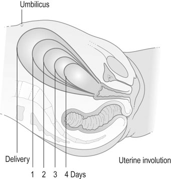
The uterus weighs 1 kg after birth, but less than 100 g by 6 weeks. Uterine muscle fibres undergo autolysis and atrophy and within 10 days the uterus is no longer palpable abdominally (Fig 13.1). By the end of the puerperium, the uterus has largely returned to the non-pregnant size. The endometrium regenerates within 6 weeks and menstruation occurs within this time if lactation has ceased. If lactation continues, the return of menstruation may be deferred for 6 months or more.

Discharge from the uterus is known as lochia. At first this consists of blood, either fresh or altered (lochia rubra) and lasts 2–14 days. It then changes to a serous discharge (lochia serosa), and finally becomes a slight white discharge (lochia alba). These changes may continue for up to 4–8 weeks after delivery. Abnormal persistence of lochia rubra may indicate the presence of retained placental tissue or fetal membranes.

The importance of breastfeeding

Colostrum

Colostrum is the first milk and is present in the breast from 12–16 weeks of pregnancy. Colostrum is produced for up to 5 days following birth before evolving into transitional milk, from 6–13 days and finally into mature milk from 14 days onwards. It is thick and yellow in colour, due to β-carotene and has a mean energy value of 67 kcal/dL, compared to 72 kcal/dL in mature milk. The volume of colostrum per feed varies from 2–20 mL in keeping with the size of the newborn’s stomach.

Linked with the importance of the baby having colostrum as its first food, is the importance of the baby being skin-to-skin with its mother after birth. This has the benefit of the baby being colonized by its mother’s bacteria. Colonizing starts during the birth process for vaginally born infants, while those born via caesarean section are more likely to colonize bacteria from the air. Early breastfeeding also promotes tolerance to antigens, thus reducing the number of food allergies in breast-fed babies. The development of healthy intestinal flora also reduces the incidence of allergic disease, inflammatory gut disease and rotavirus diarrhoea in infants.

While breastfeeding is desirable and women should be encouraged, the overall wishes of the woman should not be ignored. There are social and often emotional reasons why a woman may choose not to breastfeed. In some cases, it is not possible or even advisable, such as inverted nipples, previous breast surgery, breast implants, cracked or painful nipples or because the mother may have a condition, e.g. HIV positive mothers, or may be on medical treatment, e.g. chemotherapeutic agents that serve as a contraindication to breastfeeding.

Breastfeeding

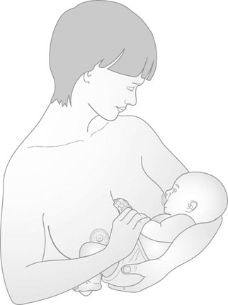
The breasts and nipples should be washed regularly. The breasts should be comfortably supported and aqueous-based emollient creams may be used to soften the nipple and thus avoid cracking during suckling. Suckling is initially limited to 2–3 minutes on each side, but subsequently this period may be increased. Once the mother is comfortably seated, the whole nipple is placed in the infant’s mouth, taking care to maintain a clear airway (Fig. 13.2). Correct attachment of the baby to the breast is essential to the success of breastfeeding. The common problems such as sore nipples, breast engorgement and mastitis usually occur because the baby is poorly attached to the breast or is not fed often enough. Most breastfeeding is given on demand and the milk flow will meet the demand stimulated by suckling. Once the baby is attached correctly to the nipple, the sucking pattern changes from short sucks to long deep sucks with pauses. It may, on occasions, be necessary to express milk and store it, either because of breast discomfort or cracked nipples or because the baby is sick. Milk can be expressed manually or by using hand or electric pumps. Breast milk can be safely stored in a refrigerator at 2–4°C for 3–5 days or frozen and stored for up to 3 months in the freezer.

In women who choose not to breastfeed, have suffered a stillbirth or intrauterine death or where there is a contraindication to breast feeding, suppression of lactation may be achieved by conservative methods or by drug therapy. Firm support of the breasts, restriction of fluid intake, avoidance of expression of milk and analgesia may be sufficient to suppress lactation. The administration of oestrogens will effectively suppress lactation but carries some risk of thromboembolic disease. The preferred drug therapy is currently the dopamine receptor agonist cabergoline. This can be given as a single dose and will inhibit prolactin release and hence suppress lactation. Bromocriptine is also effective, but the dosage necessary to produce this effect tends to create considerable side effects.

Complications of the postpartum period

Puerperal infections

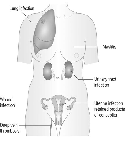
Puerperal sepsis has been reported as far back as the 5th century BC. The Centre for Maternal and Child Enquiries (CMACE 2006–2008) has highlighted the re-emergence of sepsis (in particular group A β-haemolytic streptococci) as a leading cause of maternal morbidity and mortality in the UK. Other common causes of infection are urinary tract infections, wound infections (perineum or caesarean section scar) and mastitis (Box 13.1 and Fig 13.3).

In the puerperium, the placental surface in the womb is vulnerable to infection. This is exposed to the vagina, which harbours aerobic and anaerobic bacteria. Peripartum events, such as prolonged rupture of membranes, chorioamnionitis, repeated vaginal examinations, poor personal hygiene, bladder catheterization, invasive fetal monitoring, instrumental deliveries, caesarean sections, perineal trauma and manual removal of placenta lead to introduction of pathogens into the uterus and thus contribute to puerperal infections.

Endometritis

The patient with endometritis usually presents with fever, lower abdominal pain, secondary postpartum haemorrhage and foul smelling vaginal discharge. The organisms involved are group A β-haemolytic streptococci, aerobic Gram negative rods and anaerobes. On examination, the patient often has a fever, is tachycardic and is tender on palpation of the lower abdomen. There may be foul smelling vaginal discharge, bleeding and cervical excitation. The white cell count and C-reactive protein may be raised. Vaginal or blood cultures may identify the organism responsible. Broad spectrum antibiotics are the first-line treatment and resolution should start to occur within the first 48 hours. The complications of endometritis are parametritis, peritonitis, septic pelvic thrombophlebitis, pelvic abscesses and rarer is toxic shock syndrome.

Caesarean wound infections and perineal infections

Puerperal infection is more common in caesarean sections than vaginal deliveries. Intraoperative antibiotics have helped reduce the incidence. The commonest organisms involved are S. aureus, methicillin-resistant S. aureus (MRSA), skin flora and those involved with endometritis. Complications include wound dehiscence and necrotizing fasciitis. Infection may also occur in episiotomy wounds or perineal tears, although these infections are relatively uncommon because the vascularity of the perineum provides a higher resistance to infection. The perineum becomes tender and reddened and may be seen to exude purulent discharge. Where wound breakdown occurs, the wound should be kept clean and allowed to heal by secondary intention. Resuturing should not be performed unless the wound is clean and there is no residual inflammation around the wound margins.

Thromboembolism

Thrombophlebitis

This is the commonest form of thromboembolic disease and tends to arise within the first 3–4 days after delivery. Localized inflammation, tenderness and thickening occur in the superficial leg veins. Although the condition is painful and may spread along the leg veins, it rarely leads to serious embolic disease and does not require anticoagulant treatment. Anti-inflammatory drugs and local applications of glycerine and ichthyol should be used.

Phlebothrombosis (see also Chapter 9)

Deep vein thrombosis (DVT) is a much more serious complication that tends to arise 7–10 days after delivery and is particularly likely to occur after operative delivery or prolonged immobilization. Clotting occurring in deep veins may be silent and presents only when the clot breaks loose and lodges in the lung as a pulmonary embolus, with consequent chest pain dyspnea and haemoptysis. Clinical signs include local rhonchi and pleural rub on auscultation and a pulmonary perfusion. A ventilation scan or chest CT scan should help to confirm or refute the diagnosis. Massive pulmonary embolus (PE) results in sudden death unless treated by prompt surgical management. Successful treatments with antithrombolytic agents and fragmenting the clots with percutaneous arterial catheters have been reported.

Postnatal anticoagulation

National guidelines in the UK recommend that in non-pregnant patients, anticoagulant therapy should be continued for 6 weeks for calf vein thrombosis and three months for proximal DVT or PE when venous thromboembolism (VTE) has occurred in relation to a temporary risk factor and 6 months for a first episode of idiopathic VTE. The presence of continuing risk factors and the safety of low molecular weight heparin (LWMH) have led authorities to propose that anticoagulant therapy should be continued for the duration of the pregnancy and until at least 6 weeks postpartum, and to allow a total duration of treatment of at least 3 months. Both heparin and warfarin are satisfactory for use postpartum.

Neither heparin nor warfarin is contraindicated in breastfeeding. If the woman chooses to continue with LMWH postnatally, then either the doses that were employed antenatally can be continued or the manufacturers’ recommended doses for the non-pregnant patient can be employed. If the woman chooses to commence warfarin postpartum, this should be avoided until at least the third postnatal day. Daily testing of the international normalized ratio (INR) is recommended during the transfer from LMWH to warfarin to avoid over anticoagulation. Warfarin administration should be delayed in women with risk of postpartum haemorrhage.

Postnatal clinic review for women who develop VTE during pregnancy or the puerperium should ideally be at an obstetric medicine clinic or a joint obstetric haematology clinic. At the postnatal review, the continuing risk of thrombosis should be assessed, including a review of personal and family history of VTE and any thrombophilia screen results. Advice should be given on the need for thromboprophylaxis in any future pregnancy and at other times of increased risk. Hormonal contraception should be discussed.

Anaemia

If the haemoglobin (Hb) is less than 7–8 g/dL in the postnatal period, where there is no continuing or threat of bleeding, the decision to transfuse should be made on an informed individual basis. In fit, healthy, asymptomatic patients there is little evidence of the benefit of blood transfusion. If severe bleeding was encountered and if bleeding disorders were suspected, appropriate investigations should be made. These investigations should be repeated on a non-urgent basis at least 3–6 months after delivery when pregnancy-related coagulation changes have settled.

Oral iron should be the preferred first-line treatment for iron deficiency. Parenteral iron is indicated when oral iron is not tolerated, absorbed or patient compliance is in doubt. Parenteral therapy offers a shorter duration of treatment and a quicker response than oral therapy. It is, however, more invasive and expensive to administer. Iron sucrose is given in multiple doses whereas iron dextran may be given as a single total-dose infusion. Recombinant human erythropoietin (rHuEPO) is mostly used in the anaemia of end-stage renal disease.

Maternal collapse

Maternal collapse is defined as an acute event involving the cardiorespiratory systems and/or brain, resulting in a reduced or absent conscious level (and potentially death), at any stage in pregnancy and up to 6 weeks after delivery. An obstetric early warning score chart should be used routinely for all women, to allow early recognition of the woman who is becoming critically ill. In some cases maternal collapse occurs with no prior warning, although there may be existing risk factors that make this more likely. Antenatal care for women with significant medical conditions at risk of maternal collapse should include multidisciplinary team input with a pregnancy and delivery management plan in place.

There are many causes of collapse, and these may be pregnancy-related or result from conditions not related to pregnancy and possibly existing before pregnancy. The common reversible causes of collapse in any woman can be remembered using the 4 Ts and the 4 Hs employed by the Resuscitation Council (UK) (Table 13.1). In the pregnant woman, eclampsia and intracranial haemorrhage should be added to this list.

Table 13.1

Reversible causes of collapse in pregnancy/postpartum

Reversible cause Cause in pregnancy
4 Hs Hypovolaemia Bleeding, relative hypovolaemia of dense spinal block, septic or neurogenic shock
  Hypoxia Peripartum cardiomyopathy, myocardial infarction, aortic dissection, large-vessel aneurysms
  Hypo/hyperkalaemia (and other electrolyte imbalances) No more likely
  Hypothermia No more likely
4 Ts Thromboembolism Amniotic fluid embolus, pulmonary embolus, air embolus, myocardial infarction
  Toxicity Local anaesthetic, magnesium, other
  Tension pneumothorax Following trauma/suicide attempt
  Tamponade (cardiac) Following trauma/suicide attempt
Eclampsia and pre-eclampsia   Includes intracranial haemorrhage

image

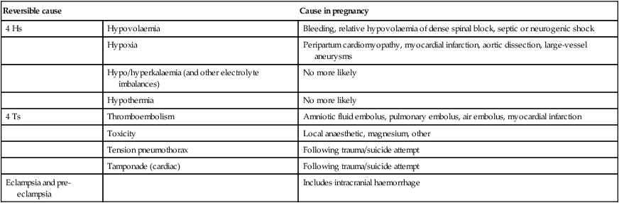
(Reproduced from Maternal Collapse in Pregnancy and the Puerperium. Royal College of Obstetricians and Gynaecology Green-top Guideline No. 56, January 2011.)

Haemorrhage is the most common cause of maternal collapse. In most cases of massive haemorrhage leading to collapse, the cause is obvious, but concealed haemorrhage should not be forgotten, including following caesarean section. Other rare causes of concealed haemorrhage include splenic artery rupture and hepatic rupture.

In the UK, thromboembolism is the most common cause of direct maternal death. Appropriate use of thromboprophylaxis has improved maternal morbidity and mortality, but improvements in clinical risk assessment and prophylaxis are still required.

Amniotic fluid embolism (AFE) presents as collapse during labour or delivery or within 30 minutes of delivery in the form of acute hypotension, respiratory distress and acute hypoxia. Seizures and cardiac arrest may occur. There are different phases to disease progression; initially, pulmonary hypertension may develop secondary to vascular occlusion either by debris or by vasoconstriction. This often resolves and left ventricular dysfunction or failure develops. Coagulopathy often occurs resulting in a massive postpartum haemorrhage. The underlying pathophysiological process has been compared to anaphylaxis or severe sepsis. Clinically, an AFE can be suspected, but a definitive diagnosis can only be made on post-mortem.

Cardiac disease was the most common overall cause of maternal death in the UK from 2006 to 2008. The majority of deaths secondary to cardiac causes occur in women with no previous history. The main cardiac causes of death are myocardial infarction, aortic dissection and cardiomyopathy. Primary cardiac arrest in pregnancy is rare and most cardiac events have preceding signs and symptoms. Aortic root dissection can present with central chest or interscapular pain and a wide pulse pressure, mainly secondary to systolic hypertension. A new cardiac murmur must prompt referral to a cardiologist and appropriate imaging. The incidence of congenital and rheumatic heart disease in pregnancy is increasing secondary to improved management of congenital heart disease and increased immigration. Other cardiac causes include dissection of the coronary artery, acute left ventricular failure, infective endocarditis and pulmonary oedema.

Bacteraemia, which can be present in the absence of pyrexia or a raised white cell count, can progress rapidly to severe sepsis and septic shock leading to collapse. The most common organisms implicated in obstetrics are the streptococcal groups A, B and D, Pneumococcus and E. coli.

Drug toxicity/overdose should be considered in all cases of collapse, and illicit drug overdose should be remembered as a potential cause of collapse outside of hospital. In terms of therapeutic drug toxicity, the common sources in obstetric practice are magnesium sulphate in the presence of renal impairment and local anaesthetic agents injected intravenously by accident. Effects initially include a feeling of inebriation and lightheadedness followed by sedation, circumoral paraesthesia and twitching; convulsions can occur in severe toxicity. On intravenous injection, convulsions and cardiovascular collapse may occur very rapidly. Local anaesthetic toxicity resulting from systemic absorption of the local anaesthetic may occur sometime after the initial injection. Signs of severe toxicity include sudden loss of consciousness, with or without tonic–clonic convulsions, and cardiovascular collapse.

Eclampsia as the cause of maternal collapse is usually obvious in the inpatient setting, as often the diagnosis of pre-eclampsia has already been made and the seizure witnessed. Intracranial haemorrhage is a significant complication of uncontrolled, particularly systolic, hypertension, but can also result from ruptured aneurysms and arteriovenous malformations. The initial presentation may be maternal collapse, but often severe headache precedes this.

Anaphylaxis causes a significant intravascular volume redistribution, which can lead to decreased cardiac output. Acute ventricular failure and myocardial ischaemia may occur. Upper airway occlusion secondary to angioedema, bronchospasm and mucous plugging of smaller airways all contribute to significant hypoxia and difficulties with ventilation. Common triggers are a variety of drugs, latex, animal allergens and foods.

Other causes of maternal collapse include hypoglycaemia and other metabolic/electrolyte disturbances, other causes of hypoxia such as airway obstruction secondary to aspiration/foreign body, air embolism, tension pneumothorax and cardiac tamponade secondary to trauma and, rarely, hypothermia.

The management of maternal collapse in the UK follows the Resuscitation Council (UK) guidelines using the standard A, B, C approach: airways, breathing and circulation. The airway should be protected as soon as possible by intubation with a cuffed endotracheal tube and supplemental oxygen should be administered. Bag and mask ventilation should be undertaken until intubation can be achieved. In the absence of breathing despite a clear airway, chest compressions should be commenced immediately. Two wide-bore cannulae should be inserted as soon as possible, to enable an aggressive approach to volume replacement. Abdominal ultrasound by a skilled operator can assist in the diagnosis of concealed haemorrhage. The same defibrillation energy levels should be used as in the non-pregnant patient. There should normally be no alteration in algorithm drugs or doses. Common, reversible causes of maternal cardiopulmonary arrest should be considered throughout the resuscitation process. If cardiac output is not restored after 3 minutes of CPR in a woman who is still pregnant the fetus should be delivered by caesarean section as this will improve the effectiveness in maternal resuscitation efforts and may save the baby. Resuscitation efforts should be continued until a decision is taken by the consultant obstetrician, and consultant anaesthetist in consensus with the cardiac arrest team. Senior staff with appropriate experience should be involved at an early stage. Accurate documentation in all cases of maternal collapse, whether or not resuscitation is successful, is essential. Debriefing is recommended for the woman, her family and the staff involved in the event. All cases of maternal collapse should generate a clinical incident form and the care should be reviewed through the clinical governance process. All cases of maternal death should be reported to CMACE.

Contraception in the postnatal period

A conversation regarding contraception is best before the woman leaves hospital, but further follow-up is essential. Discussion should ideally cover all options including lactational amenorrhoea, condoms, diaphragm, progestogen-only pills, progestogen implants or injection (Depo-Provera®), and an intrauterine contraceptive device (IUCD) such as a copper coil or levenorgestrel-releasing device (Mirena®). This consultation should include the indications, contraindications as well as the risks and benefits of each.

Condoms are a good first option. They are low cost, unlikely to have side effects and with partner compliance, have a 95% success rate in preventing pregnancy, whilst offering protection from sexual health infections. The copper IUCD is popular as it has a lifespan of 5 years. Those woman who have a history of menorrhagia may benefit from a Mirena. These are usually inserted after the uterus has involuted at 6 weeks.

The combined oral contraceptive pill can not be used in fully breastfeeding women because the oestrogen will suppress lactation. The progesterone-only pill and injectable/implantable progestogenic contraceptives can be safely given to the fully breastfeeding woman. These are normally started 6 weeks postpartum because of the potential for side effects or irregular bleeding, but where the risk of unplanned pregnancy is high can be commenced immediately after delivery.

Neonatal problems

Passage through the birth canal is a hypoxic experience for the fetus, since significant respiratory exchange at the placenta is prevented for the 50–75 seconds duration of the average contraction. Though most babies tolerate this well, the few that do not may require help to establish normal breathing at delivery. Newborn life support is intended to provide this help and comprises the following elements: drying and covering the newborn baby to conserve heat, assessing the need for any intervention, opening the airway, aerating the lung, rescue breathing, chest compression, and rarely, the administration of drugs.

If subjected to sufficient hypoxia in utero, the fetus will attempt to breathe. If the hypoxic insult is continued the fetus will eventually lose consciousness. Shortly after this the neural centres controlling these breathing efforts will cease to function because of lack of oxygen. The fetus then enters a period known as primary apnoea. Up to this point, the heart rate remains unchanged, but soon decreases to about half the normal rate as the myocardium reverts to anaerobic metabolism: a less fuel efficient mechanism. The circulation to non-vital organs is reduced in an attempt to preserve perfusion of vital organs. The release of lactic acid, a by-product of anaerobic metabolism, causes deterioration of the biochemical environment.

If the insult continues, shuddering (whole-body gasps) is initiated by primitive spinal centres. If for some reason these gasps fail to aerate the lungs, they fade away and the neonate enters a period known as secondary or terminal apnoea. Until now, the circulation has been maintained but, as terminal apnoea progresses cardiac function is impaired. The heart eventually fails and, without effective intervention, the baby dies.

Thus, in the face of asphyxia, the baby can maintain an effective circulation throughout the period of primary apnoea, through the gasping phase, and even for a while after the onset of terminal apnoea. The most urgent requirement for any asphyxiated baby at birth is that the lungs be aerated effectively. Provided the baby’s circulation is sufficient, oxygenated blood will then be conveyed from the aerated lungs to the heart. The heart rate will increase and the brain will be perfused with oxygenated blood. Following this, the neural centres responsible for normal breathing will, in many instances, function once again and the baby will recover. Merely aerating the lungs is sufficient in the vast majority of cases. Although lung aeration is still vital, in a few cases cardiac function will have deteriorated to such an extent that the circulation is inadequate and cannot convey oxygenated blood from the aerated lungs to the heart. In this case, a brief period of chest compression may be needed. In a very few cases, lung aeration and chest compression will not be sufficient, and drugs may be required to restore the circulation. The outlook in the latter group of infants is poor.

Most babies born at term need no resuscitation and they can usually stabilize themselves during the transition from placental to pulmonary respiration very effectively. Provided attention is paid to preventing heat loss and a little patience is exhibited before cutting the umbilical cord, intervention is rarely necessary. However, some babies will have suffered stresses or insults during labour and resuscitation is then required. Significantly, preterm babies, particularly those born below 30 weeks gestation, are a different matter. Most babies in this group are healthy at the time of delivery and yet all can be expected to benefit from help in making the transition. Intervention in this situation is usually limited to maintaining a baby’s health during this transition and is called stabilization.

Conducting a routine postnatal clinical review

The postnatal period marks a significant transition point in a woman’s life. The period of postnatal care extends from the hospital stay to the community and home and is provided by multiple caregivers. The objectives of care of mother and baby in the postnatal period include provision of rest and recovery following birth, supporting maternal attachment and assisting in the development of maternal self-esteem. The family unit should be supported and risks need to be identified and managed appropriately. If the mother wishes to breastfeed, this should be initiated and encouraged. Steps should be taken to prevent, identify and manage postnatal depression.

Most of a woman’s care in the community is conducted by the community midwives and general practitioners. If a woman is returning for a clinical review in the hospital, it is either for debriefing following a complication during her pregnancy, labour, delivery or postnatal period, or for the medical management of medical conditions such as diabetes or hypertension. The opportunity should be utilized to discuss family planning and contraception as well as cervical screening. Letters regarding the woman’s pregnancy and postnatal needs should be sent out in a timely fashion as they are helpful to general practitioners and in many cases are the only link between the services. An ideal model of maternity care should seek to maximize the health of women across their reproductive life rather than focus on a single pregnancy.