Postoperative hypotension

Published on 10/04/2015 by admin

Filed under Surgery

Last modified 22/04/2025

Print this page

rate 1 star rate 2 star rate 3 star rate 4 star rate 5 star
Your rating: none, Average: 5 (1 votes)

This article have been viewed 3195 times

Problem 3 Postoperative hypotension

With the various possibilities in mind, you head towards the ward.

On arrival at the ward you quickly read the patient’s notes to aid your assessment. The man had a 2 month history of dysphagia prior to his diagnosis of oesophageal adenocarcinoma. He was treated with a 6 week course of chemotherapy prior to his surgery. His past medical history included controlled hypertension and reflux, for which he was taking bendroflumethiazide and esomeprazole respectively. He is an ex-smoker with a 40 pack-year history.

He had been making an uneventful recovery from his surgery although his gut has not yet started to function. You note from his charts that a tachycardia has developed over the last 24 hours. Over the last 3 hours his urine output has dropped from over 30 mL/hour to 10 mL/hour. His temperature is 38.4°C. You read the operation record and note that the procedure was uncomplicated and a primary anastomosis was performed. A broad-spectrum antibiotic was given prophylactically and continued for 48 hours.

You attend the patient. He is overweight and you estimate his body mass index (BMI) to be 30. He is slouched in the bed with an oxygen mask on his forehead. He has a triple-lumen central line with a bag of saline attached. An epidural catheter is in situ. His chest drain contains a total of 200 mL of sero-sanguinous material, and there is 300 mL of bile-stained fluid in the nasogastric drainage bag and 400 mL in the urinary catheter bag. All the bags were emptied 12 hours earlier. A feeding jejunostomy is running at 40 mL/hour. Little history is available from the patient because of his confusion.

Before you examine the patient more closely you replace his oxygen mask over his mouth and nose and check that his intravenous fluid is running freely. He is mildly confused and is unable to answer your questions appropriately. His skin is pale and clammy and his pulse is faint but regular at 120/min. You confirm his blood pressure to be 85/65 mmHg. He has a tachypnoea of 28 breaths per minute and oxygen saturations are 87%. He already has peripheral venous access and a triple-lumen central line that was inserted during the operation. Both insertion sites look clean and dry. His JVP is not visible and his tongue is dry. His heart sounds are normal.

His lung examination reveals a dull percussion note and absent breath sounds in the lower third of both lung bases. There are associated crepitations at both mid and basal zones. The upper lung fields are clear. The thoracotomy wound looks clean. The chest drain sites are erythematous but dry (the apical drain was removed 2 days earlier). The drainage bottle contains turbid sero-sanguinous fluid and the fluid level swings with deep respiration.

His abdomen is mildly distended, but soft, with minimal wound tenderness to deep palpation. There are no localizing signs. The abdominal wound looks clean and dry, as are the drain and jejunostomy sites. Bowel sounds are absent.

His urine appears concentrated but clear. He has bilateral lower limb oedema, although his calves are not tender.

The patient is transferred to the high dependency unit for resuscitation and further management. A bolus of intravenous isotonic saline is given and blood samples collected for laboratory analysis.

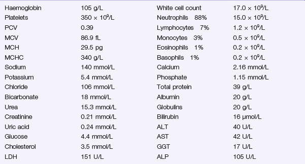
The following results become available:

Investigation 3.2 Arterial blood gas analysis on room air

pO2 52 mmHg pCO2 34 mmHg
pH 7.29 Base excess −9.5

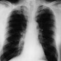
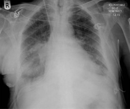
A portable chest X-ray is performed (Figure 3.1).

The patient responds well to your resuscitation measures and his hypotension corrects with intravenous fluid. He is started on broad-spectrum intravenous antibiotics, avoiding aminoglycosides due to concern about nephrotoxicity.

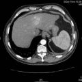
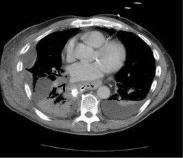
The chest effusions are further visualized with a contrast-enhanced CT scan of the chest and abdomen. One sequence is shown (Figure 3.2).

The right-sided pleural collection is drained percutaneously under radiological control. The material is sanguinous and a sample sent for culture grows enterococci, sensitive to the antibiotics you have prescribed. A chest drain is left in the cavity (and removed after a week). His condition rapidly improves over the next 48 hours. An oral contrast study is performed which does not show any evidence of an anastomotic leakage. The postoperative recovery progresses slowly, but without further mishap.

Answers

A.1 The confusion and hypotension suggest this patient is suffering from shock. Shock is defined as inadequate tissue perfusion. The common signs are related to a reduced oxygen delivery to the tissues. There is commonly a loss in effective circulating blood volume. As this occurs, physiological compensatory mechanisms are initiated, with the primary aim of maintaining oxygen delivery to the vital organs, namely the brain, heart and kidneys. As this compensation fails, so the signs of shock develop.

The causes of shock can be classified into those caused by a loss in total blood volume, or where an effective loss occurs due to volume redistribution. Loss of volume can occur with internal or external blood loss, loss in plasma volume secondary to burns, or fluid loss most commonly from the gastrointestinal tract. Volume redistribution can occur secondary to vasodilatation and increased capillary permeability, most frequently seen in sepsis, anaphylaxis or following acute spinal injury.

Cardiogenic shock occurs secondary to a primary pump failure.

There are several potential causes in this case:

Signs of shock differ according to the cause and its severity. A previously fit adult can lose 15% of their effective blood volume with minimum, transient effects such as mild tachypnoea. Physiological mechanisms will restore blood volume within 24 hours. This is equivalent to donating blood.

Loss of 30% of blood volume can result in tachycardia and narrowing of pulse pressure (the difference between systolic and diastolic blood pressure). This reduces due to compensatory vasoconstriction producing a rise in diastolic pressure. Urine output may reduce and become more concentrated, due to an increase in ADH (antidiuretic hormone).

Loss of more than 40% of effective blood volume (2000 mL) will normally result in a falling systolic blood pressure and tachycardia, as physiological mechanisms fail to compensate adequately. Patients often become confused owing to reduced cerebral perfusion. Urgent action is required as further loss in circulating volume may be fatal.

You have these thoughts in mind as you head towards the ward.

A.2 Additional information may be available from several sources:

A.3 You must do a speedy, efficient but thorough assessment of the patient looking for clues as to the underlying cause of the shock. In a methodical manner you will:

Start by taking the pulse and recheck the blood pressure. As you feel the pulse you should check the perfusion of the peripheries by observing the temperature of the hands and feet.

Consider the following:

A.4 The patient requires prompt resuscitation (A, B, C):

The type of fluid used for resuscitation is controversial. A common strategy is to aim to replace the type of fluid that has been lost i.e. transfuse blood in haemorrhagic shock, albumin in burns and crystalloid in the presence of gastrointestinal losses.

Most, units have ready access to crystalloid fluid such as Hartmann’s solution, or dextrose-saline. Both are isotonic and replace fluid and ions. They are, however, rapidly redistributed, particularly through leaky capillaries, into peripheral tissues. After high volume crystalloid transfusion, patients can become overloaded with fluid, but remain with low blood volume. This situation can ultimately hinder adequate tissue oxygen perfusion. A potential answer to this dilemma lies in the use of colloid fluids. Common colloids include Gelofusine and dextran. These fluids contain higher molecular weight molecules such as albumin or starch that slow redistribution and are designed to remain within the circulating blood volume. A Cochrane review in 2007 concluded that there was no evidence of improved outcome following resuscitation with colloid in patients with trauma, burns or postoperatively. The authors suggest that as crystalloid was cheaper and more easily available, it should continue to be the fluid of choice for resuscitation of the critically ill patient (Perel and Roberts 2007).

The rate and total volume of fluid infusion should be calculated for each individual and reassessed at regular intervals throughout the resuscitation. A fluid challenge of 1000 mL of crystalloid can be safely administered over a 30 minute period, followed by assessment of response. An improvement in cardiovascular parameters such as blood pressure would point towards hypovolaemic shock. A further drop would suggest inadequate resuscitation. A poor response to a bolus fluid challenge would suggest another cause of shock such as an acute cardiac event. In more complex situations, particularly in the presence of pre-existing cardiac dysfunction, more intensive monitoring may be required with central venous pressure measurement.

A conservative fluid replacement strategy should only rarely be considered in the presence of direct lung trauma or established acute lung injury, without associated hypoperfusion.

A.5 The results suggest the following:

The renal dysfunction is probably prerenal in origin, due to hypovolaemia and poor perfusion. The low pH, high base deficit and low bicarbonate level point to a metabolic acidosis, consistent with severe sepsis. Hypoxia could be due to a postoperative basal atelectasis, chest infection or diaphragmatic splinting secondary to surgery.

Low haemoglobin and protein levels are common after major surgery such as oesophagectomy and do not require specific treatment.

A.6 The presence of fever, tachycardia, hypotension, dehydration and confusion indicate this patient most likely has severe sepsis. The development of pyrexia, tachycardia, tachypnoea and abnormal white cell count is defined as systemic inflammatory response syndrome or SIRS. The criteria for a diagnosis of SIRS are listed in Box 3.1. When SIRS occurs in the presence of infection, this is termed sepsis.

In the more advanced cases, there may be evidence of organ dysfunction. Where two or more organ systems are affected this is termed multiple organ dysfunction syndrome or MODS. Where MODS occurs in the presence of an identified source of infection, this is termed severe sepsis or sepsis syndrome. Infection and hypotension that fail to respond to initial resuscitation is termed septic shock.

The body’s response to sepsis is mediated through inflammatory cytokines. As well as helping to fight infection, an excessive cytokine response can have a negative effect. Cytokines such as tumour necrosis factor, platelet activating factor and the interleukins can produce peripheral vasodilatation, leaking capillaries and microvascular coagulation. Specific bacterial toxins also have a detrimental effect on myocardial contractility. These actions result in reduced blood pressure, decreased tissue perfusion and reduced oxygen transfer. Excessive clotting within small vessels can lead to a consumptive coagulopathy. Clotting factors are rapidly used up, resulting in excessive bleeding.

Poor oxygen delivery to the tissues leads to anaerobic metabolism and production of lactic acid. A lactic acidosis results from its release back into the circulation. Lung function worsens as fluid leaks into the lungs, reducing oxygen transfer still further. Reduced urine output leads to accumulation of urea and nitrogen into the circulation.

A.7 There is opacification of the right lower zone. The presence of a meniscus and the preservation of the diaphragm and cardiac border suggest that this is a moderate sized pleural effusion. There is associated patchy consolidation. This could be due to a postoperative pneumonia or an underlying anastomotic leak. The mediastinum is widened secondary to the gastric pull up within the chest. There is interstitial shadowing of both lungs consistent with some pulmonary oedema. The basal chest drain and skin staples are visible.

Although an infected pleural effusion and now systemic sepsis is most likely, you still do not know exactly what is happening. The patient could have a nosocomial infection of the bloodstream from infection of his central venous, epidural or urinary catheter. As he is seriously ill, he requires broad-spectrum intravenous antibiotics along with his resuscitation.

A.8 Once the patient has been resuscitated, further imaging studies need to be performed. The preferred investigation is a thoracic and abdominal CT scan with percutaneous drainage of any identified localized collection.

If the patient’s condition worsens or there is evidence of peritonitis, surgical intervention may be required. If an anastomosis has leaked, either the anastomosis may need to be refashioned or a proximal (diverting) oesophagostomy may need to be formed. If a leak can be controlled with adequate drainage, a period of conservative treatment often allows a small leak to heal spontaneously.

A failure to respond to initial resuscitation suggests the need for management in an intensive care unit, with facilities for invasive monitoring, vasopressor support and mechanical ventilation.

Definitive treatment of sepsis includes a full screen, looking for common sites of postoperative sepsis. Central to this assessment should be blood cultures prior to any antibiotic therapy and early imaging to identify sites of infection. Infected foreign bodies such as catheters should be removed and infected collections should be drained.

Bacteraemia should be treated with appropriate antibiotics, with close involvement of the microbiologist. Appropriate antibiotic choice should follow local hospital guidelines and will depend on the patient’s age, co-morbidity, previous antibiotic history and length of time in hospital. The length of the course of antibiotic therapy should be guided by clinical response.

An appropriate choice for hospital acquired infection would be an extended spectrum beta-lactam antibiotic such as piperacillin in combination with tazobactam. This combination has activity against Gram-negative, Gram-positive and anaerobic pathogens. Alternative choices include the carbapenems (imipenem or meropenem). Vancomycin is an appropriate choice where resistant infections such as MRSA (methicillin-resistant Staphylococcus aureus) or Clostridium difficile are suspected.

Whichever antibiotic regimen is used, it must be reviewed on a regular basis and altered according to response and to the results of microbiological (particularly blood) cultures. If there is any suggestion of renal impairment, the use of aminoglycosides should be avoided. However, aminoglycosides can be used in settings where the risk of uncontrolled sepsis outweighs the risk of renal impairment or failure. In such circumstances, the dose should be adjusted to the calculated glomerular filtration rate (using the Cockroft–Gault nomogram) and the serum concentrations of the drug monitored closely. Caution should also be exercised in the prolonged use of broad-spectrum cephalosporins or quinolones to avoid the risk of developing Clostridium difficile colitis.

A.9 The scan shows a view through the lower chest. Contrast has been given to outline the oesophageal remnant and gastric tube within the posterior mediastinum. There is a small left-sided pleural effusion. There is consolidation within the right lower lung with an air bronchogram. Posterolateral to the intrathoracic stomach on the right is a loculated collection of fluid. This is likely to be an infected collection.

Revision Points

Accurate assessment and early treatment of sepsis have been emphasized by the Surviving Sepsis Campaign (Dellinger et al. 2008). Early, goal directed therapy is recommended in the form of ‘care bundles’. The immediate ‘Resuscitation’ care bundle is given within the first 6 hours, followed by the second ‘Management’ care bundle completed within 24 hours. It is recommended that broad-spectrum antibiotic therapy is commenced within 1 hour of diagnosis of sepsis, with goal directed resuscitation within the first 6 hours of diagnosis. Initial fluid and oxygen resuscitation should aim at maintaining a central venous pressure of 8–12 mmHg, a mean arterial pressure of 65 mmHg, a urine output of 0.5 mL/kg/hour and a central venous oxygen saturation of over 70%. Noradrenaline (norepinephrine) or dopamine can be used as vasopressors in septic shock as long as adequate fluid resuscitation is ensured (Table 3.1).

Table 3.1 Inotropic and vasoactive drugs in shock

Drug Action Use
Noradrenaline (norepinephrine) Alpha agonist
Vasopressor
Septic shock, neurogenic shock
Dobutamine Beta1 agonist
Inotropic
Cardiogenic shock, myocardial failure, without hypotension
Dopamine Beta1 agonist
Inotropic and chronotropic
Cardiogenic shock
?Increase renal perfusion in low doses
Dopexamine Beta2 agonist
Inotropic
Septic shock
Increase splanchnic perfusion

Numerous trials have looked at the use of steroids in severe sepsis. A recent trial failed to show any improvement in mortality following administration of hydrocortisone in septic shock. Although shock was seen to reverse more quickly in patients receiving steroids, they were more susceptible to further infections and sepsis (Sprung et al. 2008).

A key factor in the normal clotting mechanism is activated protein C. Levels of activated protein C are often low in severe sepsis, and its administration is known to have antithrombotic and anti-inflammatory effects. Two multicentre trials looking at the role of activated protein C in the management of severe sepsis have concluded that it may reduce overall mortality, but that it should not be used in patients with a low risk of death because of the associated risk of bleeding complications (Abraham et al. 2005, Bernard et al. 2001).

Other recommended management strategies in sepsis include maintaining a target haemoglobin 70–90 g/L with red cell transfusion, maintaining glycaemic control with infused insulin if required, the use of intermittent haemodialysis in renal failure, thrombosis prophylaxis with low molecular weight heparin and stress ulcer prophylaxis with a proton pump inhibitor.

Shock is defined as inadequate tissue perfusion and its causes are:

The signs of shock include:

A normal systolic blood pressure does not exclude shock. A fit young patient can lose up to 30% blood volume (∼1.5 L) before a drop in blood pressure occurs.

Treatment of a patient with shock must include:

Those at high risk of sepsis and septic complications have been defined in Chapter 2 and include the elderly and malnourished, those with diabetes and any immunocompromised patients.