[level-membership-for-opthalmology-category]11.1
Postoperative Cystoid Macular Edema
Clinical Features:
PCME has the characteristic clinical appearance of numerous small cystic cavities bunched together in a petalloid arrangement centered on the fovea (Fig. 11.1.1, left). In some cases, the optic disc will be hyperemic or even frankly edematous. Small flame, shaped superficial hemorrhages in the inner retina are not rare.
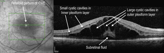
Figure 11.1.1 Infrared image (left) shows a petalloid arrangement of cystic cavities centered on the fovea, characteristic of PCME. OCT (right) shows numerous, hyporeflective cystic cavities located within the outer plexiform layer. There are also smaller hyporeflective cystic cavities located within the inner plexiform and inner nuclear layers. Subretinal fluid is present underneath the fovea. The outer nuclear layer located just above this fluid is hyper-reflective, which is likely an artifact due to the directional manner by which light traverses through the overlying large cystic cavities. (Courtesy of Jeffrey S. Heier, MD.)
OCT Features:
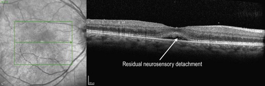
The characteristic feature on OCT is large, hyporeflective cystic spaces located in the outer plexiform layer, though there can also be additional, smaller hyporeflective spaces within the inner plexiform and nuclear layers (Fig. 11.1.1, right). In severe cases, there may be a central neurosensory detachment. Following successful treatment, the cystic spaces seen on OCT typically improve (Fig. 11.1.2).
Ancillary Testing:
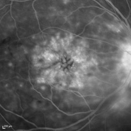
In all cases, fluorescein angiography reveals PCME, even occasionally when it is not visible clinically or by OCT. The angiographic appearance is very characteristic with late diffuse central leakage in a petalloid pattern (Fig. 11.1.3).
[/level-membership-for-opthalmology-category][not-level-membership-for-opthalmology-category]11.1
Postoperative Cystoid Macular Edema
Clinical Features:
PCME has the characteristic clinical appearance of numerous small cystic cavities bunched together in a petalloid arrangement centered on the fovea (Fig. 11.1.1, left). In some cases, the optic disc will be hyperemic or even frankly edematous. Small flame, shaped superficial hemorrhages in the inner retina are not rare.
[/not-level-membership-for-opthalmology-category]
