Posterior Staphyloma
Clinical Features:
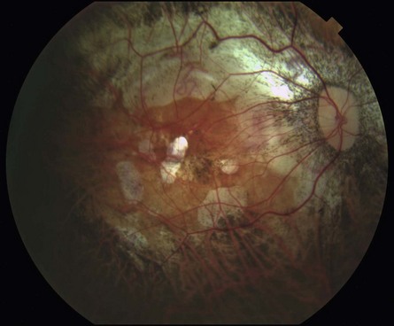
Externally, the globe itself may appear elongated, which is consistent with high myopia. On fundoscopy, there are associated atrophic changes of the retina, retinal pigment epithelium, and choroid in the posterior pole (Fig. 9.1.1). A teacup-like deformity is present, typically within the macula, but can also involve the optic nerve. The deformity can be difficult to appreciate clinically and requires stereopsis to appreciate.
OCT Features:
The appearance of a posterior staphyloma on OCT is rather striking compared to the more subtle clinical appearance. OCT reveals significant posterior bowing and curvature of the posterior eye wall, including the sclera and overlying choroid and retinal layers (Fig. 9.1.2). The choroid is typically almost imperceptible due to significant thinning.
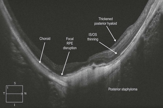
Figure 9.1.2 OCT (corresponding to Figure 9.1.1) shows dramatic posterior bowing of the eye wall. The choroid is so thin that it is barely appreciable. The RPE layer has focal disruptions. The retina takes on the same curvature as the sclera. There is also a mild dome-shaped macula present (see Chapter 9.4, Dome-Shaped Macula)


