Pompholyx
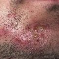
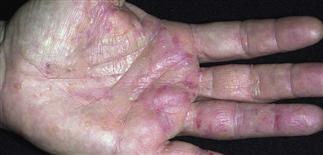
Bilateral palmar scattered vesicles that have evolved to pustules. In pompholyx, the vesicles usually erupt suddenly and in crops. Pruritus may be present or absent.
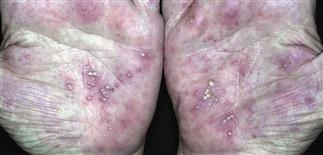
Vesicles and pustules of dyshidrotic eczema in varying stages of evolution. The differential diagnosis is pustular psoriasis and contact dermatitis. The feet often develop a similar eruption.
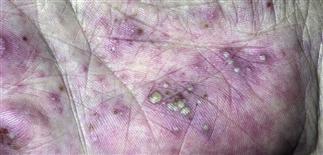
Severe hand eczema: marked erythema, vesicles, and scaling. Contact allergy should be considered, and the patient patch tested to a broad screening series and occupational allergens.
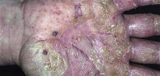
Aftermath of a bout of dyshidrotic eczema: some tender macules of healed epidermis; some diffuse scaling and erythema; and an occasional resolving, dry-appearing vesicle.
DESCRIPTION
Distinctive, chronic relapsing, vesicular eczematous dermatitis of unknown etiology. Characterized by sudden, recurrent eruptions of usually highly pruritic, symmetric vesicles on palms, lateral fingers, and/or plantar feet.
HISTORY
• Affected patients frequently have atopic background (personal or family history of asthma, allergic rhinitis, or atopic eczema). • Moderate or severe itching precedes a flare. • Hyperhidrosis (increased sweating) often aggravates or accompanies. • Vesicles resolve over 1–3 weeks. • Causes of this recurrent, sometimes disabling dermatitis are unknown; provoking factors seem heterogeneous. Consider role of atopy, occupational and/or other contact chemicals, and distant tinea infection. • Systemic contact allergens may play a role; some individuals with positive patch tests show vesicular reactions on hands when challenged orally with nickel, cobalt, or chromium. • Differential diagnosis: contact allergy, pustular psoriasis, inflammatory tinea, ‘id reaction’ on hands to tinea on feet, bullous pemphigoid, rarely cutaneous T-cell lymphoma.
PHYSICAL FINDINGS
• Vesicles 1–5 mm, monomorphic, deep-seated, filled with clear fluid. They erupt suddenly, symmetrically on palms, lateral fingers, or plantar feet. • Rings of scale and peeling follow eruption as itch diminishes. • Depending on phase of disease, clinician may see only brown spots. When acute process ends, skin peels, revealing a red, cracked base with brown macules. • Chronic eczematous changes with erythema, scaling, and lichenification may follow. • Waves of symmetrically distributed vesiculation can recur indefinitely. • For unknown reasons, the chronic recurring eruption sometimes ceases with time.
TREATMENT
• Initial treatment with cold compresses twice a day with either tap water or Burow’s solution, followed by medium- or high-potency steroid cream (group I–III). • Prednisone 0.5–1 mg/kg q.d. tapered over 1–2 weeks is prescribed, but not frequently or as maintenance. • Tacrolimus ointment (Protopic 0.1%) rotating with a topical medium-strength corticosteroid (group II–III) twice daily for cycles of 3–4 weeks can provide some relief. • Oral antihistamines can alleviate pruritus. • Topical psoralen plus ultraviolet A is a treatment option for frequent, refractory eruptions. • Elimination diets (such as a nickel-reduced diet for nickel-allergic patients) may be worth a trial in difficult cases. • If distant focus of tinea identified, treat with topical antifungal medication (econazole or terbinafine cream every day for 3 weeks) or short course of oral antifungal medication (terbinafine or itraconazole) of appropriate duration and dose. • Moderating or eliminating stress can help and is anecdotally curative for some. • For chronic or severe disabling dyshidrotic eczema, consult a dermatologist.