Pneumonia and Respiratory Infections

Published on 06/06/2015 by admin

Filed under Pediatrics

Last modified 22/04/2025

Print this page

rate 1 star rate 2 star rate 3 star rate 4 star rate 5 star
Your rating: none, Average: 0 (0 votes)

This article have been viewed 2142 times

91 Pneumonia and Respiratory Infections

This chapter discusses pneumonia and lower respiratory infections such as bacterial tracheitis. These lower respiratory infections are encountered frequently in children; although up to half of lower respiratory infections in children are viral, bacterial pneumonia is the most common serious bacterial infection in children. The number of diagnostic methods available for identification of the causative pathogen has increased dramatically over the past decade, yet amoxicillin and ampicillin remain the most appropriate first-line therapy for uncomplicated community-acquired pneumonia (CAP).

Etiology and Pathogenesis

Microbiology

The common causes of CAP in healthy children in the developed world vary by age group, although an extensive number of pathogens can cause CAP (Table 91-1). Respiratory viruses such as respiratory syncytial virus (RSV); influenza A and B; parainfluenza 1, 2, and 3; adenovirus; and human metapneumovirus (hMPV) can be identified in up to half of patients admitted to the hospital for CAP. These viral pathogens may be identified alone or as part of a co-infection with bacteria. First described in 2001, hMPV, similar to RSV infection in young children, causes a spectrum of respiratory disease ranging from mild bronchiolitis to severe pneumonia.

Table 91-1 Common Bacterial and Viral Causes of Community-Acquired Pneumonia by Age in Healthy Children in the Developed World

≤3 Months Old 3 Months to 5 Years Old ≥5 Years Old
Bacteria Bacteria Bacteria
Group B streptococcus Streptococcus pneumoniae Streptococcus pneumoniae
Enteric gram-negative bacilli Mycoplasma pneumoniae Mycoplasma pneumoniae
Streptococcus pneumoniae Chlamydophila pneumoniae* Chlamydophila pneumoniae*
Bordetella pertussis Staphylococcus aureus Staphylococcus aureus
Chlamydia trachomatis
Staphylococcus aureus
Haemophilus influenzae (nontypable)  
Lower Respiratory Viruses Lower Respiratory Viruses Lower Respiratory Viruses
Respiratory syncytial virus Respiratory syncytial virus Influenza A and B
Influenza A and B Influenza A and B  
Parainfluenza viruses 1, 2, 3 Parainfluenza viruses 1, 2, 3  
Human metapneumovirus Human metapneumovirus  
Rhinovirus Rhinovirus  
Adenovirus Adenovirus  
Bocavirus Bocavirus  
Coronaviruses Coronaviruses  

* Formerly Chlamydia pneumoniae.

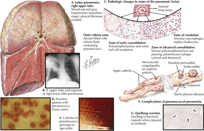
Streptococcus pneumoniae is the most common bacterial cause of childhood CAP (Figure 91-1). Randomized trials of the heptavalent pneumococcal conjugate vaccine (PCV7) demonstrated that the incidence of radiographically confirmed pneumonia was reduced by 20% in vaccine recipients compared with placebo recipients, suggesting that S. pneumoniae causes at least 20% of CAP cases. Postlicensure epidemiologic studies have shown a 39% decrease in all-cause pneumonia hospitalizations in children younger than 2 years of age but nonsignificant decreases in older children. Thus, the significant role of pneumococcus as a cause of childhood CAP drives the choice of empiric antibiotic therapy for younger children.

Staphylococcus aureus, particularly community-associated methicillin-resistant S. aureus (CA-MRSA), has been recognized with increasing frequency as a cause of severe CAP even in previously healthy children without exposure to health care settings. Mycoplasma pneumoniae, although previously described as a pathogen limited to adolescents and young adults, is also a common pathogen in school-age children and toddlers. M. pneumoniae has been associated with wheezing, identified in one study in half of patients with a first episode of wheezing and 20% of patients admitted for an exacerbation of their known prior asthma.

Less common causes of CAP include Streptococcus pyogenes, nontypable Haemophilus influenzae, enteric gram-negative pathogens (in cases of aspiration or neurologic compromise), Mycobacterium tuberculosis, herpes simplex virus (in newborns), varicella-zoster virus, Legionella pneumophila, and endemic mycoses such as Histoplasma capsulatum, Coccidioides immitis, and Blastomyces dermatitidis. Before the introduction of the conjugate H. influenzae type b (Hib) vaccine, Hib was a common cause of CAP. In countries and areas where Hib vaccine uptake is low, Hib should still be considered as a common cause of CAP.

Bacterial tracheitis is a serious respiratory infection encountered only rarely, and it is most frequently caused by S. aureus, although other organisms such as nontypable H. influenzae, Moraxella catarrhalis, and anaerobes have been implicated in its pathogenesis. As with bacterial pneumonias, tracheitis often follows an antecedent viral upper respiratory infection.

Pathogenesis

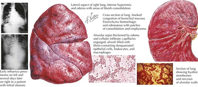
Viral illness alone, such as influenza, can cause severe and necrotizing pneumonia (Figure 91-2). Preceding viral illness may play a part in the pathogenesis of bacterial pneumonia. One study demonstrated that rates of invasive pneumococcal disease each winter season rose in close association with respiratory viral illness diagnoses (RSV, influenza, and hMPV), suggesting that the respiratory damage caused by viral respiratory illness may allow for subsequent bacterial pneumonia. Such data do not prove the direct causation of invasive bacterial pneumonia as a consequence of viral respiratory infection, however. Likewise, a randomized trial of children receiving a pneumococcal vaccine found fewer admissions for both pneumococcal pneumonia and hMPV pneumonia among vaccine recipients compared with placebo recipients, suggesting that hospitalizations for hMPV may involve co-infection with pneumococcus.

Clinical Presentation and Differential Diagnosis

Children with lower respiratory illness classically present with fever, cough, and tachypnea. The spectrum of illness among children with CAP is broad and ranges from mild, well-appearing children to those who require intubation and intensive care. The clinical manifestations of CAP are equally diverse, and few physical examination findings allow for distinction among viral, bacterial, and atypical causes.

The sensitivity of tachypnea alone is up to 74% in identifying children with radiographically confirmed pneumonia. Tachypnea alone has a low specificity for pneumonia, however, because many noninfectious causes of tachypnea exist, so most patients with tachypnea do not have pneumonia. Combinations of multiple physical examination findings, such as tachypnea, rales, and increased respiratory effort, raise the specificity for a clinical diagnosis of pneumonia dramatically but substantially lower the sensitivity, thereby potentially missing many patients with pneumonia. Likewise, the absence of rales on examination does not preclude a pneumonia diagnosis.

Many children with pneumonia also have abdominal symptoms, such as vomiting and abdominal pain. Vomiting may be posttussive, after episodes of severe coughing. Abdominal pain can at times be the most prominent complaint and occurs most commonly in patients with basilar pneumonia. Wheezing and exacerbation of underlying asthma are symptoms more typically encountered in patients with CAP caused by viruses and atypical bacteria such as M. pneumoniae and Chlamydophila pneumoniae (formerly Chlamydia pneumoniae). Children with lower respiratory tract infections caused by atypical bacteria often have mild and nonspecific symptoms such as headache, low-grade fever, pharyngitis, and cough for 5 to 7 days before their presentation with pneumonia.

Children with bacterial tracheitis are often younger than 5 years of age and most commonly present with barking cough, high fever, and significant respiratory distress, often lending these patients a “toxic” appearance. These patients will not have the drooling or inability to lie flat found in patients with epiglottitis, but they can develop life-threatening respiratory distress nonetheless.

The differential diagnosis of lower respiratory infection includes pulmonary anatomic abnormalities, foreign bodies and chemical irritants, autoimmune diseases, and malignancies, among others (Box 91-1).

Evaluation and Management

Diagnostic Methods

An etiologic diagnosis is seldom made in cases of childhood CAP because invasive diagnostic procedures such as bronchoalveolar lavage and needle thoracentesis are infrequently performed in young children and because young children cannot provide adequate sputum specimens. However, children 8 years and older with a productive cough are often able to provide adequate sputum specimens for culture after nebulized hypertonic saline treatments (so-called induced sputum cultures).

Bacterial tracheitis is typically diagnosed based on clinical presentation and through culture of sputum or the purulent exudate found in the trachea on bronchoscopy or intubation. Sputum cultures must be nevertheless interpreted cautiously because they can also capture colonizing oropharyngeal flora. A high-quality sputum specimen should have few squamous epithelial cells (≤10 per high power field [hpf]) and many white blood cells (≥25 per hpf) and should arrive within 2 hours to a microbiology laboratory for culture.

Nasopharyngeal aspirates can provide good samples on which to perform polymerase chain reaction (PCR) to identify both viral and bacterial CAP etiologies. PCR on nasopharyngeal aspirates detects respiratory viruses such as RSV and influenza A and B with a sensitivity in the high 90% range, and immunofluorescence on similar specimens has a sensitivity ranging from approximately 50% to 90%, depending on the specific kit. Likewise, PCR from a nasopharyngeal aspirate is the diagnostic method of choice for Bordetella pertussis during the first 4 weeks of illness, but the diagnosis can be confirmed with acute and convalescent serologies in patients with negative PCR results and a high suspicion for the clinical disease. After that time, B. pertussis serology can be used to make the diagnosis because the organism is cleared from the nasopharynx over time. Although B. pertussis can be cultured during the first 3 weeks of illness, results may not be available for 5 to 7 days.

Chlamydia trachomatis (in neonates) and C. pneumoniae can also be detected via PCR, although prolonged shedding can occur, causing PCR test results to remain positive after the initial period of active disease. In neonates and young infants, direct fluorescent antibody testing can also be used on conjunctival and respiratory specimens for the diagnosis of C. trachomatis. Both C. trachomatis and C. pneumoniae can also be diagnosed through use of acute and convalescent serologies. Likewise, M. pneumoniae is most reliably detected by serologic testing of paired specimens obtained 2 to 3 weeks apart; a fourfold or greater increase in the antibody titer indicates a recent or current infection. Unfortunately, paired serology testing does not provide a diagnosis quickly enough to influence clinical practice. PCR testing for M. pneumoniae has a sensitivity of approximately 80% for pharyngeal specimens and 90% for nasopharyngeal specimens, and specificity is greater than 95% when compared with acute and convalescent serologies.

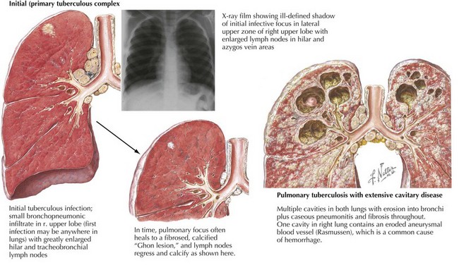
Tuberculin skin testing should be considered for all patients with appropriate tuberculosis risk factors (i.e., travel to an endemic area, contact with a person with active tuberculosis, or contact with people who work or reside in prison or health care settings) and chest radiography findings (Figure 91-3). Newer diagnostic methods that measure serum interferon-γreleased by T-cells stimulated by M. tuberculosis antigens are becoming available but are not validated in children, and they cannot distinguish latent from active disease. Of note, children younger than 2 years of age are more likely to have nonspecific chest radiography findings and disseminated or miliary tuberculosis at presentation.

A urine antigen test for L. pneumophila exists and can be considered for CAP in immunocompromised children and older adolescents. In patients with an appropriate travel history and chest radiography results, Histoplasma serology or urine antigen can be considered. Paired acute and convalescent serologies are useful in diagnosing pneumonia caused by C. immitis and B. dermatitidis.

Blood culture results are seldom positive (≈2% of cases) in outpatients with CAP and are therefore of little utility in well-appearing patients without hypoxia. Up to 10% of patients requiring admission may have positive blood culture results, and the rate of positive blood cultures may be even higher in those with pneumonia complicated by empyema. Blood cultures in these patient populations may therefore provide useful microbiologic data, including antibiotic sensitivities. Gram stain and bacterial culture of pleural fluid should always be performed in patients with pneumonia with associated pleural effusion that has been drained. Urine antigen testing for S. pneumoniae should not, however, be performed routinely because false-positive results may occur as a result of pneumococcal nasopharyngeal colonization in up to 18% of children, and the results are difficult to interpret reliably in the absence of a true gold standard.

Serum white blood cell count and inflammatory markers cannot be used to differentiate between viral and bacterial pneumonia reliably. Although very high circulating neutrophil and C-reactive protein (CRP) levels may be found more commonly in patients with bacterial pneumonia, normal neutrophil and CRP levels do not exclude its possibility. Serum inflammatory markers should therefore be used only to provide supplementary objective measures of disease resolution when tracking the course of a severe pneumonia.

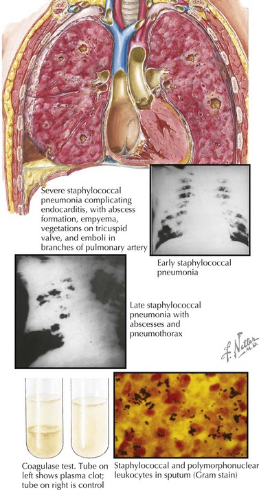
Chest radiography should be considered in highly febrile patients without another identifiable source, especially those with tachypnea or a peripheral blood leukocytosis. Specific chest radiography findings in patients with lower respiratory infection can suggest the etiology. Lobar pneumonia is most common with bacterial pneumonia, although atypical bacterial pathogens occasionally cause lobar infiltrates. Patchy bronchopneumonia with or without effusion or empyema can be seen with S. aureus infection (Figure 91-4). Interstitial infiltrates on chest radiography are most frequently seen with viral and atypical bacterial pathogens such as M. pneumoniae or L. pneumophila. Simple parapneumonic effusions can be seen with almost any pathogen, and severe bacterial pneumonias can also be complicated by complex pleural effusions or empyemas. Hilar lymphadenopathy and nodular disease suggest unusual etiologies such as M. tuberculosis; P. jiroveci; and endemic mycoses such as Histoplasma, Coccidioides, or Blastomyces spp. Pneumatoceles are air-filled cavities caused by alveolar rupture that can be visualized on chest radiography and can be seen in S. aureus pneumonia (and occasionally in pneumonia caused by enteric gram-negative bacilli). More detailed imaging with chest computed tomography (CT) should be sought if an underlying pulmonary malformation (e.g., a congenital cystic adenomatoid malformation) is being considered. Bilateral decubitus chest radiographs are also useful in diagnosing aspiration of radiolucent foreign bodies: the foreign body will act as a ball-valve mechanism, trapping air in the affected lung and allowing it to remain paradoxically inflated when it is in the dependent (and therefore typically deflated) position.
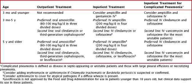
Because follow-up chest radiograph results can remain abnormal long after complete clinical recovery, such studies are typically unwarranted in patients with uncomplicated recoveries and seldom change the management of these patients. Chest radiography should be considered for patients who have not improved on empiric treatment and who have not yet had any chest imaging. Patients with increasing effusions and worsening symptoms should be referred for diagnostic and therapeutic drainage of the effusion. The findings of hilar lymphadenopathy, cystic or nodular disease, or pulmonary anatomic abnormalities should prompt further diagnostic procedures to identify possible tuberculosis, endemic mycoses, and atypical pathogens; to diagnose certain autoimmune or neoplastic conditions; and to consider underlying immune deficiencies (e.g., chronic granulomatous disease).

Treatment

Distinguishing between viral and bacterial pneumonia clinically can be extremely difficult. It is therefore reasonable to provide empiric antimicrobial therapy to outpatients with a clinical diagnosis of pneumonia. Amoxicillin remains the drug of choice for outpatient pediatric pneumonia (Table 91-2); there are no data to support the choice of empiric standard-dose amoxicillin versus high-dose amoxicillin. If chosen, high-dose amoxicillin or ampicillin should have continued efficacy against resistant S. pneumoniae, whose resistance is mediated by alterations in penicillin-binding proteins and can be overcome at higher drug concentrations.

A randomized trial in Pakistan demonstrated fewer failures with amoxicillin treatment compared with trimethoprim−sulfamethoxazole (TMP-SMX) treatment for CAP. Amoxicillin was also specifically more effective in patients under one year of age and those with severe pneumonia, as defined by cough or difficulty breathing with lower chest retractions. Most notably, no patients with pneumococcal bacteremia failed amoxicillin treatment, but almost 30% of such patients treated with TMP-SMX failed treatment. Despite increasing reports of in vitro β-lactam (i.e., amoxicillin or ampicillin) resistance among S. pneumoniae isolates, no significant increase in clinical failures for patients treated with amoxicillin or ampicillin has occurred, underscoring the notion that amoxicillin or ampicillin should be the initial therapy of choice for pediatric CAP. Additionally, since the introduction of PCV7 in 2000, the proportion of pneumococcal isolates resistant to penicillins has decreased.

The incidence of community-acquired MRSA has increased in recent years, and community-acquired MRSA has been recognized as a cause of severe, necrotizing pneumonia. MRSA should be considered as a possible causative pathogen in severely ill hospitalized patients. Although local patterns can vary, community-acquired MRSA is typically susceptible to clindamycin; clindamycin should be considered for use in severe or necrotizing pneumonia in hospitalized patients unless local clindamycin resistance is high among MRSA isolates (>15% of isolates is a common threshold). In such cases, vancomycin should be used. TMP-SMX can also be considered as an alternate treatment for CAP in which MRSA is isolated or suspected, given the high proportion of community-acquired MRSA isolates that are susceptible.

The duration of CAP treatment is unclear but is typically 10 days (or 5 days if azithromycin is used). A randomized, controlled trial of 3 versus 5 days of oral amoxicillin for 2- to 59-month-old patients in Pakistan with nonsevere CAP showed no difference in failure rates or relapse between the two groups, even when the results were limited to those with radiographically confirmed pneumonia. This study’s results may be less applicable in the developed world, where there may be greater access to laboratory testing for viral pathogens: this study used the WHO definition of pneumonia and therefore likely included patients with viral illnesses (e.g., 20% of patients in one of these studies had laboratory-confirmed RSV).

Clindamycin is an appropriate alternative treatment choice in patients with penicillin allergies given its excellent pneumococcal coverage. Oral third-generation cephalosporins or macrolides such as azithromycin can also be considered in these patients. These antibiotics are not as effective antipneumococcal agents as the penicillins, however, and there is increasing macrolide resistance among pneumococcal strains. Levofloxacin is a fluoroquinolone effective against most resistant pneumococcal strains and with a broad range of activity in general (including atypical pathogens such as Legionella spp. or M. pneumoniae). Fluoroquinolones are not recommended in children younger than 18 years of age based on safety concerns for tendon rupture and other musculoskeletal injuries, but data support its safety in these patients, and levofloxacin may be useful in patients with drug allergies or known resistant isolates.

Hospital admission should be considered for any patients younger than 3 months of age, with oxygen saturations less than 92% on room air, severe respiratory distress or grunting, or dehydration or inability to take in oral fluids and oral antibiotics or when providers are unable to monitor patients closely for clinical decompensation.

Patients with persistent symptoms or failure to improve after 48 hours on appropriate empiric therapy should have chest radiography performed or repeated, partly to detect a new or evolving pleural effusion or empyema. Although some patients can have simple parapneumonic effusions requiring no intervention, others require drainage of a significant pleural bacterial infection to speed recovery. Viral diagnostics such as rapid respiratory viral PCR panels should also be considered because positive viral test results often enable providers to discontinue antibiotics. Patients who fail treatment with oral amoxicillin and who have no effusion may receive inpatient treatment with ampicillin, with or without a macrolide. Those admitted to the hospital with an empyema or necrotizing pneumonia should receive broad-spectrum antibiotics that provide coverage against highly resistant S. pneumoniae and MRSA isolates (e.g., a combination of clindamycin and cefotaxime).

Suggested Readings

Ampofo K, Bender J, Sheng X, et al. Seasonal invasive pneumococcal disease in children: role of preceding respiratory viral infection. Pediatrics. 2008;122:229-237.

Black SB, Shinefield HR, Ling S, et al. Effectiveness of heptavalent pneumococcal conjugate vaccine in children younger than five years of age for prevention of pneumonia. Pediatr Infect Dis J. 2002;21:810-815.

Cardoso MR, Nascimento-Carvalho CM, Ferrero F, et al. Penicillin-resistant pneumococcus and risk of treatment failure in pneumonia. Arch Dis Child. 2008;93:221-225.

Grant GB, Campbell H, Dowell SF, et al. Recommendations for treatment of childhood non-severe pneumonia. Lancet Infect Dis. 2009;9:185-196.

Grijalva CG, Nuorti JP, Arbogast PG, et al. Decline in pneumonia admissions after routine childhood immunisation with pneumococcal conjugate vaccine in the USA: a time-series analysis. Lancet. 2007;369:1179-1186.

Haider BA, Saeed MA, Bhutta ZA: Short-course versus long-course antibiotic therapy for non-severe community-acquired pneumonia in children aged 2 months to 59 months. Cochrane Database Syst Rev 2:CD005976, 2008.

Kabra SK, Lodha R, Pandey RM: Antibiotics for community acquired pneumonia in children. Cochrane Database Syst Rev 3:CD004874, 2006.

Kyaw MH, Lynfield R, Schaffner W, et al. Effect of introduction of the pneumococcal conjugate vaccine on drug-resistant Streptococcus pneumoniae. N Engl J Med. 2006;354:1455-1463.

Madhi SA, Ludewick H, Kuwanda L, et al. Pneumococcal coinfection with human metapneumovirus. J Infect Dis. 2006;193:1236-1243.

McIntosh K. Community-acquired pneumonia in children. N Engl J Med. 2002;346:429-437.

Michelow IC, Olsen K, Lozano J, et al. Epidemiology and clinical characteristics of community-acquired pneumonia in hospitalized children. Pediatrics. 2004;113:701-707.