Pleural Effusion and Empyema

Published on 23/05/2015 by admin

Filed under Pulmolory and Respiratory

Last modified 22/04/2025

Print this page

rate 1 star rate 2 star rate 3 star rate 4 star rate 5 star
Your rating: none, Average: 0 (0 votes)

This article have been viewed 3295 times

Pleural Effusion and Empyema

Anatomic Alterations of the Lungs

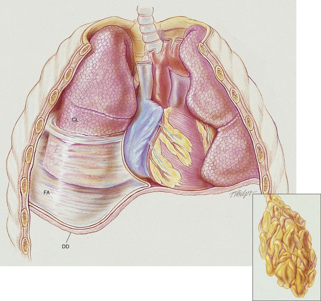
A number of pleural diseases can cause fluid to accumulate in the pleural space; this fluid is called a pleural effusion, or if infected, an empyema (see Figure 23-1). Similar to free air in the pleural space, fluid accumulation separates the visceral and parietal pleura and compresses the lungs. In severe cases, atelectasis will develop, the great veins may be compressed, and cardiac venous return may be diminished. Pleural effusion and empyema produce a restrictive lung disorder.

The major pathologic or structural changes associated with significant pleural effusion are as follows:

Etiology and Epidemiology

Pleural effusion affects approximately 1.3 million people each year in the United States. Early signs and symptoms include pleuritic chest pain, “chest pressure,” dyspnea, and cough. Chest pain can occur early when there is intense inflammation of the pleural surfaces. “Chest pressure” usually develops until the effusion is in the moderate (500 to 1500 mL) to large (>1500 mL) category. Dyspnea rarely occurs in small effusions unless significant pleurisy is present. A cough is usually directly related to the degree of atelectasis caused by the effusion.

A pleural effusion may be transudative or exudative. A transudate develops when fluid from the pulmonary capillaries moves into the pleural space. The fluid is thin and watery, containing a few blood cells and little protein. The pleural surfaces are not involved in producing the transudate. In contrast, an exudate develops when the pleural surfaces are diseased. The fluid has a high protein content and a great deal of cellular debris. Exudate is usually caused by inflammation, infection, or malignancy. Transudative pleural effusions and exudative pleural effusions are differentiated by comparing the chemistries of the pleural fluid with those of the blood. The pleural effusion is classified as exudative when one or more of the following is found in the pleural fluid:

Common Causes of Transudative Pleural Effusion

Congestive Heart Failure

Congestive heart failure is the most common cause of pleural effusion. Both right- and left-sided heart failure can result in pleural effusion. In general, left-sided heart failure is more likely to produce pleural effusion than right-sided heart failure. In right-sided heart failure (cor pulmonale), an increase in the hydrostatic pressure in the systemic circulation can (1) increase the rate of pleural fluid formation and (2) decrease lymphatic drainage from the pleural space because of the elevated systemic venous pressure. In left-sided heart failure, an increase in hydrostatic pressure in the pulmonary circulation can (1) decrease the rate of pleural fluid absorption through the visceral pleura and (2) cause fluid movement through the visceral pleura into the pleural space.

Common Causes of Exudative Pleural Effusion

Malignant Pleural Effusions

About two thirds of malignant pleural effusions occur in women. Malignant pleural effusions are highly associated with breast and gynecologic malignancies.

Other Pathologic Fluids That Separate the Parietal from the Visceral Pleura

In addition to transudate and exudate, other pathologic fluids can separate the parietal pleura from the visceral pleura.

Hemothorax

The presence of blood in the pleural space is known as a hemothorax. Most of these are caused by penetrating or blunt chest trauma. An iatrogenic hemothorax may develop from trauma caused by the insertion of a central venous or pulmonary artery catheter.

Blood can gain entrance into the pleural space from trauma to the chest wall, diaphragm, lung, or mediastinum. A hematocrit of the pleural fluid should always be obtained if the pleural fluid looks like blood. A hemothorax is said to be present only when the hematocrit of the pleural fluid is at least 50%.

image OVERVIEW of the Cardiopulmonary Clinical Manifestations Associated with Pleural Effusion and Empyema

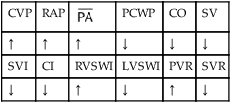
The following clinical manifestations result from the pathologic mechanisms caused (or activated) by Atelectasis (see Figure 9-7)—the major anatomic alteration of the lungs associated with pleural effusion (see Figure 24-1).

CLINICAL DATA OBTAINED AT THE PATIENT’S BEDSIDE

CLINICAL DATA OBTAINED FROM LABORATORY TESTS AND SPECIAL PROCEDURES

Pulmonary Function Test Findings (Restrictive Lung Pathology)

LUNG VOLUME AND CAPACITY FINDINGS

VT IRV ERV RV  
N or ↓  
VC IC FRC TLC RV/TLC ratio
N

image

RADIOLOGIC FINDINGS

Chest Radiograph

The diagnosis of a pleural effusion is generally based on the chest x-ray film. A pleural effusion of less than 300 mL usually cannot be seen on an upright chest x-ray film. In moderate pleural effusion (>1000 mL) in the upright position, an increased density usually appears at the costophrenic angle. The fluid first accumulates posteriorly in the most dependent part of the thoracic cavity between the inferior surface of the lower lobe and the diaphragm. As the fluid volume increases, it extends upward around the anterior, lateral, and posterior thoracic walls in the so-called “meniscus sign” (see Figure 23-3). Interlobar fissures are sometimes highlighted as a result of fluid filling.

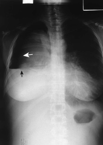
As nicely illustrated in the chest radiograph of a pleural effusion shown in Figure 23-2, the lateral costophrenic angle is usually obliterated, and the outline of the diaphragm on the affected side is lost. In severe cases the weight of the fluid may cause the diaphragm to become inverted (concave). Clinically this inversion is seen only in left-sided pleural effusions; the gastric air bubble is pushed downward, and the superior border of the left diaphragmatic leaf is concave. In addition, the mediastinum may be shifted to the unaffected side, and the intercostal spaces may appear widened.

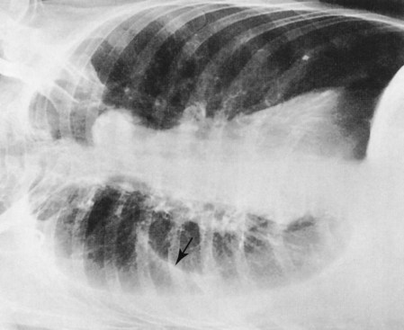
Pleural effusion, atelectasis, and parenchymal infiltrates can obliterate one or both diaphragms. Therefore when a posteroanterior or lateral chest radiograph suggests pleural effusion, additional radiographic studies are generally necessary to document the presence of pleural fluid or other pathology. The lateral decubitus radiograph is recommended because free fluid gravitates to the most dependent part of the pleural space and layers out there (Figure 23-3).

General Management of Pleural Effusion

The management of each patient with a pleural effusion must be individualized. Questions to be asked include the following: Should a thoracentesis be performed? Can the underlying cause be treated? What is the appropriate antibiotic? Should a chest tube be inserted? When it is determined that a chest tube should be inserted, it is normally placed in the fourth or fifth intercostal space at the midaxillary line. Typically, a No. 28 to No. 36 French gauge thoracostomy tube is used for adults, with a smaller size used for children.

The best way to resolve a pleural effusion is to direct the treatment at what is causing it, rather than treating the effusion itself. For example, if the heart failure is reversed or the lung infection is cured by antibiotics, the effusion usually resolves. When the cause of the pleural effusion is not readily evident, microscopic and chemical examination of pleural fluid may determine whether the effusion is a transudate or an exudate. If the fluid is a transudate, treatment is directed to the underlying problem (e.g., congestive heart failure, cirrhosis, nephrosis).

When an exudate is present, a cytologic examination may identify a malignancy. The fluid also may be examined for its biochemical makeup (e.g., protein, sugar, various enzymes) and for the presence of bacteria. Examination of the effusion may reveal blood after trauma or surgery, pus in empyema, or milky fluid in chylothorax. The presence of blood in the pleural fluid in the absence of trauma or surgery suggests malignant disease or pulmonary embolization or infarction.

Respiratory Care Treatment Protocols

Oxygen Therapy Protocol

Oxygen therapy is used to treat hypoxemia, decrease the work of breathing, and decrease myocardial work. The hypoxemia that develops in pleural effusion is mostly caused by the atelectasis and pulmonary shunting associated with the disorder. Hypoxemia caused by capillary shunting is often refractory to oxygen therapy (see Oxygen Therapy Protocol, Protocol 9-1).

CASE STUDY

Pleural Effusion and Empyema

Admitting History

Against her doctor’s advice, a 38-year-old white woman had discharged herself from the hospital about 2 months before the admission discussed here. She had originally been admitted for severe right lower lobe pneumonia. After 5 days of treatment, she became angry because she was not allowed to smoke. She was a longtime, three-pack-per-day smoker. When a nurse found her smoking in her hospital bed while on a 2 L/min oxygen nasal cannula, the nurse quickly confiscated her cigarettes and matches.

The woman became upset. She told her doctor that this was the last straw and that she was going to leave the hospital on her own. Her doctor wanted her to remain so that a thorough follow-up could be performed for what was described as a “spot” on her lower right lung. The woman promised that she would make an appointment at the doctor’s office the next week. She then got dressed and left. However, 2 days after she left the hospital, she felt so much better that she decided the spot on her lung was not an issue for concern. The woman told her friends that smoking one pack of cigarettes made her feel better than 5 days’ worth of nurses, doctors, and hospitals.

On the day of the admission discussed here, the woman appeared at the doctor’s office without an appointment. She told the receptionist that something was very wrong. She thought that she had the flu and that it had been getting progressively worse over the previous 4 days. At the time of the office visit, she could speak in short sentences only and was unable to inhale deeply. Seeing that the woman was in obvious respiratory distress, the nurse interrupted the doctor. Within 5 minutes, the doctor had the woman transported and admitted to the hospital a few blocks away.

Physical Examination

The woman appeared malnourished, exhibited poor personal hygiene, and had yellow tobacco stains around her fingers. She appeared to be in moderate to severe respiratory distress. Her nails and mucous membranes were cyanotic, and her shirt was wet from perspiration. She demonstrated an occasional hacking, nonproductive cough. She stated that she could not take a deep breath and that maybe the problem stemmed from “that spot” on her lung.

Her vital signs were as follows: blood pressure 130/60, heart rate 112 bpm, and respiratory rate 36/min with shallow respirations. She was slightly febrile, with an oral temperature of 37.7° C (99.8° F). Palpation showed that the trachea was shifted slightly to the left. Dull percussion notes were found over the right middle and right lower lobes. Auscultation revealed normal vesicular breath sounds over the left lung fields and upper right lobe. No breath sounds could be heard over the right middle and right lower lobes.

The patient’s chest x-ray film showed a large, right-sided pleural effusion. The right costophrenic angle demonstrated blunting, the right hemidiaphragm was depressed, and the right middle and lower lung lobes were partially collapsed and showed changes consistent with pneumonia. The arterial blood gas values (ABGs) on a 3 L/min oxygen nasal cannula were as follows: pH 7.48, Paco2 24, image 17, and Pao2 37. The oxygen saturation measured by pulse oximetry (Spo2) was 72%. The doctor, assisted by the respiratory therapist, performed a thoracentesis on the patient at the bedside. Slightly more than 2 L of yellow fluid was withdrawn. The patient then was started on intravenous antibiotics. A portable chest x-ray examination was ordered, and a respiratory care consultation was requested. On the basis of these clinical data, the following SOAP was documented.

Respiratory Assessment and Plan

S “I can’t take a deep breath.”

O Malnourished appearance with poor personal hygiene; cyanosis with an occasional hacking, nonproductive cough; vital signs: BP 130/60, HR 112, RR 36 and shallow, T 37.7° C (99.8° F); trachea slightly shifted to the left; dull percussion notes over the right middle and right lower lobes; normal vesicular breath sounds over the left lung fields and right upper lobe; no breath sounds over the right middle and right lower lobes; CXR: large, right-sided pleural effusion, right middle and right lower lobes partially collapsed and consolidated; about 2 L of yellow fluid obtained via thoracentesis; ABGs (on 3 L/min O2 by nasal cannula): pH 7.48, Paco2 24, image 17, Pao2 37, Spo2 72%

A 

P Begin Lung Expansion Therapy Protocol (incentive spirometry q2h) and Oxygen Therapy Protocol (Fio2 = 0.50 per HAFOE mask). Monitor vital signs carefully and reevaluate.

3 Hours after Admission

At this time the patient was sitting up in bed. She stated that although she was feeling better, she did not feel great. She still had an occasional dry-sounding, nonproductive cough. Her skin appeared pale. She was still cyanotic. She was no longer perspiring, as she was when she was first admitted. Her vital signs were as follows: blood pressure 135/85, heart rate 100 bpm, respiratory rate 24/min, and temperature normal. Her respiratory efforts, however, no longer appeared shallow. Palpation of the chest was not remarkable. Dull percussion notes were found over the right middle and right lower lobes. Normal vesicular breath sounds were heard over the left lung and upper right lung. Loud bronchial breath sounds were audible over the right middle and right lower lobes.

The patient’s chest x-ray showed a small, right-sided pleural effusion. Increased opacity was still present in the right middle and lower lung, consistent with pneumonia. The patient’s trachea and mediastinum were in their normal positions. On an Fio2 of 0.50, her ABGs were as follows: pH 7.52, Paco2 29, image 22, and Pao2 57. Her Spo2 was 92%. At this time, the following SOAP was charted.

Respiratory Assessment and Plan

S “I’m feeling better but not great yet.”

O Cyanotic and pale appearance; occasional dry, nonproductive cough; vital signs: BP 135/85, HR 100, RR 24, T normal; dull percussion notes over right middle and right lower lobes; normal vesicular breath sounds over left lung and over right upper lobe; bronchial breath sounds over right middle and lower lobes; CXR: small right-sided pleural effusion; right middle and right lower lobe consolidation; ABGs: pH 7.52, Paco2 29, image 22, Pao2 57; Spo2 92% on an Fio2 of 0.50.

A 

P Up-regulate Lung Expansion Therapy Protocol (CPAP mask at 10 cm H2O q2h for 15 minutes). Up-regulate Oxygen Therapy Protocol (Fio2 = 0.60 per HAFOE mask). Monitor and reevaluate.

5 Hours after Admission

The patient was sitting in a semi-Fowler’s position. She appeared relaxed and alert. She stated that she had finally caught her breath. Although she still appeared pale, she did not look cyanotic. No spontaneous cough was observed at this time.

Her vital signs were as follows: blood pressure 128/79, heart rate 88 bpm, respiratory rate 16/min, and temperature normal. Palpation of the chest was unremarkable. Dull percussion notes were found over the right middle and right lower lobes. Normal vesicular breath sounds were heard over the left lung and right upper lobe. Bronchial breath sounds were audible over the right middle and right lower lobes. No current chest x-ray was available. The patient’s ABG values on an Fio2 of 0.60 were as follows: pH 7.45, Paco2 36, image 24, and Pao2 77. Her Spo2 was 95%. On the basis of these clinical data, the following SOAP was documented.

Respiratory Assessment and Plan

S “I’ve finally caught my breath.”

O Relaxed, alert appearance, in semi-Fowler’s position; paleness but no cyanosis; no spontaneous cough; vital signs: BP 128/79, HR 88, RR 16, T normal; dull percussion notes in right middle and right lower lung lobes; normal vesicular breath sounds over left lung and right upper lobe; bronchial breath sounds over right middle and right lower lobes; ABGs: pH 7.45, Paco2 36, image 24, Pao2 77; Spo2 95%

A 

P Maintain present level of Lung Expansion Therapy Protocol and Oxygen Therapy Protocols. Monitor and reevaluate each shift.

Discussion

This case illustrates a patient with postpneumonic pleural effusion, one of the pleural diseases that generally can be improved with appropriate therapy (in this case a 2-L thoracentesis).

During the first assessment, the respiratory care practitioner recognizes that the patient has significant respiratory morbidity. Indeed, the patient has an extensive right-sided pneumonia and pleural effusion and partially collapsed right middle and lower lobes. Clearly the patient is in respiratory distress. The patient’s acute alveolar hyperventilation and severe hypoxemia are a direct result of the partial collapse of the lung lobes. Because of the extremely low Pao2 noted on the initial arterial blood gas, the presence of lactic acid is very likely. In fact, this was confirmed by the respiratory practitioner with the Pco2/image/pH nomogram. Understanding that Atelectasis is the main pathophysiologic mechanism operating in this case (see Figure 9-8), the practitioner correctly assesses the situation as one that requires careful monitoring and begins the Lung Expansion Therapy Protocol (Protocol 9-3) (with incentive spirometry) and the Oxygen Therapy Protocol (Protocol 9-1) (with a high concentration of oxygen).

A trial of bronchopulmonary hygiene therapy would not be unwarranted in this case, given the patient’s cigarette smoking history alone or the degree of severity of the condition. Admittedly, the physical findings in this patient (no sputum production) did not indicate such therapy. Given the patient’s history, the respiratory care practitioner also would be interested in the results of the cytologic studies for malignancy in both the sputum and thoracentesis fluid. Frequently, blood gases do not improve immediately after a thoracentesis, despite the fluid removal, because the atelectasis under the pleural effusion takes some time (hours or days) to dissipate. For this reason, the Lung Expansion Therapy Protocol, after thoracentesis, is appropriate.

At the time of the second assessment, the patient was beginning to improve, although she still had signs of right middle and lower lobe Consolidation (Figure 9-9). Good breath sounds were heard over the left lung and upper right lung, although bronchial breath sounds reflecting consolidation were still noted on the right. The respiratory care practitioner was appropriately concerned that atelectasis was still present, and in such a case he or she should increase the Lung Expansion Therapy Protocol (Protocol 9-3). In this case, the practitioner selected a continuous positive airway pressure (CPAP) mask at 10 cm H2O every 2 hours for 15 minutes. The practitioner could have also intensified use of incentive spirometry, carefully used intermittent positive-pressure breathing (IPPB) or extended the amount of time the patient was using the CPAP mask.

In the last assessment the patient continued to do fairly well, although she was far from returning to baseline values. The pneumonitis, atelectasis, and mild hypoxemia, which persisted despite supplemental oxygen therapy, suggested the need for continued significant (though unchanged) therapy. This case demonstrates that in-place therapy often does not need to be changed at each assessment. Indeed, this guide may apply to as many as 50% to 60% of accurately performed seriatim assessments. For pedagogic reasons, this option has not been exercised often in this text. However, this third assessment (in a patient with pleural effusion and underlying atelectasis and pneumonia) is a good case in point.

Share this: