Placental and fetal growth and development

Published on 09/03/2015 by admin

Filed under Obstetrics & Gynecology

Last modified 22/04/2025

Print this page

rate 1 star rate 2 star rate 3 star rate 4 star rate 5 star
Your rating: none, Average: 0 (0 votes)

This article have been viewed 1925 times

Placental and fetal growth and development

E. Malcolm Symonds

Early placental development

After fertilization and egg cleavage, the morula is transformed into a blastocyst by the formation of a fluid-filled cavity within the ball of cells.

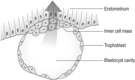
The outer layer of the blastocyst consists of primitive cytotrophoblast and, by day 7, the blastocyst penetrates the endometrium as a result of trophoblastic invasion (Fig. 4.1). The outer layer of trophoblast becomes a syncytium. In response to contact with the syncytiotrophoblast, the endometrial stromal cells become large and pale, a process known as the decidual reaction. Some endometrial cells are phagocytosed by the trophoblastic cells.

The nature and function of the decidual reaction remain uncertain, but it seems likely that the decidual cells both limit the invasion of trophoblastic cells and serve an initial nutritional function for the developing placenta.

During development of the placenta, cords of cytotrophoblast or Langhans cells grow down to the basal layers of decidua and penetrate some of the endometrial venules and capillaries. The formation of lacunae filled with maternal blood presages the development of the intervillous space.

The invading cords of trophoblast form the primary villi, which later branch to form secondary villi and, subsequently, free-floating tertiary villi.

The central core of these villi is penetrated by a column of mesoblastic cells that become the capillary network of the villi. The body stalk attaching the developing fetus to the placenta forms the umbilical vessels, which advance into the villi to join the villous capillaries and establish the placental circulation.

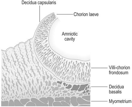
Although trophoblastic cells surround the original blastocyst, the area that develops into the placenta becomes thickened and extensively branched and is known as the chorion frondosum. However, in the area that subsequently expands to form the outer layer of the fetal membranes or chorion laeve, the villi become atrophic and the surface becomes smooth (Fig. 4.2). The decidua underlying the placenta is known as the decidua basalis and the decidua between the membranes and the myometrium as the decidua capsularis.

Further placental development

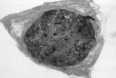
By 6 weeks after ovulation, the trophoblast has invaded some 40–60 spiral arterioles. Blood from the maternal vasculature pushes the free-floating secondary and tertiary capillaries into a tent-shaped maternal cotyledon. The tents are held down to the basal plate of the decidua by anchoring villi, and the blood from arterioles spurts towards the chorionic plate and then returns to drain through maternal veins in the basal plate. There are eventually about 12 large maternal cotyledons and 40–50 smaller ones (Fig. 4.3).

The villus

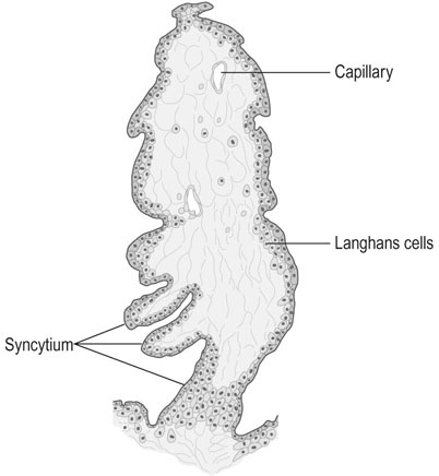
Despite the arrangement of villi into maternal cotyledons, the functional unit of the placenta remains the stem villus or fetal cotyledon. The end unit of the stem villus, sometimes known as the terminal or chorionic villus is shown in Figure 4.4. There are initially about 200 stem villi arising from the chorion frondosum. About 150 of these structures are compressed at the periphery of the maternal cotyledons and become relatively functionless, leaving a dozen or so large cotyledons and 40–50 smaller ones as the active units of placental function.

The estimated total surface area of the chorionic villi in the mature placenta is approximately 11 m2. The surface area of the fetal side of the placenta and of the villi is enlarged by the presence of numerous microvilli. The core of the villus consists of a stroma of closely packed spindle-shaped fibroblasts and branching capillaries. The stroma also contains phagocytic cells known as Hofbauer cells. In early pregnancy, the villi are covered by an outer layer of syncytiotrophoblast and an inner layer of cytotrophoblast. As pregnancy advances, the cytotrophoblast disappears until only a thin layer of syncytiotrophoblast remains. The formation of clusters of syncytial cells, known as syncytial knots, and the reappearance of cytotrophoblast in late pregnancy are probably the result of hypoxia. There is evidence that the rate of apoptosis of syncytial cells accelerates towards term and is particularly increased where there is evidence of fetal growth impairment.

Structure of the umbilical cord

The umbilical cord contains two arteries and one vein (Fig. 4.5). The two arteries carry deoxygenated blood from the fetus to the placenta and the oxygenated blood returns to the fetus via the umbilical vein. Absence of one artery occurs in about 1 in 200 deliveries and is associated with a 10–15% incidence of cardiovascular anomalies. The vessels are surrounded by a hydrophilic mucopolysaccharide known as Wharton’s jelly and the outer layer covering the cord consists of amniotic epithelium. The cord length varies between 30 and 90 cm.
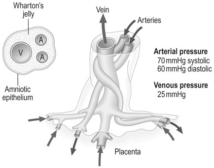
The vessels grow in a helical shape. This configuration has the functional advantage of protecting the patency of the vessels by absorbing torsion without the risk of kinking or snarling of the vessels.

The few measurements that have been made in situ of blood pressures in the cord vessels indicate that the arterial pressure in late pregnancy is around 70 mmHg systolic and 60 mmHg diastolic, with a relatively low pulse pressure and a venous pressure that is exceptionally high, at approximately 25 mmHg. This high venous pressure tends to preserve the integrity of the venous flow and indicates that the pressure within the villus capillaries must be in excess of the cord venous pressures.

The cord vessels often contain a false knot consisting of a refolding of the arteries; occasionally, blood flow is threatened by a true knot, although such formations are often seen without any apparent detrimental effects on the fetus.

In the full-term fetus, the blood flow in the cord is approximately 350 mL/min.

Uteroplacental blood flow

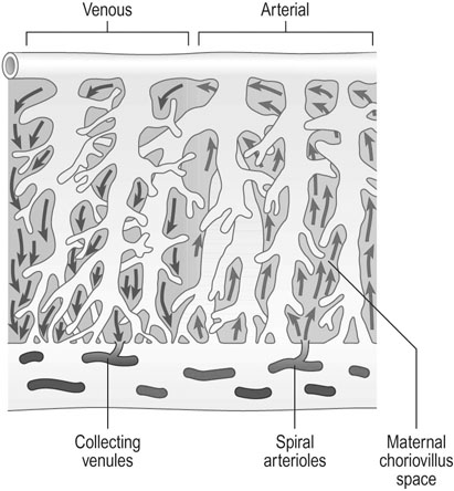
Trophoblastic cells invade the spiral arterioles within the first 10 weeks of pregnancy and destroy some of the smooth muscle in the wall of the vessels which then become flaccid dilated vessels. Maternal blood enters the intervillous space and, during maternal systole, blood spurts from the arteries towards the chorionic plate of the placenta and returns to the venous openings in the placental bed. The intervillous space is characterized by low pressures, with a mean pressure estimated at 10 mmHg and high flow. Assessments of uterine blood flow at term indicate values of 500–750 mL/min (Fig. 4.6).

Factors that regulate fetoplacental and uterine blood flow

The fetoplacental circulation is effected by the fetal heart and aorta, the umbilical vessels and the vessels of the chorionic villi, so factors that affect these structures may affect the fetal circulation. Such factors as oedema of the cord, intramural thrombosis and calcification within the large fetal vessels or acute events such as acute cord compression or obstruction of the umbilical cord may have immediate and lethal consequences for the fetus. However, the more common factors that influence the welfare of the fetus arise in the uteroplacental circulation. Access to these factors by the use of Doppler ultrasound has greatly improved our understanding of the control mechanisms of uterine blood flow.

The regulation of uterine blood flow is of critical importance to the welfare of the fetus. The uteroplacental blood flow includes the uterine arteries and their branches down to the spiral arterioles, the intervillous blood flow and the related venous return.

Impairment of uterine blood flow leads to fetal growth impairment and, under severe circumstances, to fetal death. Factors that influence uteroplacental blood flow acutely include maternal haemorrhage, tonic or abnormally powerful and prolonged uterine contractions and substances such as noradrenaline (norepinephrine) and adrenaline (epinephrine). Angiotensin II increases uterine blood flow at physiological levels, as it has a direct effect on the placental release of vasodilator prostaglandins, but in high concentrations it produces vasoconstriction.

At the simplest level, acute fetal asphyxia can be produced by the effect of the mother lying in the supine position in late pregnancy, causing compression of the maternal inferior vena cava and hence a sudden reduction in blood flow through the uteroplacental bed.

In terms of chronic pathology, the main causes of impaired uteroplacental circulation are inadequate trophoblast invasion and acute atherosis affecting the spiral arterioles; resulting in placental ischaemia, advanced maturation and placental infarction.

Placental transfer

The placenta plays an essential role in growth and development of the fetus and in regulating maternal adaptation to pregnancy. The placenta is an organ of fetal nutrition, excretion, respiration, and of hormone synthesis.

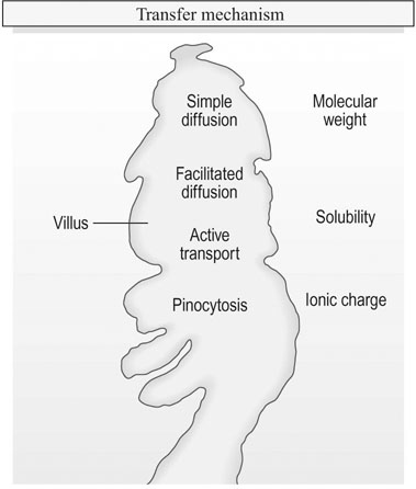
Transfer of materials across the placental membrane is governed by molecular mass, solubility and the ionic charge of the substrate involved. Actual transfer is achieved by simple diffusion, facilitated diffusion, active transport and pinocytosis (Fig. 4.7).

Simple diffusion

Transfer between maternal and fetal blood is regulated by the trophoblast, and it must be remembered that the layer separating fetal from maternal blood in the chorionic villus is not a simple semipermeable membrane but a metabolically active cellular layer. However, with regard to some substances, it does behave like a semipermeable membrane and substances pass by simple diffusion.

Although there are some exceptions, small molecules generally cross the placenta in this way and movement is determined by chemical or electrochemical gradients. The quantity of solute transferred is described by the Fick diffusion equation:

< ?xml:namespace prefix = "mml" />Qt=KA(C1C2)L

image

where image is the quantity transferred per unit of time, K is a diffusion constant for the particular substance, A is the total surface area available, C1 and C2 indicate the difference in concentrations of solute and L represents the thickness of the membrane.

This method is applicable particularly to transfer of gases, although the gradient of oxygen, for example, is exaggerated by the fact that oxygen is extracted by the villous trophoblast.

Water and electrolyte transfer

Water passes easily across the placenta and a single pass allows equilibrium. The driving forces for movement of water across the placenta include hydrostatic pressure, colloid osmotic pressure and solute osmotic pressure.

Placental function

The placenta has three major functions:

Gaseous exchange

As the transfer of gases occurs by simple diffusion, the major determinants of gaseous exchange are the efficiency and flow of the fetal and maternal circulation, the surface area of the placenta that is available for transfer and the thickness of the placental membrane.

Oxygen transfer

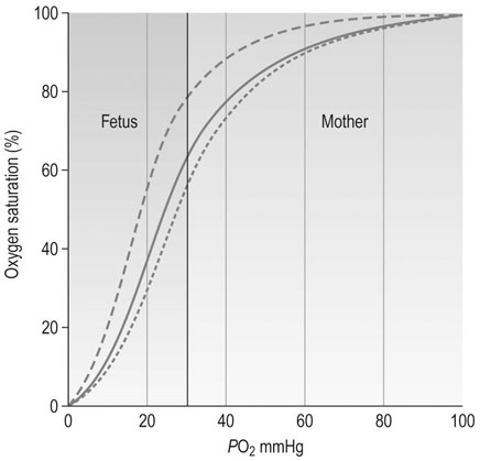
The average oxygen saturation of maternal blood entering the intervillous space is 90–100% at a PO2 of 90–100 mmHg and these high levels of oxygen favour transfer to the fetal circulation. After the placenta itself has utilized some of this oxygen, the remainder is available to the fetal circulation. Fetal haemoglobin has a higher affinity for oxygen than does adult haemoglobin and haemoglobin concentration is higher in the fetus. All of these factors favour the rapid uptake of oxygen by the fetus at PO2 levels as low as 30–40 mmHg. The extent to which haemoglobin can be saturated by oxygen is affected by hydrogen ion concentration. The increase that occurs in deoxygenated blood arriving in the placental circulation from the fetus favours the release of maternal oxygen in the fetoplacental bed. The oxygen dissociation curve is shifted to the right by the increase in H+ concentration, PCO2 and temperature and this is known as the Bohr effect (Fig. 4.8). Oxygen is predominantly transported in the form of oxyhaemoglobin as there is little free oxygen in solution.

Fetal nutrition and removal of waste products

Carbohydrate metabolism

Glucose transferred from the maternal circulation provides the major substrate for oxidative metabolism in the fetus and placenta and provides 90% of the energy requirements of the fetus. Facilitated diffusion ensures that there is rapid transfer of glucose across the placenta. In late pregnancy, the fetus retains some 10 g/kg body weight and any excess glucose is stored as glycogen or fat. Glycogen is stored in the liver, muscle, the placenta and the heart, whereas fat is deposited around the heart and behind the scapulae.

Animal studies have shown that the transfer of sugars is selective. Generally, glucose and the monosaccharides cross the placenta readily, whereas it is virtually impermeable to disaccharides such as sucrose, maltose and lactose. The placenta is also impermeable to the sugar alcohols such as sorbitol, mannitol, deleitol and meso-inositol.

In the fasting normal pregnant woman, blood glucose achieves a concentration of approximately 4.0 mmol/L in the maternal venous circulation and 3.3 mmol/L in the fetal cord venous blood. Infusion of glucose into the maternal circulation results in a parallel increase in both maternal and fetal blood until the fetal levels reach 10.6 mmol/L, when no further increase occurs regardless of the values in the maternal circulation.

The hormones that are important in glucose homeostasis – insulin, glucagon, human placental lactogen and growth hormone – do not cross the placenta and maternal glucose levels appear to be the major regulatory factor in fetal glucose metabolism. The placenta itself utilizes glucose and may retain as much as half of the glucose transferred to the fetoplacental unit.

In mid-pregnancy, approximately 70% of this glucose is metabolized by glycolysis, 10% via the pentose phosphate pathway, and the remainder is stored by glycogen and lipid synthesis. By full term, the rate of placental glucose utilization has fallen by 30%.

Glycogen storage in the fetal liver increases steadily throughout pregnancy and by full term is twice as high as the storage in the maternal liver. A rapid fall to adult levels occurs within the first few hours of life.

Fetal glycogen reserves are particularly important in providing an energy source in the asphyxiated fetus when anaerobic glycolysis is activated.

Protein metabolism

Fetal proteins are synthesized by the fetus from free amino acids transported across the placenta against a concentration gradient. The concentration of free amino acids in fetal blood is higher than in the maternal circulation.

The placenta takes no part in the synthesis of fetal proteins, although it does synthesize some protein hormones that are transferred into the maternal circulation: chorionic gonadotrophin and human placental lactogen. By full term, the human fetus has accumulated some 500 g of protein.

Immunoglobulins are synthesized by fetal lymphoid tissue and IgM first appears in the fetal circulation by 20 weeks gestation, followed by IgA and finally IgG.

IgG is the only gamma-globulin to be transferred across the placenta and this appears to be selective for IgG. There is no evidence of placental transfer of growth-promoting hormones.

Placental hormone production

The placenta plays a major role as an endocrine organ and is responsible for the production of both protein and steroid hormones. The fetus is also involved in many of the processes of hormone production and in this capacity the conceptus functions as a unit involving both fetus and placenta.

Protein hormones

Chorionic gonadotrophin

Human chorionic gonadotrophin (hCG) is produced by trophoblast and has a structure that is chemically very similar to that of luteinizing hormone. It is a glycoprotein with two non-identical α and β subunits and reaches a peak in maternal urine and blood between 10 and 12 weeks gestation. A small sub-peak occurs between 32 and 36 weeks. The β subunit of hCG can be detected in maternal plasma within 7 days of conception.

The only known function of the hormone appears to be the maintenance of the corpus luteum of pregnancy, which is responsible for the production of progesterone until such time as this production is taken over by the placenta.

The hormone is measured by agglutination inhibition techniques using coated red cells or latex particles and this forms the basis for the standard modern pregnancy test (see Chapter 18). This will be positive in urine by 2 weeks after the period is missed in 97% of pregnant women. Home pregnancy test kits are able to detect 25-50iu/L of βhCG.

Human placental lactogen

Human placental lactogen (hPL), or chorionic somatomammotrophin, is a peptide hormone with a molecular weight of 22 000 that is chemically similar to growth hormone. It is produced by syncytiotrophoblast and plasma hPL levels rise steadily throughout pregnancy. The function of the hormone remains uncertain. It increases levels of free fatty acids and insulin. The level tends to rise steeply in the third trimester and is linked to higher blood sugars and abnormal glucose tolerance tests, i.e. helping to unmask the late onset diabetes.

Plasma hPL levels have been extensively used in the assessment of placental function as the levels are low in the presence of placental failure. In the last 2 weeks of gestation the levels in the serum fall in normal pregnancy. However, the use of these measurements as placental function tests has largely fallen into disfavour because of their low discriminant function. The hormone is measured by immunoassay.

Steroid hormones

Oestrogens

Over 20 different oestrogens have been identified in the urine of pregnant women, but the major oestrogens are oestrone, oestradiol-17β and oestriol. The largest increase in urinary oestrogen excretion occurs in the oestriol fraction. Whereas oestrone excretion increases 100-fold, urinary oestriol increases 1000-fold.

The ovary makes only a minimal contribution to this increase as the placenta is the major source of oestrogens in pregnancy. The substrate for oestriol production comes from the fetal adrenal gland. Dehydroepiandrosterone (DHEA) synthesized in the fetal adrenal cortex passes to the fetal liver where it is 16-hydoxylated. Conjugation of these precursors with phosphoadenosyl phosphosulphate aids solubility and active sulphatase activity in the placenta results in the release of free oestriol.

Oestradiol and oestrone are directly synthesized by the syncytiotrophoblast. Urinary and plasma oestriol levels increase progressively throughout pregnancy until 38 weeks gestation, when some decrease occurs.

The use of oestriol measurements has now largely been replaced by the use of various forms of ultrasound assessment.

Fetal development

Growth

Up to 10 weeks gestation, a massive increase in cell numbers occurs in the developing embryo but the actual gain in weight is small. Thereafter a rapid increase in weight occurs, until the full-term fetus reaches a final weight of around 3.5 kg.

Protein accumulation occurs in the fetus throughout pregnancy. However, the situation is very different as far as fetal adipose tissue is concerned. Free fatty acids are stored in brown fat around the neck, behind the scapulae and the sternum and around the kidneys. White fat forms the subcutaneous fat covering the body of the full-term fetus. Fat stores in the fetus between 24–28 weeks gestation make up only 1% of the body weight whereas by 35 weeks it makes up 15% of body weight.

The rate of fetal growth diminishes towards term. Actual fetal size is determined by a variety of factors, including the efficiency of the placenta, the adequacy of the uteroplacental blood flow and inherent genetic and racial factors in the fetus.

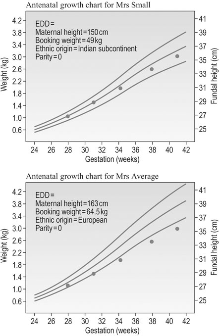
Fetal birth weight is determined by gestational age, race, maternal height and weight and parity. Thus, the projected normal birth weight for an infant is determined by a combination of all of these factors (Fig. 4.9). The normal growth curve therefore varies in each infant and can only be determined by taking into account the history of each individual mother. From all these factors, a nomogram for growth can be calculated. Figure 4.9 has the nomogram constructed for Mrs Small that shows the 5th and 95th centile to be between 35 and 39 cm in fundal height at 41 to 42 weeks, whilst it is 37 to 42 cm for Mrs Average. Hence the same growth trajectory plotted in the Mrs Small’s nomogram shows the fetus to be growing within normal limits and the last plot shows the estimated fetal weight to be about 3.0 kg, but the same plots in Mrs Average’s nomogram results in a fetus that shows growth restriction with progress of gestation.

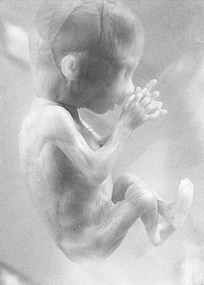
The characteristic appearance of the fetus at 12 weeks gestation is shown in Figure 4.10. The skin is translucent and there is virtually no subcutaneous fat so that the blood vessels in the skin are easily seen, but even at this stage the fetus reacts to stimuli. The upper limbs have already reached their final relative length and the external genitals are distinguishable externally but remain undifferentiated.

By 16 weeks gestation (Fig. 4.11), the crown–rump length is 122 mm and the lower limbs have achieved their final relative length. The external genitalia can now be differentiated.

By 24 weeks gestation (Fig. 4.12), the crown–rump length is 210 mm. The eyelids are separated, the skin is opaque but wrinkled because of the lack of subcutaneous fat, and there is fine hair covering the body. By 28 weeks, the eyes are open and the scalp is growing hair.

The cardiovascular system

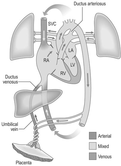
The heart develops initially as a single tube and, by 4–5 weeks gestation, a heartbeat is present at a rate of 65 beats/min. The definitive circulation has developed by 11 weeks gestation and the heart rate increases to around 140 beats/min. In the mature fetal circulation, about 40% of the venous return entering the right atrium flows directly into the left atrium through the foramen ovale (Fig. 4.13). Blood pumped from the right atrium into the right ventricle is expelled into the pulmonary artery, where it passes either into the aorta via the ductus arteriosus or into the pulmonary vessels.

In the mature fetus, the fetal cardiac output is estimated to be 200 mL−1min−1kg body weight. Unlike the adult circulation, fetal cardiac output is entirely dependent on heart rate and not on stroke volume. Autonomic control of the fetal heart rate matures during the third trimester and parasympathetic vagal tonus tends to reduce the basal fetal heart rate.

The respiratory system

Fetal respiratory movements can be detected from as early as 12 weeks gestation and, by mid-trimester, a regular respiratory pattern is established. By 34 weeks gestation, respiration occurs at a rate of 40–60 movements/min with intervening periods of apnoea. These respiratory movements are shallow, with movement of amniotic fluid only into the bronchioles. There are occasional larger flows of fluid into the bronchial tree, but this does not extend into the alveoli because of the high pressure maintained in the developing alveoli from the secretion of alveolar fluid. An exception to this situation may result from episodes of hypoxia, when gasping may lead to the inhalation of amniotic fluid deeper into the alveoli. This fluid may often, under these circumstances, be meconium-stained.

Fetal breathing is stimulated by hypercapnia and by raised maternal glucose levels, as in the post-prandial state, whereas hypoxia reduces the number of respiratory movements, as does maternal smoking.

The occurrence of fetal apnoea increases towards term, when breathing movements may be absent for as long as 120 minutes in a normal fetus.

The fetal pulmonary alveoli are lined by two main types of alveolar epithelial cell. Gaseous exchange occurs across the type I cells and the type II cells secrete a surface-active phospholipid surfactant that is essential in maintaining the functional patency of the alveoli. The principal surfactants are sphingomyelin and lecithin; production of lecithin reaches functional levels by 32 weeks gestation, although it may begin as early as 24 weeks. In some circumstances, such as in the diabetic pregnancy, the production of surfactant may be delayed and the process can be accelerated by the administration of corticosteroids to the mother.

The measurement of lecithin concentration in the amniotic fluid provides a useful method of assessing functional fetal lung maturity.

The kidney

Functional renal corpuscles first appear in the juxtaglomerular zone of the renal cortex at 22 weeks gestation and filtration begins at this time. The formation of the kidney is completed by 36 weeks gestation. Glomerular filtration increases towards term as the number of glomeruli increases and fetal blood pressure rises.

In the fetus, only 2% of the cardiac output perfuses the kidney as most of the excretory functions normally served by the kidney are met by the placenta.

The fetal renal tubules are capable of active transport before any glomerular filtrate is received and thus some urine may be produced within the tubules before glomerular filtration starts. The efficiency of tubular reabsorption is low and glucose in the fetal circulation spills into fetal urine at levels as low as 4.2 mmol−1L.

Fetal urine makes a significant contribution to amniotic fluid.

The special senses

The external ear can be visualized using ultrasound from 10 weeks onwards. The middle ear and the three ossicles are fully formed by 18 weeks, when they also become ossified; the contents of the inner ear, including the cochlear and the membranous and bony labyrinth, are all fully developed by 24 weeks gestation. The perception of sound by the fetus has to be gauged by behavioural responses and it is generally agreed that the first responses to acoustic stimuli occur at 24 weeks gestation, although some observations have suggested that there may be perception as early as 16 weeks. In view of the developmental timetable of the inner ear, this seems unlikely.

Visual perception is much more difficult to assess but it seems likely that some perception to light through the maternal abdominal wall does develop in late pregnancy. Certainly, fetal eye movements can be observed during pregnancy and form an important part of the observations made concerning various fetal behavioural states, a subject that is discussed in Chapter 10.

Amniotic fluid

Formation

The amniotic sac develops in early pregnancy and has been identified in the human embryo as early as 7 days. The first signs of the development of the amniotic cavity can be seen in the inner cell mass of the blastocyst.

Early in pregnancy, amniotic fluid is probably a dialysate of the fetal and maternal extracellular compartments and therefore is 99% water. It does have a cellular and protein content as well. There is evidence that up to 24 weeks gestation when keratinization of fetal skin begins, significant transfer of water may occur by transudation across the fetal skin. In the second half of pregnancy after the onset of kidney function, fetal urine provides a significant contribution to amniotic fluid volume. Certainly, when the kidneys are missing, as in renal agenesis, the condition is invariably associated with minimal amniotic fluid volume, a condition known as oligohydramnios.

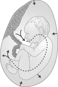
The role of the fetus in the regulation of amniotic fluid volume in normal pregnancy is poorly understood but the fetus swallows amniotic fluid, absorbs it in the gut and, in later pregnancy, excretes urine into the amniotic sac (Fig. 4.14).

It must be noted that this is a highly dynamic state, as the total volume of water in the amniotic sac is turned over every 2–3 hours. Any factor that interferes with either formation or removal of amniotic fluid may therefore result in a rapid change in amniotic fluid volume.

Congenital abnormalities that are associated with impaired ability to ingest amniotic fluid are commonly associated with excessive amniotic fluid volume, a condition known as polyhydramnios.

In summary, amniotic fluid is formed by the secretion and transudation of fluid through the amnion and fetal skin and from the passage of fetal urine into the amniotic sac. Circulation of amniotic fluid occurs by reabsorption of fluid through the fetal gut, skin and amnion.

Clinical significance of amniotic fluid volume

Oligohydramnios

The diminution of amniotic fluid volume is most commonly associated with impaired secretion of fluid and therefore is a sign of the impairment of placental function, with the exception of the effect of post-maturity. It may be associated with the preterm rupture of the membranes with chronic loss of amniotic fluid.

Oligohydramnios is commonly associated with intrauterine fetal growth restriction and is therefore an important sign of fetal jeopardy.

It is also associated with congenital abnormalities such as renal agenesis where there is no production of fetal urine.

Oligohydramnios is associated with various structural and functional problems in the fetus. It may be associated with pulmonary hypoplasia and respiratory difficulties at birth. It may also cause physical deformities such as club foot, skull deformities and wry neck. In labour it has been associated with abnormal cord compression during contractions and hence with fetal hypoxia. Amniotic fluid infusions are used in some units to try and avoid these problems but the efficacy of these techniques remains in doubt.

Polyhydramnios

The presence of excessive fluid commonly arises as a chronic condition but may on occasions be acute.

Acute polyhydramnios is a rare condition that tends to arise in the second trimester or the early part of the third trimester and commonly results in the onset of preterm labour. The condition is painful for the mother and may cause dyspnoea and vomiting. The uterus becomes acutely distended and it may be necessary to relieve the pressure by amniocentesis. However, this only gives short-term relief and nearly always requires repeated procedures. There is often an underlying congenital abnormality. A rare cause is congenital diabetes insipidus. It could also be managed medically with indomethacin to the mother at a dose of 1–3 mg/kg body weight. Indomethacin over a prolonged period may cause renal and pulmonary arterial vasoconstriction and hence should be a thin space used only for a few days.

Chronic hydramnios may arise in those pregnancies where there is a large placenta, such as occurs in multiple pregnancy, chorioangioma of the placenta or a mother with diabetes. It may also be idiopathic, with no obvious underlying cause, and the fetus may be entirely normal. However, in approximately 30% of all cases, there is a significant congenital anomaly. The distribution of such anomalies is as follows in order of frequency:

Hydramnios itself is associated with certain complications and these include:

Clinical value of tests on amniotic fluid

Both the biochemical and cytological components of amniotic fluid can be used for a variety of clinical tests. However, many of the tests previously used have been replaced by ultrasonography and procedures such as cordocentesis and chorionic villus biopsy.

Amniotic fluid contains two distinct types of cell. The first group is derived from the fetus and the second from the amnion. Cells of fetal origin are larger and more likely to be anucleate, whereas those derived from the amnion are smaller, with a prominent nucleolus contained within the vesicular nucleus, and are found in proportionately greater numbers prior to the 32nd week of gestation.

Cells that stain with eosin are most prominent in early gestation and are derived from the amnion. After 38 weeks gestation, numbers of these cells fall to less than 30% of the total cell population.

Basophilic cells increase in number as pregnancy progresses but also tend to decrease after 38 weeks. The presence of large numbers of these cells has been related to the presence of a female fetus; the fetal vagina is thought to be the possible source.

After 38 weeks, a large number of eosinophilic anucleate cells appear. These cells stain orange with Nile blue sulphate and are thought to be derived from maturing sebaceous cells.

These cells have been used in the past as a method of assessing gestational age but this has now been replaced as a method by ultrasound imaging and the assessment of fetal growth.

Amniocentesis

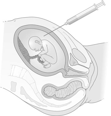
Amniotic fluid is obtained by the procedure of amniocentesis. This procedure involves inserting a fine-gauge needle under aseptic conditions through the anterior abdominal wall of the mother under local anaesthesia. The procedure, when used for diagnostic testing for chromosomal abnormalities, is commonly performed at 14–16 weeks gestation but can be performed as early as 12 weeks in some circumstances. The procedure must be performed under ultrasound control in order to identify the best and most accessible pool of amniotic fluid and, where possible, to avoid the placenta and the fetus. Up to 10 mL of fluid is withdrawn and the presence of a fetal heart beat is checked both before and after the procedure (Fig. 4.15).

Indications for amniocentesis

Chromosomal abnormalities and sex-linked diseases

The fetal karyotype can be determined by the culture of fetal cells obtained from amniotic fluid. This can reveal chromosome abnormalities such as those found in Down’s syndrome, Turner’s syndrome and various mosaics. It also allows the determination of fetal sex and hence may be useful in the management of sex-linked disorders such as thalassaemia, haemophilia and Duchenne’s muscular dystrophy.

Estimation of fetal lung maturity

The estimation of lecithin or the lecithin/sphingomyelin ratio in amniotic fluid has been used to measure functional lung maturity in the fetus after 28 weeks gestation and prior to premature delivery, and where there is a significant risk of the child developing the respiratory distress syndrome. However, it is now routine practice to give the mother corticosteroids under these circumstances. Such is the efficacy of this procedure that it has reduced the need to use the test. Other tests for fetal maturity based on amniotic fluid have now been abandoned in favour of ultrasound techniques.

image   Essential information