Pituitary function

Published on 01/03/2015 by admin

Filed under Basic Science

Last modified 22/04/2025

Print this page

rate 1 star rate 2 star rate 3 star rate 4 star rate 5 star
Your rating: none, Average: 0 (0 votes)

This article have been viewed 1313 times

42

Pituitary function

The pituitary gland

Pituitary function is regulated by the hypothalamus, to which it is connected via the pituitary stalk, which comprises portal blood capillaries and nerve fibres. The anterior pituitary is influenced by a variety of stimulatory and inhibitory hormones through these capillaries. The posterior pituitary is a collection of specialized nerve endings that derive from the hypothalamus.

Anterior pituitary hormones

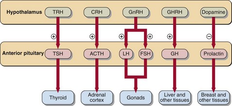
The hypothalamic factors that control anterior pituitary hormone secretion are shown in Figure 42.1.

Hyperprolactinaemia

Hyperprolactinaemia is common and can cause infertility in both sexes. An early indication in women is amenorrhoea and galactorrhoea, whereas in men there may be no early signs and the first indication of the presence of a prolactinoma may be when a large growing tumour begins to interfere with the optic nerves. Causes of hyperprolactinaemia include:

If these causes are excluded, the differential diagnosis is between:

Differentiating between these, after exclusion of stress, drugs and other disease, is by detailed pituitary imaging together with dynamic tests of prolactin secretion. A rise in serum prolactin following administration of TRH or metoclopramide is observed in idiopathic hyperprolactinaemia but not in the presence of a pituitary tumour.

In a small portion of cases a raised prolactin is due to the presence of macroprolactin, an immune complex.

Posterior pituitary hormones

Hypothalamic neurons synthesize arginine vasopressin (AVP) and oxytocin, which pass along axonal nerve fibres in the pituitary stalk to the posterior pituitary where they are stored in granules in the terminal bulbs of nerves in close proximity to systemic veins.

Secretion of AVP, also known as antidiuretic hormone (ADH), is stimulated by:

The role of AVP in fluid and electrolyte regulation is discussed on pages 14–15. A pituitary tumour arising in the anterior gland may cause impaired secretion of this posterior pituitary hormone, with consequent diabetes insipidus.

Oxytocin is released in response to suckling of the breast and uterine contraction at the onset of labour.

Pituitary tumours

Diagnosis

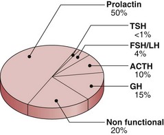
Pituitary tumours may be either functional (that is they secrete hormones) or non-functional. The incidence of different tumour types is shown in Figure 42.2. Local effects include headaches, papilloedema and visual field defects. There may be specific signs of hormone excess particularly in acromegaly, Cushing’s syndrome and prolactinoma. There may be signs of hypopituitarism in skin, hair and musculature.

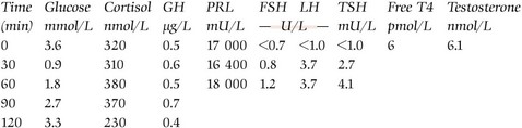
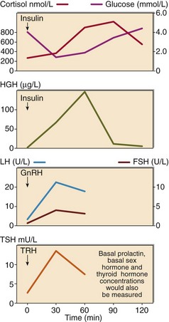
The impact of the tumour on pituitary function requires formal assessment by dynamic function tests. GH and ACTH-secreting cells are most vulnerable, and an insulin stress test (see p. 82) may suffice. However, comprehensive assessment of anterior pituitary reserve requires a combined anterior pituitary function test (Fig 42.3). TRH, GnRH and insulin are administered. All hormones are assessed at 0, 30 and 60 minutes, and GH additionally at 90 and 120 minutes. It is usual also to assess basal thyroid (thyroxine) and gonadal (testosterone or oestradiol) function.

The administration of insulin is contraindicated if there is established coronary disease or epilepsy. A clinician must be on hand throughout the test, and intravenous dextrose and hydrocortisone must be readily available in the event of severe or prolonged hypoglycaemia resulting in neuroglycopenia, e.g. loss of consciousness or fits.

Because of its associated dangers, insulin-induced hypoglycaemia is being replaced by administration of GHRH and CRH to investigate GH and cortisol secretion.

Hypopituitarism

There are many causes of hypopituitarism, a relatively uncommon condition in which there is failure of one or more pituitary functions. These causes include tumour, infarction, trauma, congenital malformation, infection and hypothalamic disorder.

The clinical presentation of hypopituitarism depends on the age of the patient. In infancy, short stature or impaired development may point to the condition. In the reproductive years, women may present with amenorrhoea or infertility. Men may present with decreased libido or a lack of male secondary sex characteristics. Elderly patients may complain of symptoms relating to ACTH or TSH deficiency such as hypoglycaemia or hypothermia.